Collaborative Advanced Microscopy Laboratories of Dentistry
| An Imaging facility under UofT which provides researchers with Microscopy & Image analysis 🔬🔎
Discover how Image Scanning Microscopy (ISM) can enhance your research by providing superior image quality without compromising on acquisition time or illumination intensity. Check out the post https://t.co/LKJ9BW1r9H
#newblogpost
Calling all AFM enthusiasts! 📢 Join the BioAFM virtual user meeting on April 18th at 1pm (EDT) with Bruker experts and elevate your AFM knowledge.
#AFMWebinar#LearnFromExperts#NetworkingEvent
https://t.co/a3OThSihSE
Don't miss out the Bruker webinar focusing on AFM probe selection! Learn from experts in the field! For details, check out our post https://t.co/hUmvOzubL7
#newblogpost
We had an engaging and enlightening 'Lunch & Learn' presentation by Dr. Anthony Choo on emerging technologies, including Stimulated Raman Scattering #Microscopy, Correlative Cryo-confocal, and Aivia AI Imaging Software. Thank you #Leica Microsystems for sponsoring the seminar!!
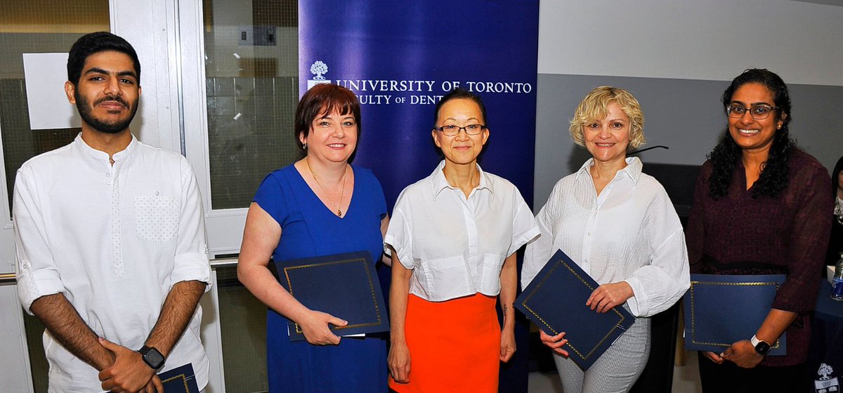
Every year #UofTDentistry recognizes staff members who have demonstrated excellence in their work through their professionalism, teamwork and contribution to morale.
🌟Meet the 2023 recipients of the Mary Choi Staff Excellence Awards: https://t.co/XxwX7voTPo
The Nikon Spatial Array Confocal (NSPARC) detector brings the power of #SuperResolution#ImageScanningMicroscopy#ISM to our AX / AX R #Confocal system, providing a photon budget-friendly option for deep superresolution imaging in 3D samples. Learn more: https://t.co/duvnxvbBWZ
✅ Autonomous Microscopy powered by Aivia on Stellaris increases the reproducibility of your experiments.
✅ Sequences of the rare event detection workflow can be restored at any time & run again with exactly the same settings.
✅ Be ready for new cutting-edge experiments!
Unleash the Power of EM Imaging for Fuel Cell Research. Join us tomorrow for a webinar to Reveal the Invisible. It’s not too late to register to join us live or on demand!
👉 https://t.co/RBMsUQWXQi
#EM#fuelcellresearch#samplepreparation#electronmicroscopy
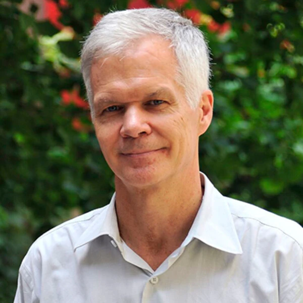
Drs. Carneiro and McCulloch at @UofTDentistry and team describe the use of a DNA hydrogel that promotes bone repair in defects that would otherwise not heal. This approach could overcome some of the inherent disadvantages of the currently used #BoneGrafts.
https://t.co/M3eK4JrTKG
Fantastic news, Dr. Carneiro! Congratulations to all the collaborators. Yet another milestone for CAMiLoD! We are very happy to support your #cellmechanics#research and #imaging.
Excited to share our latest article on DNA hydrogels for bone regeneration!! @DimitraAthan Wonderful collaboration with @OkamotoRoberta!!
https://t.co/f7vBgoePQZ
The crosslinking of extracellular matrix in cirrhotic liver tissue is mediated by advanced glycation end-products and can be inhibited by rosmarinic acid.
https://t.co/SI3mF1ODGm
Check out this combination of #SuperResolution radial fluctuations (SRRF) and expansion #Microscopy#ExM for #Pathology, providing 25 nm lateral resolution using a standard widefield microscope and applicable to archival paraffin-embedded tissue: https://t.co/XxeK9VsAjx
➡ Leica YouTube tutorial of the week: How to optimize your acquisition using Mica’s Intelligent imaging.
👉 https://t.co/ing8ZFwdvn
➡ Meet Mica 💯🙌
👉 https://t.co/6Qb0f4oeYn
#Mica#Microhub#Tutorial
📣
New funding of 20 training initiatives for students and postdoctoral fellows via #NSERC Collaborative Research and Training Experience (CREATE) program.
Details ▶️ https://t.co/iD8tasFIXj
#NSERCSupport#CdnSci#NSERC_CREATE
You can still sign up for our free online workshop on microscopy image deconvolution, visualization, and analysis on April 25th and 26th. Registration is open until Friday 21st 7pm CEST: https://t.co/ZEOVq4z8kg. Join to get the best out of your images! #microscopy#imaging