How to semi-quantitatively grade Mitral Regurgitation (MR) on echocardiography?
A quick guide using practical Doppler-based parameters:
1️⃣ Color Flow Jet Area (Apical 4-Chamber View):
➡️ Jet area as % of LA area:
🔵 Mild: <20%
🔵 Moderate: 20–39%
🔵 Severe: ≥40% or very eccentric jet (wall-hugging = increase grade by 1)
2️⃣ Vena Contracta Width (PLAX view):
➡️ Measures the narrowest neck of the MR jet
🔴 Mild: <0.3 cm
🔴 Moderate: 0.3–0.69 cm
🔴 Severe: ≥0.7 cm
3️⃣ Continuous-Wave (CW) Doppler of MR Jet:
- Dense, triangular jet = Severe MR
- Sparse or faint jet = likely Mild MR
4️⃣ Pulmonary Vein Flow (PW Doppler in Right Upper PV):
- Systolic flow reversal (S wave below baseline) = Severe MR
- Normal S>D pattern = Mild MR
- Blunted S wave = Moderate MR
Tip: Always combine multiple views and parameters. No single value defines severity.
#CardioTwitter #EchoFirst #Echocardiography #MitralRegurgitation
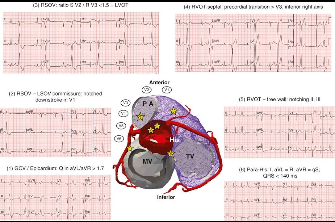
PVC localization on ECG.
Premature ventricular complexes (PVCs) are early depolarizations originating from the ventricles, bypassing the His-Purkinje system.
This powerful image illustrates PVCs from different anatomical origins➡️outflow tracts (RVOT/LVOT) and the tricuspid annulus, mapped with characteristic ECG signatures:
ECG Features & Localizing Clues:
1️⃣ GCV/Epicardium: Look for a Q in aVL/aVR > 1.7 ➡️suggests epicardial origin from the great cardiac vein.
2️⃣ RSOV - LSOV Commissure: Notched downstroke in V1 ➡️ points to aortic valve origin.
3️⃣ LVOT (RSOV): S in V2 / R in V3 < 1.5 ➡️supports a left ventricular outflow tract site.
4️⃣ RVOT Septal: Transition > V3 with inferior right axis ➡️ typical of septal RVOT origin.
5️⃣ RVOT Free Wall: Notching in II, III ➡️ consistent with free wall RVOT PVCs.
6️⃣ Para-Hisian region: R in I, aVL = R; aVR shows qS with QRS < 140 ms ➡️near the His bundle.
Why This Matters:🤔
- RVOT PVCs are the most common idiopathic form and often benign.
- LVOT or epicardial PVCs may require a retroaortic or epicardial ablation approach.
- Para-Hisian PVCs pose an ablation challenge due to proximity to the conduction system.
- Accurate localization via ECG helps plan safer and more effective ablation strategies.
Source: Braunwald’s Heart Disease: A Textbook of Cardiovascular Medicine, 12th ed.
#CardioTwitter #EPeeps #PVCs #Electrophysiology #ECG #MedEd #Cardiology
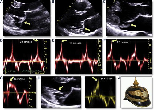
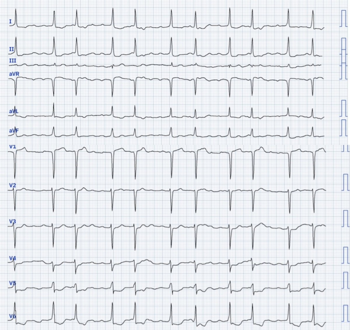
1/⚡️ Ever heard of the Pickelhaube sign? It’s not just history — it’s cardiology! A quick 🧵 on how this spiked helmet shows up in ECGs and echocardiograms.
New Clinical Consensus on Left Atrium & Left Atrial Appendage Imaging 🚨
The #EACVI with EHRA has released updated guidance on #MMI for the assessment of the Left Atrium (LA) & Left Atrial Appendage (LAA).
This evidence-based document is essential for improving diagnosis, management, and prognosis of conditions like #AF #HF & cardioembolism. A must-read for all cardiologists and #CVImaging specialists!
Read the full consensus https://t.co/ylNDBlEyOZ
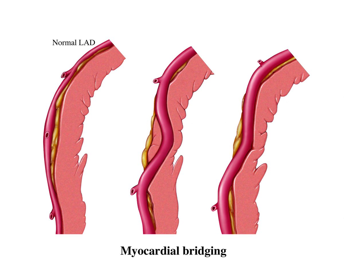
Myocardial Bridging: A Hidden Cause of Angina 🫀 Myocardial bridging occurs when a segment of a coronary artery, most commonly the left anterior descending (LAD), tunnels through the heart muscle instead of running on the surface.
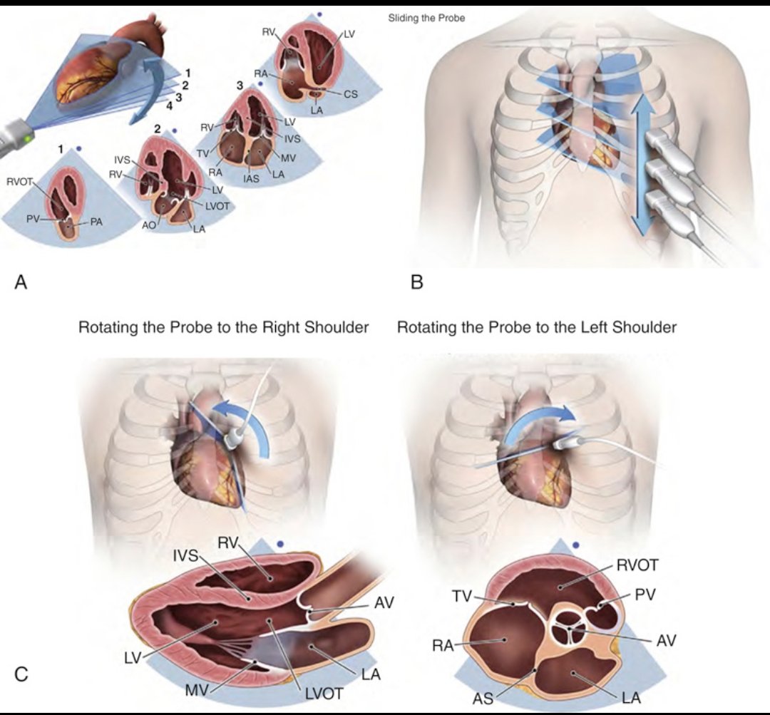
Understanding Transducer Movements in Transthoracic Echo (TTE)
Accurate echocardiographic imaging depends heavily on how the transducer is manipulated on the chest wall. This image illustrates 3 essential movements:
(A) Tilting in place changes the angle of the ultrasound beam, capturing different cardiac structures through the same acoustic window.
(B) Sliding the probe superiorly or inferiorly helps align the beam with different heart levels—useful for transitioning between base, mid, and apical views.
(C) Rotating the probe clockwise or counterclockwise changes the imaging plane—e.g., from long axis to short axis.
The blue dot in each image shows the orientation of the probe’s index marker, crucial for maintaining anatomical consistency.
Mastering these movements is foundational to performing a comprehensive TTE.
Reference: Mitchell C, et al. Guidelines for Comprehensive TTE in Adults. JASE 2019;32:1–64.
#Cardiology #Echo #TTE #Ultrasound #CardiacImaging
A 48 year old male presented with effort angina, TMT +ve, CT CAG was done, invasive CAG not done yet. I am desperately trying to demonstrate the likely cause for angina in TEE which is already beautifully demonstrated in CTCAG.
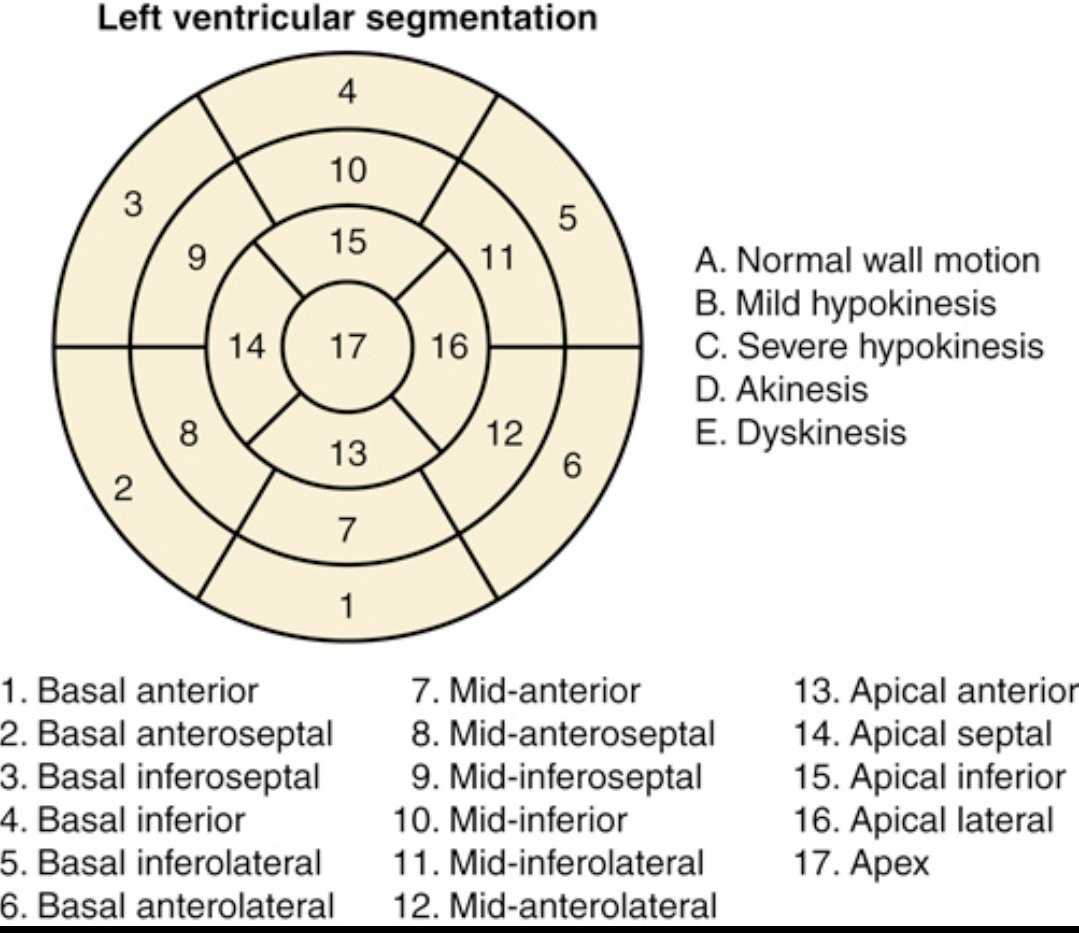
Understanding regional wall motion abnormalities (RWMA) starts with this: the 17-segment LV model.
It divides the left ventricle into basal, mid, apical segments for precise localization of dysfunction.
Why does it matter?
RWMA refers to areas of the LV wall that contract abnormally—key in diagnosing ischemia, infarction, cardiomyopathies, and evaluating viability.
Wall motion grades: A. Normal
B. Mild hypokinesis (↓ motion)
C. Severe hypokinesis
D. Akinesis (no motion)
E. Dyskinesis (paradoxical motion)
Clinical significance:
- Helps identify territory of ischemia/infarct (e.g., LAD vs RCA vs LCx).
- Essential for assessing outcomes post-MI or revascularization.
- Guides decisions in cardiac surgery, ICD implantation, and heart failure management.
@TrackYourHeart
#Cardiology #Echocardiography #MedTwitter #LVFunction #RWMA
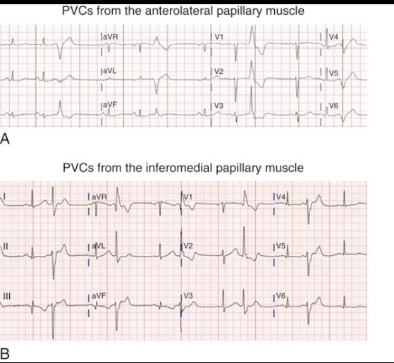
Recognizing PVCs from the Papillary Muscles on ECG – Key to Targeted Therapy
Premature Ventricular Complexes (PVCs) can arise from the anterolateral or inferomedial papillary muscles of the left ventricle—both mid-cavity structures. The ECG features help localize the origin and guide ablation therapy.
ECG Characteristics:
A) Anterolateral Papillary Muscle PVCs:
- Right bundle branch block (RBBB)-like morphology
- rS configuration in V3–V4
- Inferior axis: Positive QRS in II, III, aVF
B) Inferomedial Papillary Muscle PVCs:
- Also show RBBB-like pattern
- rS in V3–V4, but with
- Superior axis: Negative QRS in II, III, aVF
How to Determine Axis:
- Inferior Axis: Positive QRS in leads II, III, aVF (activation moves downward)
- Superior Axis: Negative QRS in II, III, aVF and positive in aVR, aVL (activation moves upward)
Why It Matters Clinically:
Papillary muscle PVCs are often idiopathic, but can trigger ventricular tachycardia (VT) or contribute to PVC-induced cardiomyopathy in high burden.
They are known for being challenging to ablate due to deep intramyocardial or mobile origins—often requiring intracardiac echocardiography (ICE) and 3D mapping.
Accurate ECG localization allows for strategic procedural planning, minimizing risks and improving outcomes.
Reference:
Braunwald’s Heart Disease, 14th Edition,
#CardioTwitter #EPeeps #ECG #PVC #VT #Electrophysiology #Cardiology #Braunwalds #