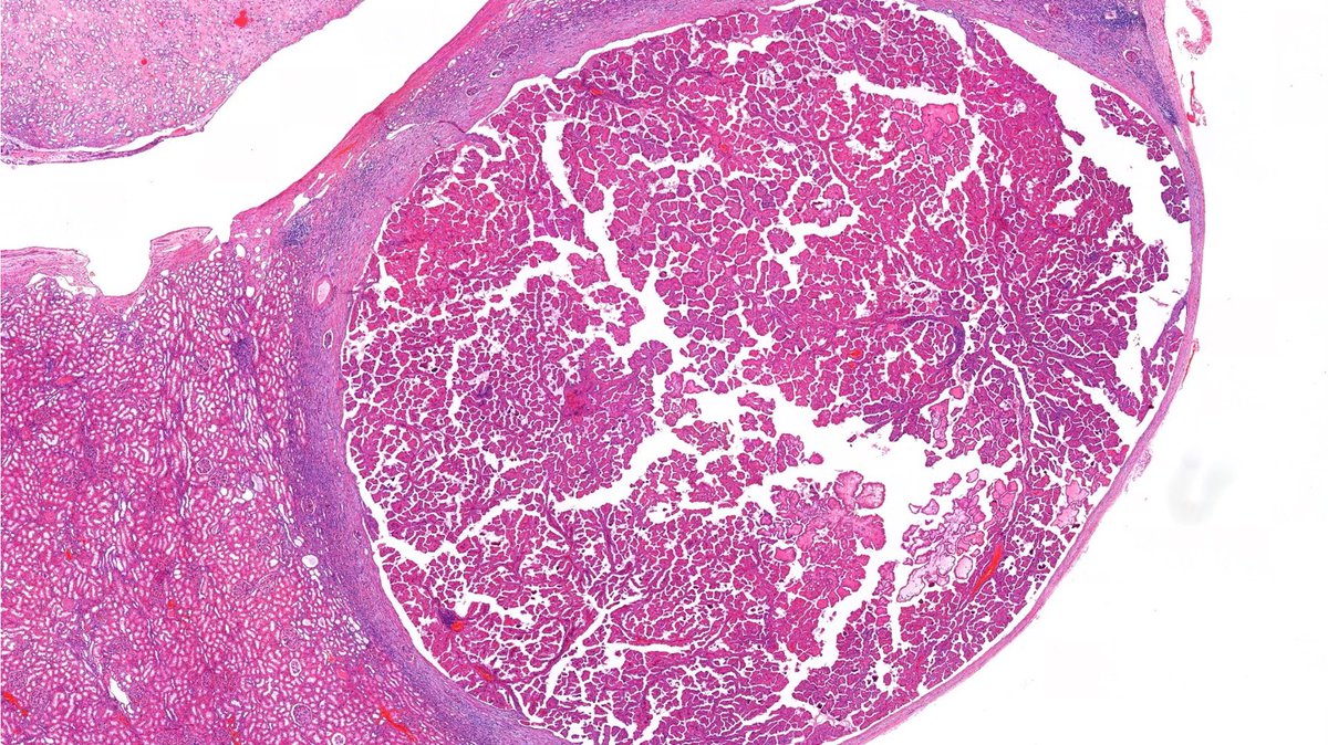
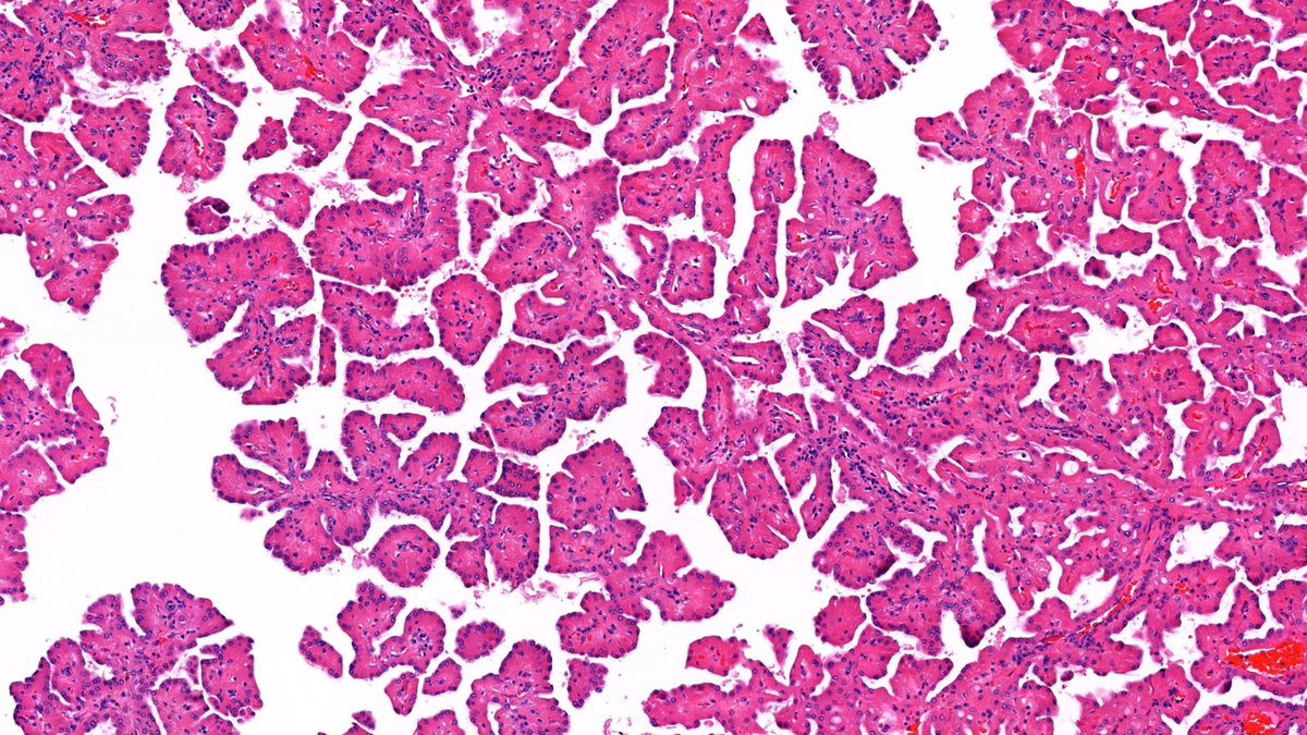
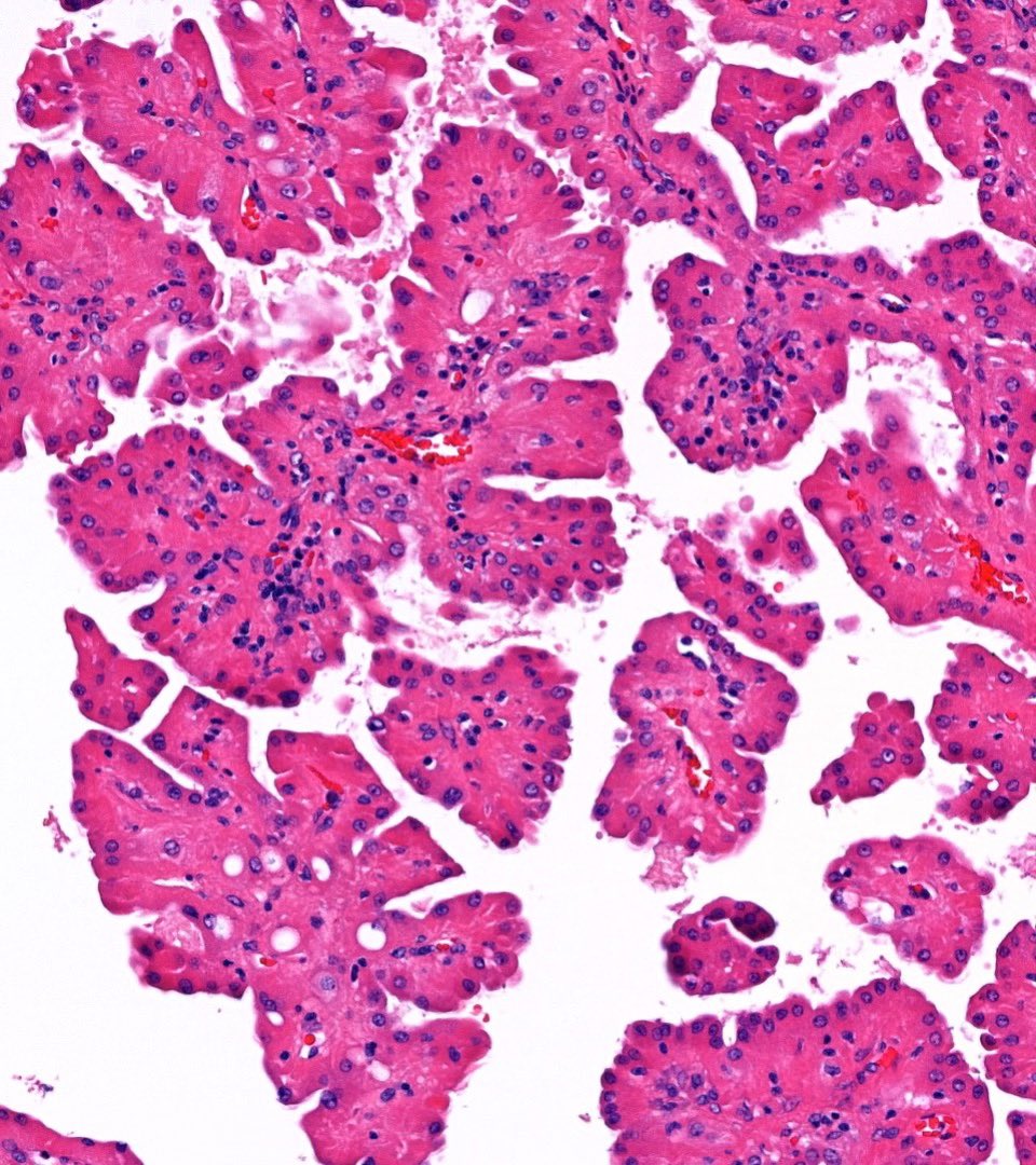
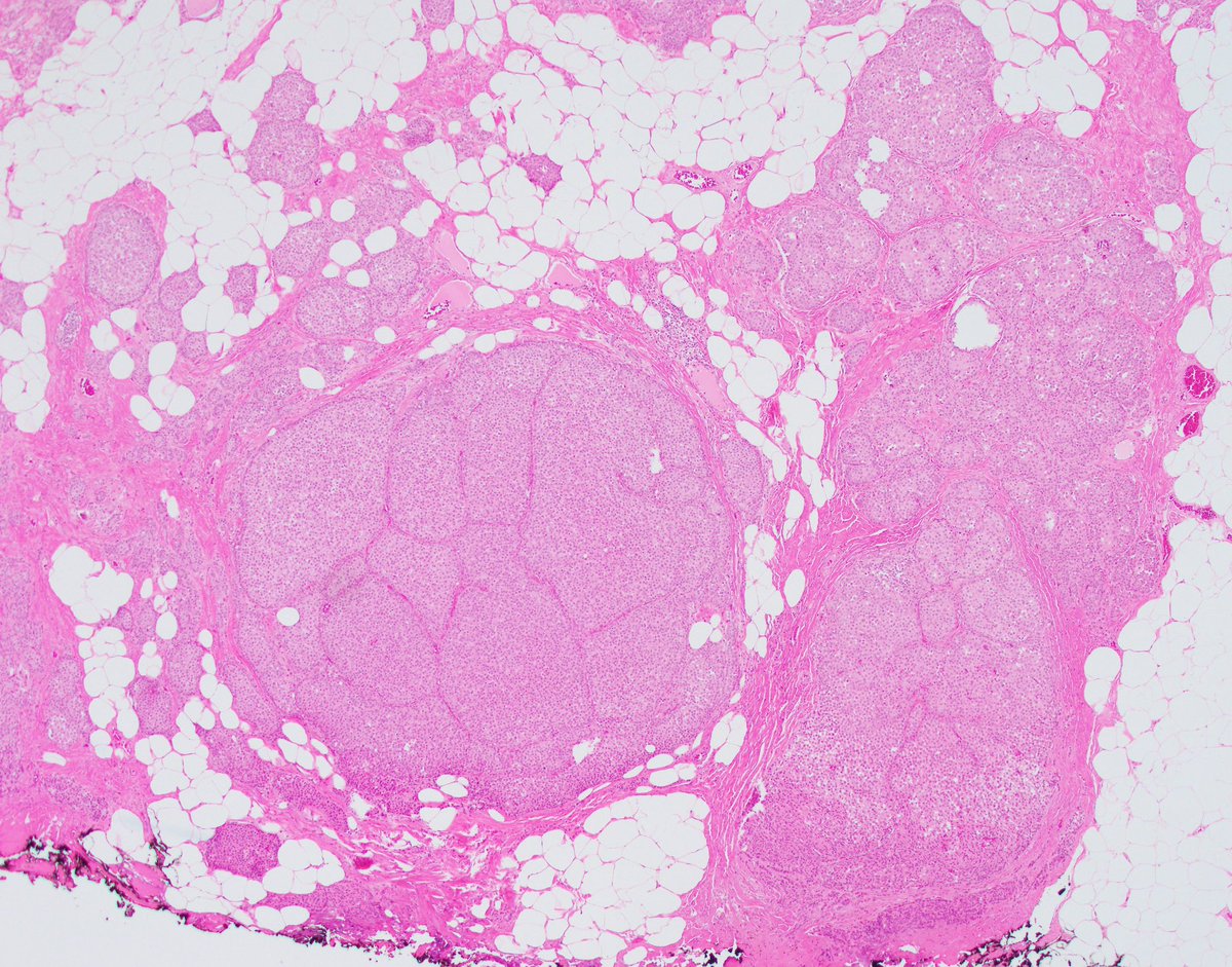
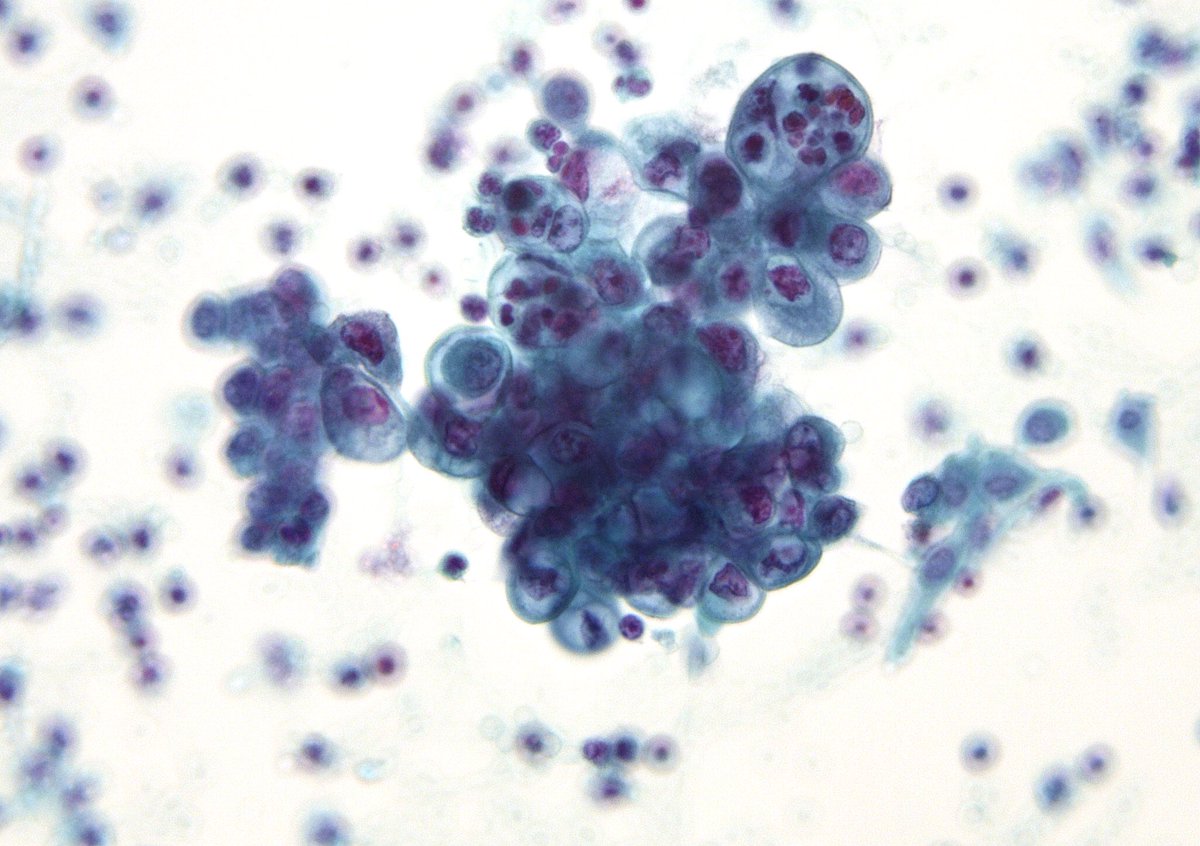
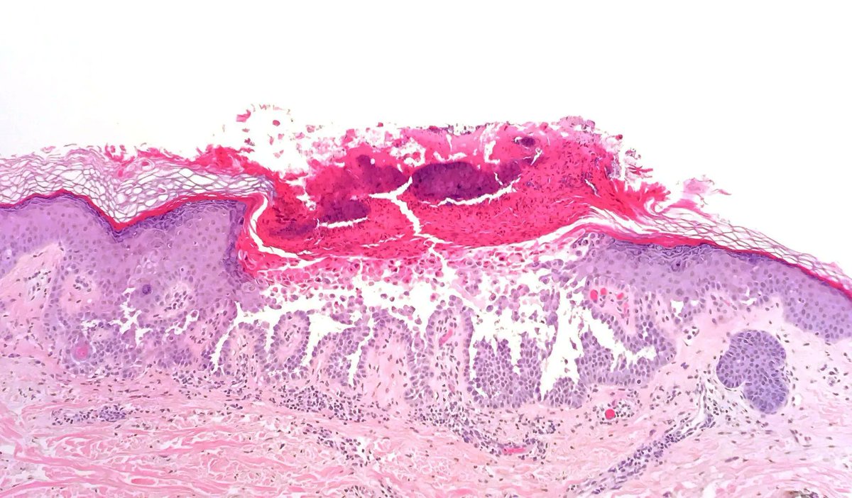
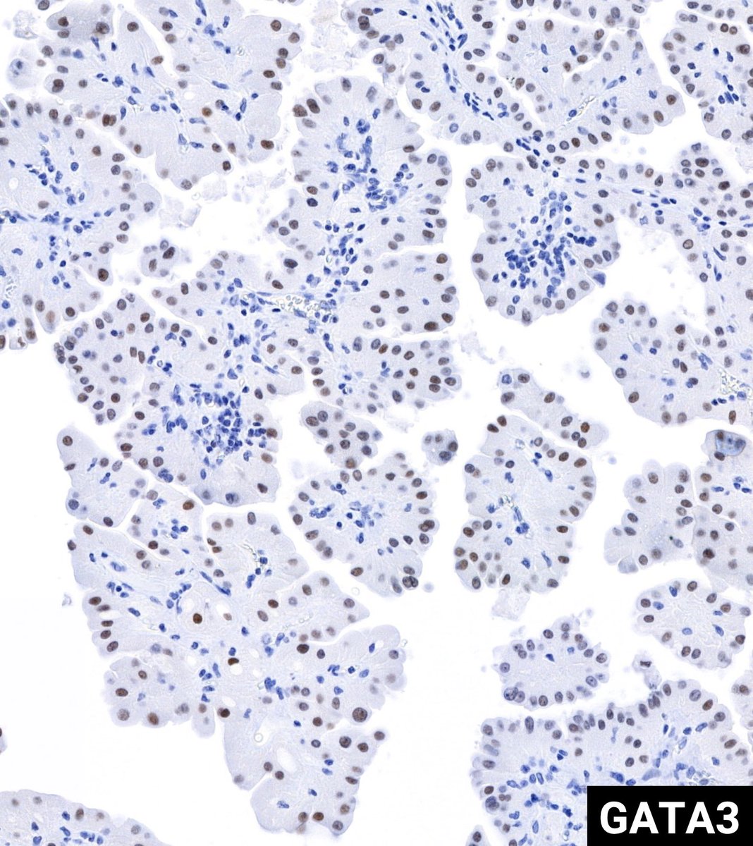
Ok friends, let's vote! Here is a transanal excision of a 20 mm polypoid mass. I will let you take polls and subsequently feed you with additional information. Don't cheat, do them sequentially - you might be surprised. Follow the thread!
My approach to those bits of lung pathologists receive after surgical treatment of pneumothorax . . .
We commonly get wedges of lung generated during surgical treatment of pneumothorax.
Overwhelming I sign these out as 'PULMONARY APICAL CYSTIC DISEASE' by which I mean the sort of blebs and bullae that tall/lean young people sometimes get and cause spontaneous pneumothorax (and sometimes older smokers get too). It's just a neat four word summary for this phenomenon that can go on the bottom line and means there are no other important underlying pathologies.
However there are a few underlying pathologies that are very important not to miss.
These include:
1. Langerhans Cell Histiocytosis.
2. Pneumocystis pneumonia (pretty rare now to present as pneumothorax but in the olden days before widespread uptake of HAART we used to see quite a bit)
3. What we called 'Bong Lung' in 2005 in Am J Surg Path:https://t.co/GaJynAIsAB
(clue to the diagnosis of Bong Lung - they often present late in the evening to the emergency department)
4. Birt-Hogg-Dube syndrome (good luck diagnosing this on pathology alone, but on imaging look from the proximal/non-apical cysts and other disease associations (renal tumours and facial skin lesins)
5. Personally I've never seen a vasculitis or connective tissue disease presenting with pneumothorax (or at least if I've seen it then I have missed it) - but people say it happens
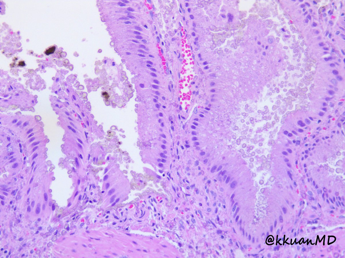
6. And of course - this entity illustrated below. What do you think?
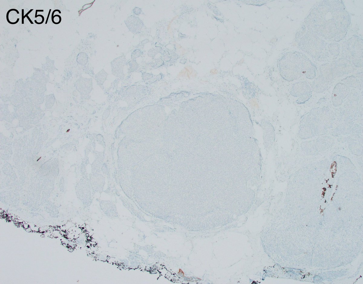
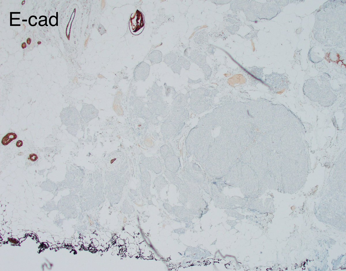
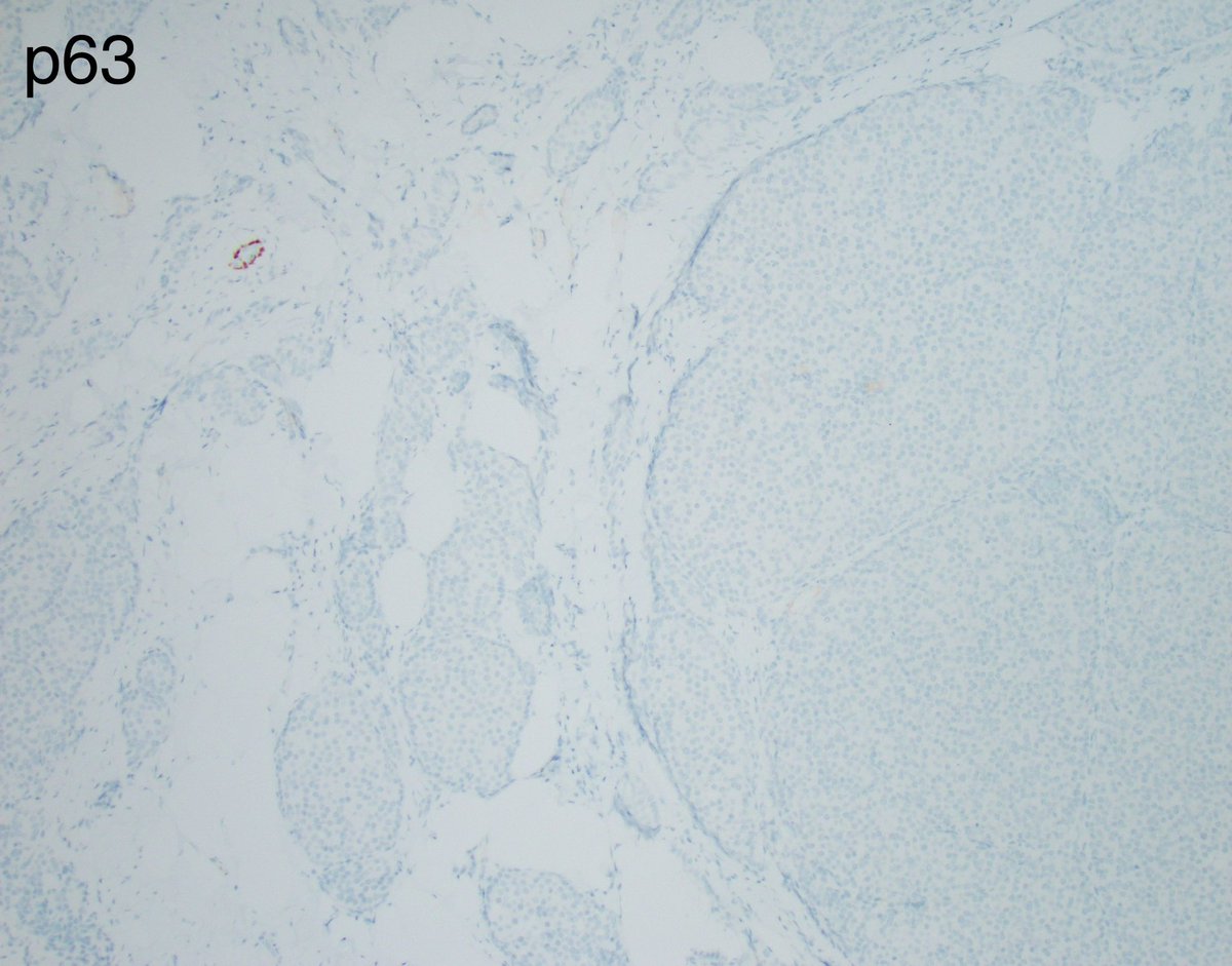
The case I posted yesterday is a resection on a biopsy that was called LCIS.
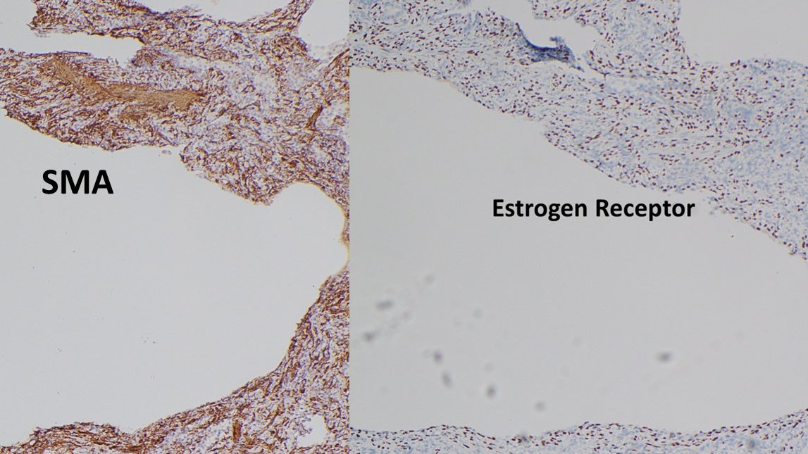
The key in this case of lobular neoplasia is to remember that E-cadherin, though often weaker than in epithelial cells, is usually retained in myoepithelial cells and its complete absence should make you pause and order some myoepithelial markers.
Here, myoepithelial cells are completely absent in all nests of lobular neoplasia, and positive internal controls are surrounded by tumor in an infiltrative pattern.
This serves as a reminder that invasive lobular carcinoma can adopt unusual morphologies like solid and alveolar. This LCIS-like pattern is highly unusual in my experience. I shall call it the large nested variant of invasive lobular carcinoma 🙂
Happy weekend everyone!
@wusm_pathology @washupathedu #PathTwitter #PathX #breastpath
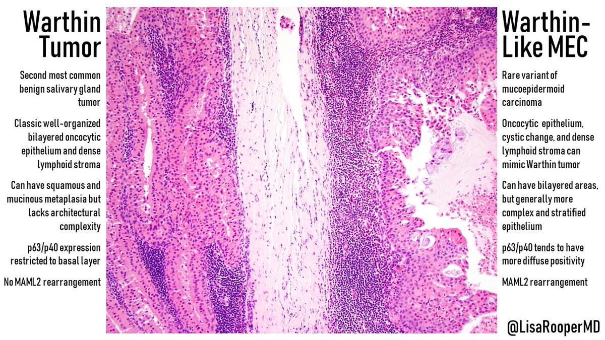
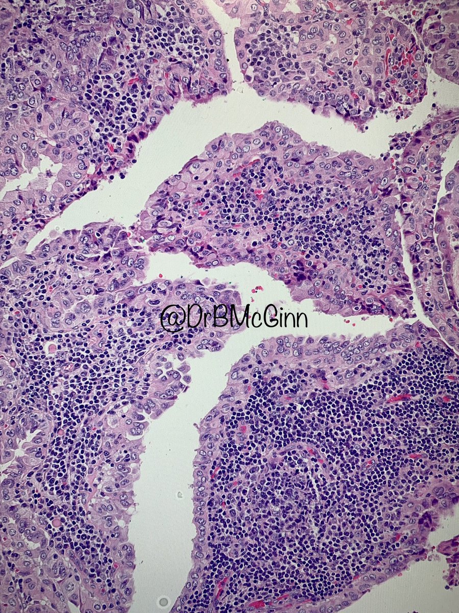
This Warthin-like mucoepidermoid carcinoma very conveniently arose next to a benign Warthin tumor, letting us compare their features side by side! Architecture is key for this differential, and MAML2 confirms the diagnosis. #ENTPath#OralPath#SurgPath
My “CASE of the week” for inhouse cyto signout, illustrating the remarkable phenomenon of how the “bag of polys” are recruited by the “endometrioid endometrial adenocarcinoma”, even at metastatic sites. This case is an abdominal fluid involved by the pt’s known EM CA.
Endoscopic fine needle aspiration of a mass in the head of the pancreas. What is the diagnosis? Answer: https://t.co/mrsQk63IUT
#PathArt#PathTwitter#Cytopath#Pathology
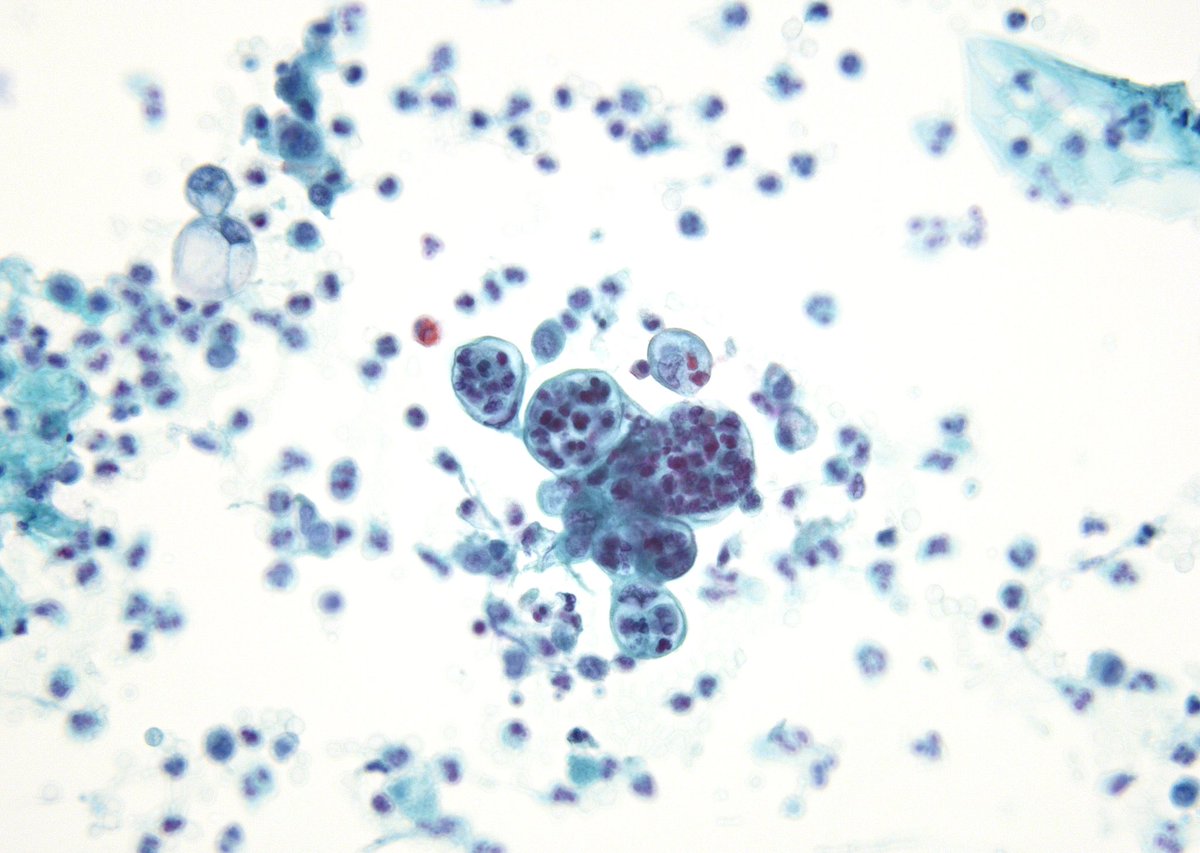
Metastatic prostate carcinoma in a pleural fluid. This particular plane highlights a larger three dimensional spherule structure as well as some smaller gland-like formations on its surface. #cytology#cytopath
Cyto-histological correlations: Invasive mucinous adenocarcinoma of the lung. (The term lepidic mucinous carcinoma is obsolete).
Cytology:
🔬Mild atypia
🔬Drunken-honeycomb pattern
🔬No cilia
🔬Abundant intracytoplasmic mucin
🔬A mucinous background may be present
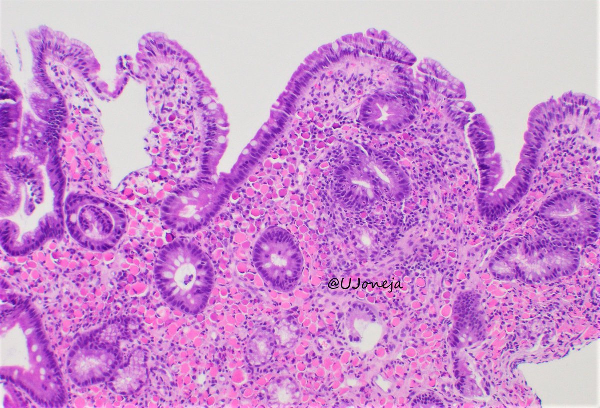
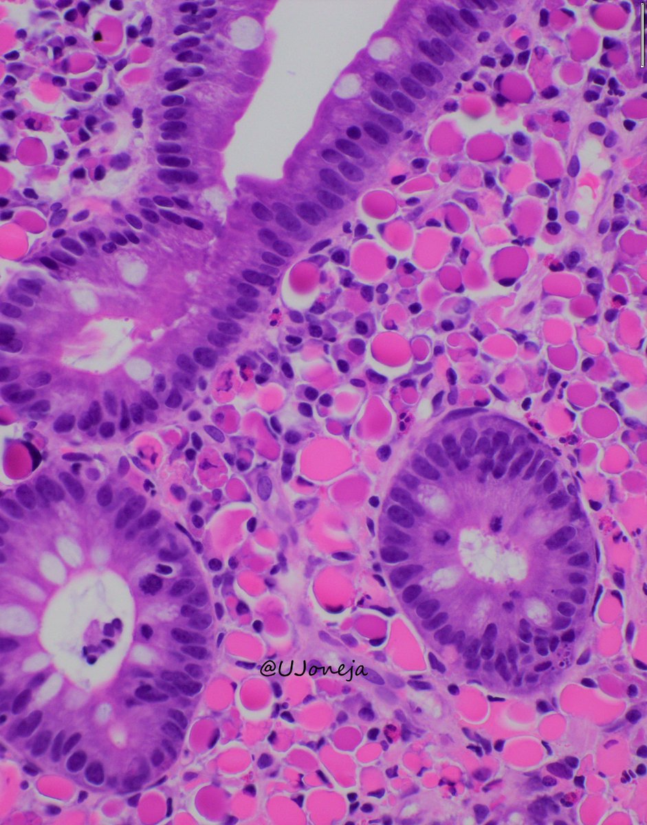
RUSSELL BODY DUODENITIS
- Less commonly seen than russell body gastritis
- Disturbed secretion of immunoglobulins that accumulate within plasma cells
- Can be seen in association with H. pylori infection and/or hematopoeitic malignancy
#pathology#PathTwitter
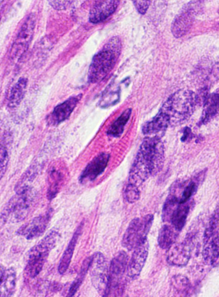
What are the pink cells? What tumors can you find them in?
More pics & Answer ✅ https://t.co/Wi6GKxe2Zq
Another example: https://t.co/hhWikyVE6T
Videos ⏯️ https://t.co/UNeMkqiUX5 & https://t.co/5zbepxcRsu
#BSTpath#pathologists#pathology#pathTwitter