We have a cure definition for myeloma.
Requires:
1) Off all therapy and sustained MRD negative for 5 years
2) Negative imaging studies
Presented by @NikhilMunshiMD#IMS26@Myeloma_Society
Note:
1) We need to see how this definition performs. Relapse rate for patients meeting these cure criteria must ideally be less than 5%. If not, we may have to adjust the criteria.
2) I hope the cure criteria will be the impetus to stop doing trials with endless therapy. You cannot claim cure with continuous therapy. I’m glad the field is mostly aligned on this.
Cure for patients and public in any disease has a deep meaning. Cure means cure. We cannot dilute it.
The world is unfair and inequitable. We know this impacts patient outcomes.
But what can we do about it?
Read our paper Strategies to overcome inequities in patients with hematologic malignancies for free here: https://t.co/MrE8HuuIs1 @cadelacruzd
🚨🩸 Acute GVHD: ASTCT 2026 recommendations
From diagnosis to steroid failure—practical pearls for transplant practice 🧵👇
1️⃣ 🔍 Diagnosis is clinical
Biopsy supports acute GVHD but is not mandatory for diagnosis.
Always assess competing causes: infection, drug toxicity, conditioning injury and other transplant complications.
⚠️ GVHD and infection can coexist. A nondiagnostic biopsy does not exclude GVHD.
2️⃣ 🔬 When to biopsy?
🔹 Lower GI symptoms: endoscopy with biopsies, especially with significant diarrhea, abdominal pain, ileus or bleeding.
🔹 Persistent unexplained upper GI symptoms: consider upper endoscopy.
🔹 Isolated hepatic involvement: biopsy is recommended because isolated liver GVHD is uncommon.
⏳ Do not delay clinically indicated treatment while awaiting histology.
3️⃣ 📊 Grade consistently
ASTCT endorses MAGIC as the consensus staging/grading system.
Document skin, liver, upper GI and lower GI involvement separately, then assign the overall grade.
📌 Acute manifestations beyond D+100 can represent late acute GVHD; timing alone does not establish chronic GVHD.
4️⃣ 🧬 Biomarkers: prognostic value
MAGIC algorithm probability—MAP—uses ST2 + REG3α to estimate risk, particularly nonrelapse mortality.
Validated risk tools include:
🔹 Minnesota risk score
🔹 Manhattan risk system
🔹 MAGIC Composite Score
⚠️ These tools do not diagnose GVHD and are not yet validated to direct routine treatment selection.
5️⃣ 💊 First-line systemic therapy
Corticosteroids remain standard.
🔴 Grade III–IV: methylprednisolone 2 mg/kg/day, or equivalent.
🟡 Selected limited skin/upper GI presentations requiring systemic treatment: 0.5–1 mg/kg/day may be appropriate.
⚠️ Do not extrapolate lower-dose strategies to severe lower GI or hepatic disease. Doses >2 mg/kg/day have not demonstrated added benefit.
6️⃣ 🧴 Local therapy matters
Topical corticosteroids are useful for limited skin involvement.
Oral beclomethasone or budesonide may complement systemic therapy; selected isolated upper GI cases may receive them alone, although supporting evidence is limited.
⚠️ Systemic absorption can increase with triazole antifungals.
7️⃣ 🚫 Routine upfront combinations?
ASTCT does not endorse routinely:
🔹 Replacing systemic corticosteroids with another systemic agent.
🔹 Adding another systemic agent to initial corticosteroids.
Promising trial findings have not established a broadly applicable new first-line standard.
8️⃣ ⏱️ Recognize steroid failure early
Consensus criteria discussed include:
🔴 Progression after 3 days of methylprednisolone 2 mg/kg/day.
🔴 No improvement after 7 days at that dose.
🔴 New organ involvement despite 1 mg/kg/day for skin/upper GI disease.
🔴 Recurrence during or after taper.
📌 Taper-associated recurrence can overlap with steroid dependence or flare; reassess the clinical context and alternative causes.
9️⃣ 🎯 Second-line standard: ruxolitinib
ASTCT gives ruxolitinib its strongest treatment recommendation: Grade A.
REACH2:
✅ D28 overall response: 62.3% vs 39.4% with investigator-selected therapy.
✅ Improved durable response and failure-free survival.
⚠️ Monitor cytopenias and infections closely. Severe disease remains challenging despite treatment.
🔟 👶 Pediatric alternative: remestemcel-L
An allogeneic bone marrow-derived mesenchymal stromal cell therapy.
🇺🇸 FDA-approved for steroid-refractory acute GVHD in children aged ≥2 months.
The single-arm pediatric study reported D28 overall response of 70.4%.
⚠️ No head-to-head comparison with ruxolitinib—cross-trial response rates cannot establish superiority.
1️⃣1️⃣ 🧩 After second-line failure
No preferred third-line agent has consensus endorsement.
Options discussed include extracorporeal photopheresis, mesenchymal stromal cells, α1-antitrypsin, vedolizumab and microbiota-based approaches.
Selection depends on involved organs, comorbidities, infection risk, availability and trial access. Evidence remains limited.
Diagnostic Approach to Large B-Cell Lymphoma
Large B-cell lymphoma (LBCL) is the most common type of aggressive mature B-cell lymphoma. In the 5th edition of the WHO Classification of Haematolymphoid Tumors, LBCL has been further refined into as many as 17 distinct specific subtypes. Cases that do not fit into any defined subtype are classified as diffuse large B-cell lymphoma, not otherwise specified (DLBCL, NOS). The increasing precision of LBCL classification requires pathologists to adopt more accurate and comprehensive diagnostic strategies. Meanwhile, the continuous development of novel therapeutic agents and the diversification of treatment approaches have placed higher demands on precise LBCL subclassification and the identification of clinically relevant molecular alterations.
The diagnostic workup typically includes an immunohistochemical panel consisting of CD20, PAX5, CD3, CD5, CD10, BCL2, BCL6, MUM1, CD138, Cyclin D1, CD30, p53, MYC, Ki-67, and Epstein–Barr virus (EBV)-encoded RNA (EBER) in situ hybridization. In addition, fluorescence in situ hybridization (FISH) analysis is performed to detect genetic rearrangements involving MYC, BCL2, BCL6, and IRF4, among other relevant molecular abnormalities.
💊📉Cómo Retirar los Corticoesteroides de Forma Segura?
⏱️Duración del Tratamiento
💉Reducción Rápida-Gradual-Lenta
🧠Supresión del Eje HPA
🚨Insuficiencia Suprarrenal
📖Indian Journal of Endocrinology and Metabolism
Artículo Completo👇🏼✅🆓
https://t.co/aR5SFK2xfj
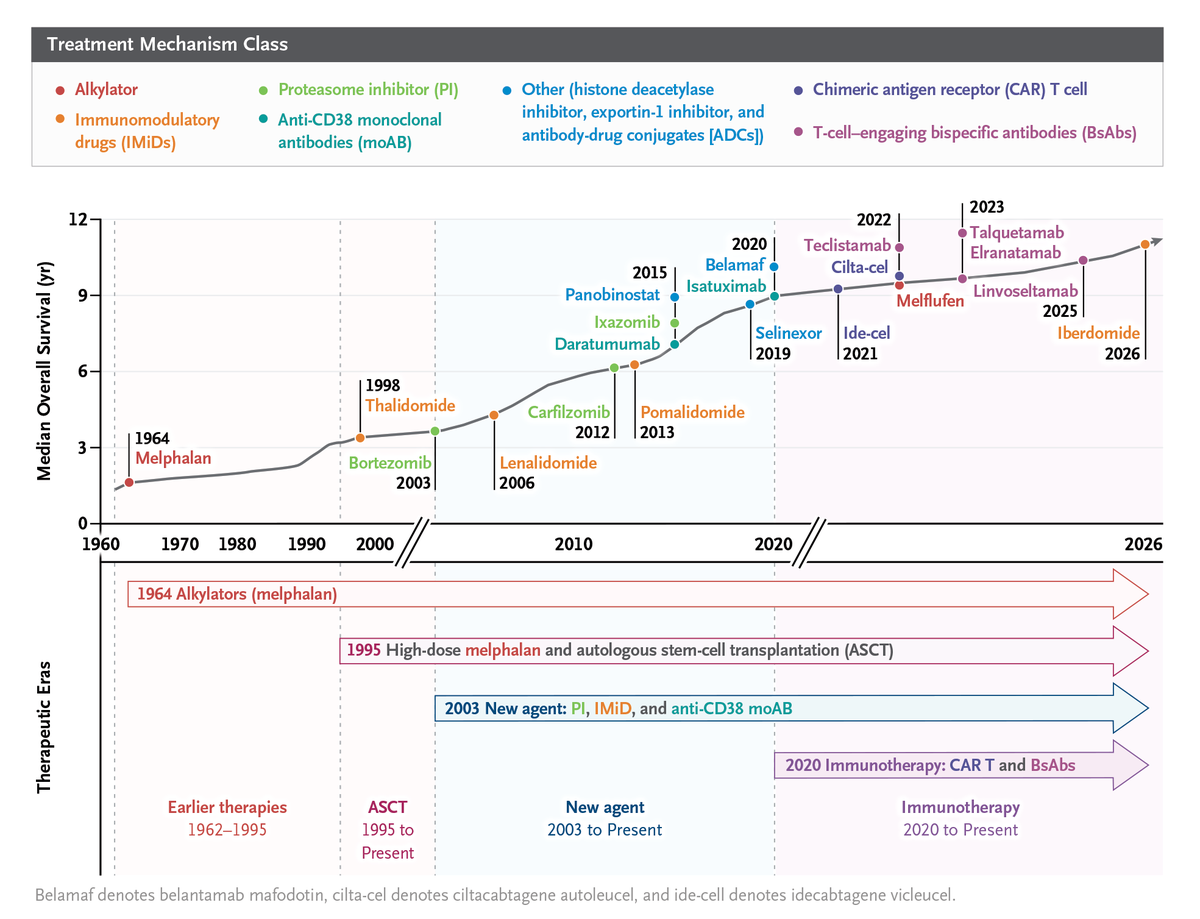
Revolutions in transplantation and therapies have transformed multiple myeloma from a disease with an associated survival of a few years into one for which functional cure is an emerging goal. This abundance of effective therapies has created complexity. Learn more: https://t.co/rtFssp5gYf
🩸 Anemia in lower-risk MDS: choosing between ESAs, luspatercept, imetelstat and lenalidomide
By Prof. Valeria Santini
📸 A proposed 2026 treatment algorithm—selection depends on del(5q), serum EPO, transfusion burden, prior therapy and other cytopenias.
1️⃣ First, define the patient’s anemia
🧬 del(5q) or non-del(5q)?
🩸 Transfusion-dependent or non-transfusion-dependent?
📊 How many RBC units over 8 weeks?
🧪 Serum erythropoietin level?
💊 Previous ESA exposure and response?
⚠️ Transfusion dependence is defined by actual transfusion requirements, not Hb ≤8 g/dL alone. The slide’s Hb values are illustrative, not universal transfusion triggers.
2️⃣ Non-transfusion-dependent disease
For symptomatic anemia, the slide favors:
🟢 Lower serum EPO—particularly <200 U/L: an erythropoiesis-stimulating agent.
🟡 EPO 200–500 U/L: individualized ESA consideration.
🔴 EPO ≥500 U/L: lower expected ESA benefit; consider clinical trials and alternative strategies.
The goal is meaningful symptom improvement and avoidance of transfusions.
3️⃣ Non-del(5q), transfusion-dependent disease
📉 Low transfusion burden + low EPO: ESAs remain an option.
🎯 Luspatercept: an established option for ESA-naïve patients who require regular transfusions. Its first-line US indication does not require ring sideroblasts.
COMMANDS directly compared luspatercept with epoetin alfa in ESA-naïve, transfusion-dependent lower-risk MDS. COMMANDS trial, FDA indication
4️⃣ Imetelstat: match the indication, not EPO alone
🧬 Telomerase inhibitor for adults with low- to intermediate-1-risk MDS requiring ≥4 RBC units/8 weeks, after ESA failure, loss of response or ESA ineligibility.
📊 IMerge:
✅ ≥8-week transfusion independence: 39.8% vs 15%
✅ ≥24-week transfusion independence: 28% vs 3.3%
⚠️ EPO ≥500 U/L alone is insufficient to establish eligibility.
🩸 Neutropenia and thrombocytopenia are major treatment-selection and monitoring considerations. FDA imetelstat approval
5️⃣ del(5q) with transfusion-dependent anemia
🎯 The slide directs this group toward lenalidomide.
Genetic context, baseline counts and treatment tolerance remain important when planning therapy.
6️⃣ After inadequate response
The slide places oral hypomethylating agents and possible transplantation later in the pathway.
📌 These require individualized reassessment of disease risk, cytopenias, fitness and treatment goals—not automatic escalation based on transfusion burden alone.
🧠 There is no head-to-head trial establishing luspatercept versus imetelstat superiority. Their separate trial response rates should not be used to rank them directly.
#MDS #Anemia #Luspatercept #Imetelstat #Lenalidomide #Hematology #ASH26 #SOHO26
🩸 Cytopenias during azacitidine + venetoclax in AML: lessons from VIALE-A
🔑 Management depends on marrow response—not the CBC alone.
1️⃣ Before remission
Cytopenias may reflect active AML. Generally, avoid interrupting venetoclax solely for low counts before remission. Bone marrow assessment guides the decision. Prescribing information
2️⃣ Blast clearance achieved, but counts remain low
📌 The slide’s VIALE-A protocol approach:
• BM blasts <5% with incomplete count recovery.
• Interrupt venetoclax from D29 and delay the next azacitidine cycle.
• Allow recovery to ANC ≥0.5 ×10⁹/L, with an initial delay of up to 14 days.
• Resume both agents together for the next cycle.
• Failure to recover by D42 required further review—not automatic restart.
3️⃣ First prolonged severe cytopenia after remission
⏸️ Delay the next cycle and monitor counts.
The current US label recommends resuming at the same venetoclax dose after grade 4 neutropenia/thrombocytopenia lasting ≥7 days resolves to grade 1–2. These recovery criteria differ from the trial thresholds shown on the slide. Prescribing information, Table 7
4️⃣ Recurrent prolonged cytopenias
📅 Shorten venetoclax exposure by 7 days in subsequent cycles—for example, 28 → 21 days—after recovery.
💊 Maintain the appropriate daily dose, including required adjustments for interacting drugs such as azole antifungals. Prescribing information
5️⃣ Persistent delayed recovery
🔬 Reassess marrow disease status and cellularity before attributing cytopenias to treatment.
📉 The slide also describes azacitidine dose adjustment for prolonged recovery, guided by marrow cellularity.
🦠 Consider antimicrobial support and G-CSF as clinically indicated. Prescribing information
🎯 Practical pearl: After blast clearance, cycle delays and shorter venetoclax duration are planned tools for managing myelosuppression.
📷 VIALE-A protocol details summarized from the shared slide.
#AML #Venetoclax #Azacitidine #Hematology #ASH26 #SOHO26
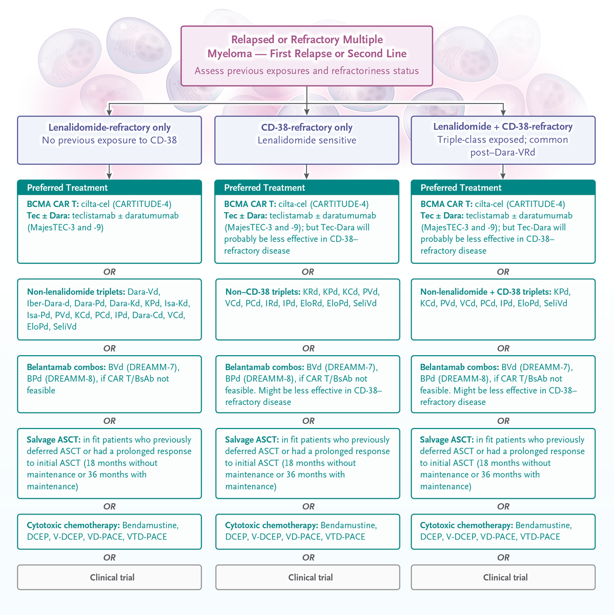
Drs. Tarek H. Mouhieddine and Kenneth C. Anderson summarize treatment for multiple myeloma, highlighting four-drug induction therapy, evolving transplantation strategies, tailored maintenance therapy, and in relapse, earlier use of BCMA-directed immunotherapy.
Read the Review Article “Treatment Decisions in Multiple Myeloma”: https://t.co/rtFssp5gYf
New clinical guidelines: American Society of Hematology 2026 guidelines for the diagnosis and management of severe and very severe acquired aplastic anemia. Read in Blood Advances ➡️ https://t.co/jsSN62TTMl
✅ GUÍA INTERNACIONAL DE PRÁCTICA CLÍNICA PARA EL MANEJO DE LA TVP. 2026.
PDF libre acceso‼️👇
📖 International Clinical Practice Guidelines for Acute Deep Vein Thrombosis: A Comparative Review of Recommendations, Evidence Gaps, and Emerging Trends https://t.co/eVORBFv8Ti
🧬 How to present a newly diagnosed lymphoma patient at the MDT meeting
Use this sequence:
Patient → Presentation → Urgency → Pathology → Stage → Risk → Fitness → Plan → MDT question
1️⃣ Patient identity 👤
Age, sex, functional status, relevant occupation/social context, treating team and date of diagnosis.
2️⃣ Clinical presentation 🩺
Presenting symptoms and duration:
🔹 Lymphadenopathy or mass
🔹 Fever, drenching night sweats and weight loss
🔹 Pruritus, fatigue or pain
🔹 Compressive symptoms
🔹 Extranodal, neurological, gastrointestinal, skin or testicular manifestations
Mention examination findings: nodal regions, largest mass, hepatosplenomegaly, performance status and extranodal disease.
3️⃣ Immediate clinical status 🚨
State whether the patient is stable and identify lymphoma emergencies:
⚠️ Airway or superior vena cava obstruction
⚠️ Spinal-cord compression
⚠️ Tumor lysis syndrome
⚠️ Bowel obstruction or perforation
⚠️ CNS involvement
⚠️ Hemophagocytic lymphohistiocytosis
⚠️ Cytopenia, bleeding or severe infection
4️⃣ Diagnostic tissue 🔬
Specify:
📍 Biopsy site and date
📍 Excisional/core biopsy and adequacy
📍 Histological architecture and morphology
📍 Immunohistochemistry
📍 Flow cytometry, when applicable
📍 EBER in-situ hybridization
📍 Cytogenetic/molecular studies
Avoid presenting lymphoma as “CD20-positive disease.” State the complete pathological entity according to WHO-HAEM5 or ICC.
5️⃣ Final lymphoma diagnosis 🧬
Clarify:
➡️ Hodgkin or non-Hodgkin lymphoma
➡️ B-cell, T-cell or NK-cell lineage
➡️ Indolent or aggressive behavior
➡️ Exact histological subtype
➡️ Relevant molecular subgroup
For aggressive large B-cell lymphoma, report cell-of-origin classification, MYC and BCL2 protein expression, and MYC/BCL2/BCL6 rearrangement testing when indicated.
Mention whether expert hematopathology review is complete or pending.
6️⃣ Baseline laboratory assessment 🧪
CBC and differential, blood film, renal and liver profiles, calcium, phosphate, uric acid, LDH, albumin, coagulation profile and pregnancy testing when relevant.
Include HIV, hepatitis B and hepatitis C screening before immunochemotherapy. Document quantitative immunoglobulins and other infection tests when clinically indicated.
7️⃣ Imaging and disease distribution 🖥️
Present the baseline PET/CT or contrast-enhanced CT systematically:
📌 Nodal regions involved
📌 Largest lesion and bulky disease
📌 Above/below diaphragm
📌 Spleen, liver and bone marrow
📌 Extranodal sites
📌 Highest FDG-avid lesion/SUV when clinically useful
📌 Potential site for re-biopsy if pathology is discordant
Do not list every lesion—describe the disease pattern that determines stage and management.
8️⃣ Stage 🎯
Give the formal Lugano/Ann Arbor stage: I–IV, with:
🔹 A/B symptoms when relevant
🔹 Bulky disease
🔹 Extranodal involvement
🔹 Bone-marrow involvement
Bone-marrow biopsy and lumbar puncture should be performed selectively according to lymphoma subtype, PET findings and CNS risk—not automatically for every patient.
9️⃣ Prognostic assessment 📊
Use the score appropriate to the lymphoma subtype:
🔸 DLBCL: IPI/NCCN-IPI ± CNS-IPI
🔸 Hodgkin lymphoma: early-stage favorable/unfavorable or advanced-stage IPS
🔸 Follicular lymphoma: FLIPI/FLIPI2
🔸 Mantle-cell lymphoma: MIPI
🔸 Peripheral T-cell lymphoma: IPI/PIT
State the individual adverse factors—not only the final score.
🔟 Patient fitness and treatment readiness ❤️
ECOG performance status, frailty when relevant, cardiac/renal/hepatic function, active infection, comorbidities, previous malignancy or immunosuppression, fertility needs and psychosocial support.
Before an anthracycline-containing regimen, report baseline cardiac assessment. Document hepatitis B prophylaxis planning before anti-CD20 therapy when indicated.
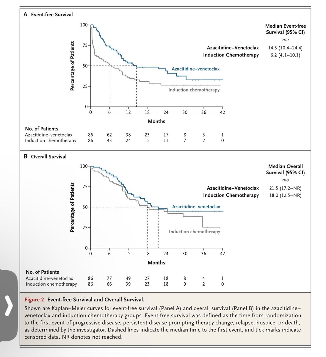
PARADIGM trial — brief summary
* Design: Randomized phase II trial; 172 fit, untreated AML patients—72% ELN adverse risk.
* Comparison: Azacitidine–venetoclax vs intensive induction (7+3 or CPX-351).
* Primary outcome: Median EFS 14.5 vs 6.2 months; HR 0.57, P=0.002.
* Responses: Composite CR 78% vs 53%; transplantation 60% vs 40%.
* Overall survival: 21.5 vs 18.0 months—no clear significant difference; trial not powered for OS.
* Safety: Fewer grade ≥3 infections (28% vs 41%), bleeding (2% vs 12%), early deaths, ICU admissions, and hospital days with aza–ven.
* Clinical message: Aza–ven is a compelling induction-to-transplant strategy for fit patients with nonfavorable-risk, FLT3-wild-type AML.
* *Caution: Phase II academic-center study; excluded CBF AML, FLT3-mutated AML, younger NPM1-mutated AML, and prior HMA exposure.* Phase III confirmation is needed before broadly replacing intensive induction.
NEJM article · Attached PDF
3 September 2026
Immunohistochemical Marker Selection in the Diagnosis and Classification of Lymphomas
When lymphohematopoietic neoplasms (primarily lymphomas) cannot be readily distinguished from other undifferentiated small-cell tumors of non-lymphohematopoietic origin, an initial antibody panel that includes leukocyte common antigen (LCA/CD45), S-100, and cytokeratin (CK) should be applied to establish the tissue of origin.
When the histologic findings are already sufficient to support a diagnosis of lymphoma, lineage-associated markers should be used to determine the neoplastic cell type. A practical first-line panel consists of two pan-T-cell markers (CD3 and CD43) and two pan-B-cell markers (CD20 and PAX5), together with CD15 and CD30. This combination usually permits a straightforward separation of Hodgkin lymphoma from non-Hodgkin lymphoma and, within the latter group, of B-cell from T-cell neoplasms.
For lesions that display a follicular or nodular growth pattern, markers such as CD10, BCL6, CD21, CD23, BCL2, and Ki-67 are useful for delineating the relationship between the nodules and residual lymphoid follicles.
When Hodgkin lymphoma is favored morphologically, the recommended panel includes CD30, CD15, LCA, PAX5, MUM1, and EBV-encoded RNA (EBER) in situ hybridization. If anaplastic large-cell lymphoma enters the differential diagnosis, EMA, ALK, and CD43 should be added. If T-cell/histiocyte-rich large B-cell lymphoma must be excluded, BOB.1, OCT2, CD20, and CD3 should be included.
Small B-cell lymphomas require simultaneous consideration of small lymphocytic lymphoma/chronic lymphocytic leukemia (SLL/CLL), marginal-zone lymphoma (MZL), follicular lymphoma (FL), and mantle-cell lymphoma (MCL). An appropriate panel comprises CD20, PAX5, CD19, CD5, CD23, CD10, BCL2, cyclin D1, SOX11, LEF1, and MNDA. For neoplasms showing clear-cut immature (blastic) differentiation, the differential diagnosis includes acute lymphoblastic leukemia/lymphoblastic lymphoma (ALL/LBL) and acute myeloid leukemia (AML). In addition to lymphoblastic markers (TdT, CD99, CD10, CD34), myeloid antigens such as myeloperoxidase (MPO) and CD117 should be assessed to exclude lymphoblastic lymphoma with myeloid or bilineage differentiation. Judicious selection of these markers greatly facilitates accurate diagnosis and subclassification.
In plasma-cell-rich lesions, useful markers include CD38, CD138, immunoglobulin light chains (κ and λ), MUM1, and cyclin D1. Demonstration of light-chain restriction (κ or λ) helps distinguish neoplastic from reactive plasma-cell proliferations.
For lesions suspected to represent highly aggressive mature B-cell neoplasms—including the majority of diffuse large B-cell lymphomas, Burkitt lymphoma, B-cell lymphomas with features intermediate between these two entities, high-grade B-cell lymphoma, and high-grade follicular lymphoma—the recommended panel includes CD10, BCL6, BCL2, MUM1, MYC, CD5, and p53, supplemented by EBER in situ hybridization.
In diagnostically difficult cases, additional markers, sometimes applied in successive rounds, may be required; however, restraint in panel size remains important. Because of known antigenic cross-reactivity, two independent T-cell markers and two independent B-cell markers are advisable to provide more reliable evidence of lineage. Combined morphologic evaluation and immunophenotyping allow accurate diagnosis and classification of the great majority of lymphomas.
Molecular studies. When the above investigations still leave the diagnosis unresolved, ancillary molecular techniques—including antigen-receptor gene-rearrangement analysis, fluorescence in situ hybridization (FISH), and gene-expression profiling—should be employed.