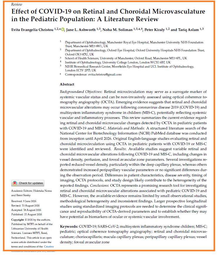
Effect of COVID-19 on Retinal and Choroidal Microvasculature in the Pediatric Population: A Literature Review
🚨UK Ophthalmology waking up?
➡️“Emerging evidence suggests that SARS-CoV-2 may lead to microcirculation alterations in the macular and the peripapillary capillary plexus in children.”
➡️“OCTA characteristics may represent potential biomarkers for the long-term effects of hypoxia on retinal and choroidal tissue in the context of COVID-19 and MIS-C.”
➡️“Currently available data are limited; therefore, further research is warranted to clarify the role of microvasculature alterations in the underlying pathophysiology of ocular manifestations associated with COVID-19 infection.”
‼️So, they’re finally starting to concede: SARSCoV2 can alter kids’ retinal and choroidal microvasculature after COVID-19 and MIS-C. Possible link to the virus’s systemic vascular damage and inflammation. But watch the goalposts move, they immediately bury it under “limited evidence,” “small studies,” “inconsistent findings,” and “OCTA is only a research tool, not a clinical biomarker.”
Starting admission of harm to children… but with maximum reserve.
More rigorous studies “needed” is of course for everyone a valid request!
Let’s just repeat, protect children: #AvoidSars2 #AvoidReinfections while others catch up!
https://t.co/QyBi79UCUZ
New study out of TUM Munich looked at long COVID through the retina. It is the one place in the body where you can see CNS nerve tissue and CNS blood vessels directly, non invasively. A window into the brain. And this window showed something most retinal COVID threads miss🧵
🔴 “Long COVID came out of nowhere.”
No, it did not.
Before SARS-CoV-2. Before COVID vaccines. Before today’s debates about Spike, mRNA, or vaccine injury.
Another coronavirus had already been followed by persistent long-term symptoms.
It was called SARS-CoV-1.
And almost everyone forgot what happened next.
(1/33) 🧵
What happens to Plasymacytoid Dendritic Cells (pDCs) when you are infected with COVID19/Omicron?
What are pDCs and what do they do?
Can a cash reward help those who have been affected?
- observations of someone with ‘too much time on their hands’….
Incidence of new-onset pulmonary fibrosis 4.5 years post SARS-CoV-2 infection in an academic urban medical center
🚨IMPORTANT STUDY with the expected BAD NEWS!
🚨Even mild COVID roughly doubles your risk of new pulmonary fibrosis lasting 4.5+ years, and the fibrosis that follows brings far more pneumonia, respiratory failure & exacerbations. #LongC0VID
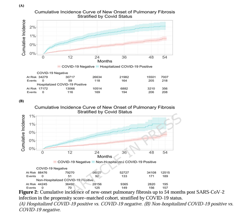
➡️This large retrospective cohort study from Montefiore Health System( March 2020–August 2024) examined new-onset clinical pulmonary fibrosis (PF, via ICD-10 codes) up to 4.5 years after SARSCoV2 infection,
➡️Follow-up runs from index date until new-onset PF, death, or the end of observation (August 2024), whichever came first,
- Only the earliest infections (spring/summer 2020) reach the full ~4.5 years / 54 months of follow-up,
- Patients infected in 2021–2024 have progressively shorter observation times.
- Average follow-up in the matched cohorts was roughly 600–630 days (~1.6–1.7 years),
➡️Hospitalized and non-hospitalized COVID-19 patients without prior PF were propensity-score matched 1:2 to COVID-19-negative controls,
➡️After matching, hospitalized patients had a subdistribution hazard ratio (sHR) of 2.05 (95% CI 1.69–2.49) and non-hospitalized patients an sHR of 2.33 (95% CI 1.88–2.88) for new-onset PF versus controls,
➡️Cumulative incidence remained elevated through ~54 months,
➡️Among those who developed PF, prior COVID-19 (especially hospitalization) linked to substantially higher odds of downstream complications:
- Pneumonia (OR up to 4.84),
- Respiratory failure (OR 3.76),
- Bronchiolitis, asthma/COPD exacerbations, plus elevated risks of pulmonary hypertension and embolism in subgroups,
➡️Vaccination status was collected and included in propensity-score matching to balance covariates (SMDs <0.1 post-match). It was not analysed as an effect modifier or outcome predictor,
➡️Possible reinfections(4 years) aren’t mentioned?🤔
➡️Conclusion:
“In a diverse cohort with up to 4.5 years follow-up, we found both hospitalized and non-hospitalized COVID-19 patients have a higher long-term risk of new-onset PF and higher burden of subsequent downstream respiratory complications compared to matched non-COVID-19 controls. These findings demonstrate post-COVID-19 PF as a relevant and clinically significant outcome and suggest the need for long-term surveillance, risk stratification, and targeted intervention.”
‼️So, according to this study, both mild and severe COVID-19 roughly double the long-term risk of clinically diagnosed new-onset pulmonary fibrosis for years afterward, and the fibrosis that does occur carries a markedly heavier burden of further life-limiting respiratory events. Post-COVID PF is a durable, clinically meaningful sequela requiring ongoing surveillance.
#AvoidSars2 #AvoidReinfections
https://t.co/tTjXtdYV47
@saabelieves The California Dept of Public Health literally writes their social media posts using AI and encourages handwashing to avoid breathing the virus deep in the lungs.
An insider tells we there is almost no communication between experts and the social media team.
SARS2 has more than structural proteins - also carries accessory ones. One of them, ORF7a, can flip a cell’s energetics on its own, without the rest of the virus. It dampens mito respiration and reroutes metabolism. And it seems to do this from a distance - it never even touches the mito.🧵
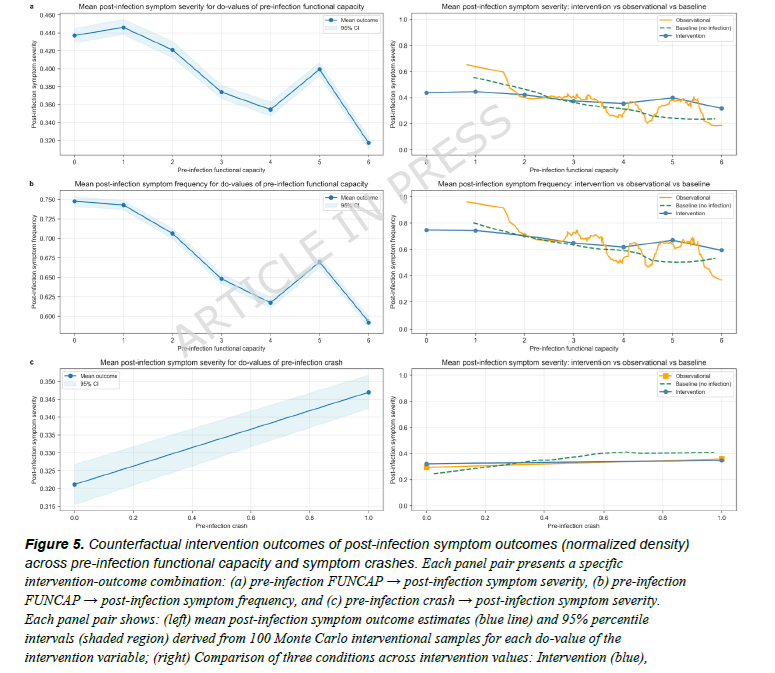
Each additional COVID reinfection measurably worsens symptoms and elevates resting heart rate in Long COVID and ME/CFS patients.
🚨Interesting Swiss study underscoring my ongoing #AvoidReinfection call!
➡️One of the first to apply causal AI to longitudinal reinfection data from a non-hospitalized LongC0VID/ME/CFS cohort.
➡️Manage your functional capacity (FUNCAP),
➡️Avoid reinfections and crashes(PEM),
➡️Or watch the damage accumulate!
#AvoidSars2 #AvoidReinfections
https://t.co/NCnYvS2pUz
In the 7th year of the pandemic there are 490,861 publications on Covid your doctor won't read.
https://t.co/eXTIqGDJBE
Like this comprehensive review of 49 MRI studies
https://t.co/AkhIZuQtjP
showing brain damage after acute and long term infection.
Billions affected. 275 views.
In the 7th year of the pandemic there are 490,861 publications on Covid your doctor won't read.
https://t.co/eXTIqGDJBE
Like this comprehensive review of 49 MRI studies
https://t.co/AkhIZuQtjP
showing brain damage after acute and long term infection.
Billions affected. 275 views.
I saw another study yesterday I wanted to expand on and explain because it's pretty important for vascular health in LC.
Side note: You guys may have seen weird videos how there are "worms" coming out of people in autopsies. 🙃 Well it's actually the lining of people endothelium. It's time we talk about how SC2 infected bone marrow leads to pMLKL activation. Check this out.
The connection between bone marrow infection, activated platelets, and pMLKL (phosphorylated Mixed Lineage Kinase Domain-Like protein) brings us right to the heart of necroptosis, a form of programmed, highly inflammatory cell death that plays a major role in chronic tissue damage and persistent post-viral/post-infectious pathologies.
Here is the step-by-step biological cascade of how an infected bone marrow niche and hyper-reactive platelets drive pMLKL expression:
1. The Core Trigger: PAMPs, DAMPs, and Inflammatory Cytokines:
When bone marrow progenitor cells or the stromal niche harbour persistent pathogens (viral reservoirs, bacterial products, or viral proteins like SARS-CoV-2 Spike):
TLR Activation:
Pathogen-Associated Molecular Patterns (PAMPs) bind to Toll-Like Receptors specifically TLR3 and TLR4 on megakaryocytes (platelet precursor cells in the bone marrow) and local immune cells.
Pro-Inflammatory Storm:
This triggers a sustained release of TNF-alpha, IFN-gamma, and IL-1beta directly within the bone marrow microenvironment.
2. The Molecular Switch to Necroptosis
(RIPK1 > RIPK3 > pMLKL)
Under normal conditions, TNF-alpha signaling leads to apoptosis (non-inflammatory programmed cell death). However, in a chronic, highly inflammatory, or viral-infected bone marrow environment
Caspase-8 Inhibition:
Viral proteins or intense inflammatory stress often inhibit or override Caspase-8 (the enzyme that normally keeps necroptosis shut off).
Necrosome Assembly:
With Caspase-8 blocked or overwhelmed, RIPK1 recruits and phosphorylates RIPK3.
MLKL Phosphorylation (pMLKL):
Active RIPK3 phosphorylates MLKL, converting it into pMLKL.
Pore Formation:
pMLKL oligomerizes and translocates directly to the plasma membrane, punching physical pores into the cell wall. This causes cell swelling, rupture, and the explosive release of intracellular contents (DAMPs), creating a self-amplifying loop of tissue destruction.
3. How Platelets Drive and Accelerate This Cascade
Platelets are not just clotting agents; they are primary immune responders born directly from bone marrow megakaryocytes. When the bone marrow itself is infected or inflamed:
Primed/Infected Megakaryocytes:
Megakaryocytes produce "pre-activated" or dysfunctional platelets carrying viral remnants, elevated pro-inflammatory signals, and pro-thrombotic molecules directly into circulation.
Platelet-Leukocyte Aggregates (PLAs):
Hyper-reactive platelets bind aggressively to monocytes and neutrophils via P-selectin (CD62P) and PSGL-1. This physical binding activates the immune cells, triggering them to produce even more TNF-alpha and upstream signals that drive MLKL phosphorylation.
NETosis Coupling:
Platelets activated in an infected microenvironment trigger neutrophils to undergo NETosis (releasing Neutrophil Extracellular Traps). NETs directly induce necroptotic pathways (pMLKL activation) in nearby endothelial and parenchymal cells.
Microvascular Hypoxia:
Platelet microclots (microthrombi) choke off microvascular blood flow. The resulting local hypoxia/ischemia is a massive secondary stimulus for RIPK3 activation and subsequent MLKL phosphorylation.
Summary of the Pathological Feedback Loop:
This loop explains why targeting single inflammatory pathways often fails in complex post-infectious states.
As long as the bone marrow reservoir keeps pumping out primed megakaryocytes/platelets and inflammatory signals, the RIPK3–pMLKL pathway stays switched on, driving continuous, systemic microvascular and cellular damage.
I know this is too complex for many. See next part. ❤️🩹🫂
"Extreme heat is killing hundreds of thousands of people every year — and the annual toll is growing.
An estimated 210,000 more people died of heat-related causes each year from 2012 to 2021 than from 1990 to 19991, on average..
[However,] Creating an accurate tally is not straightforward.. death certificates rarely cite heat as the cause of death..
Most public-health agencies estimate heat-related mortality using the ‘excess deaths’ method..
but it can’t distinguish between excess deaths caused by the heatwave and those caused by other factors.
The method also can’t be used until after a heatwave has passed, limiting its effectiveness in forecasting the death toll for an upcoming heatwave."
The COVID wave has a similar nature, although no one cares.
'Heatwaves have killed millions. Here’s how scientists tally lives lost'
https://t.co/IAOnWkZVOC
Virus reactivation in acute and long COVID-19
🚨Hidden viruses reactivate in nearly half of hospitalized COVID patients, tied to severity, inflammation & LongC0VID physical symptoms.
➡️Let’s revisit this very interesting and revealing study in which multi-omic longitudinal data from 1,154 unvaccinated hospitalized COVID-19 patients (IMPACC cohort, 2020–2021) were analysed, with a personal interpretation!
➡️Key findings:
- Significant reactivation of Herpesviridae (EBV, CMV, HSV1/2, HHV6) and Anelloviridae occurred in ~47.9% of patients during acute illness, with distinct temporal patterns (e.g, EBV earlier, CMV/HSV later),
- Reactivation correlated with greater disease severity, systemic inflammation (elevated cytokines such as IL-6, CXCL10), metabolomic shifts, immune changes, and poorer outcomes including mortality risk in critical cases,
- It frequently occurred in immunocompetent individuals, not primarily driven by immunosuppression or medications,
- Viral transcripts persisted into convalescence. Anelloviridae in PBMCs associated with physical deficits in LongC0VID, independent of age, severity, and immunosuppression,
- External validation supported the dynamics. No causation was claimed, but immune/transcriptomic/metabolomic signatures of reactivation were identified.
➡️The findings challenge the view that chronic virus reactivation is mainly an immunosuppression consequence and highlight its prevalence alongside inflammation in severe COVID-19.
➡️Interpretation:
‼️So, this intriguing study shows data that are consistent with a model in which:
1. SARSCoV2 directly drives immune dysregulation and inflammation, and
2. This “stress situation” reactivates other viruses that then secondarily MAGNIFY and/or PROLONG those same processes.
‼️This virus reactivation does not replace the direct SARSCOV2 effects, it adds a plausible secondary layer.
‼️These found signatures are at least partly independent of SARS-CoV-2 viral load and severity in some analyses, raising further the possibility that the secondary reactivated viruses actively contribute rather than being a pure side-effect/consequence of Covid-19.
‼️This hidden virus’s reactivation study further suggests that persistent Anelloviridae reactivation into convalescence may contribute to physical long-COVID deficits via ongoing inflammation and immune dysregulation, suggesting that monitoring or targeting such reactivations could help mitigate long-term sequelae( future for specific new antivirals?).
‼️Causality in either direction (or both) remains, as the authors state, a hypothesis and will need interventional and/or mechanistic follow-up studies.
‼️Nonetheless, this study is an important step forward in the exploration of SarsCoV2 and its broad and long-term (post-)infection impact! #Bookmark
#AvoidSars2 #AvoidReinfection
https://t.co/tUh0EluQjo
From an observational study of 227 adults (19–79 y/o) who were previously hospitalized for COVID-19,
"Several neurological symptoms increased in prevalence over 36 months following hospitalization..
Fatigue remained most common (53–65%), followed by memory/concentration difficulties (47–61%) and sleep disturbances (40–48%).
Dizziness had a modest increase (30–49%), while coordination impairment more than doubled (18–41%)..
Symptom co-occurrence strengthened over time, with fatigue frequently clustering with other symptoms (up to 45% at 36 months).
Overall BrainCheck composite scores were not independently associated with neurological symptoms, but significant associations emerged for executive function, memory, and attention, including time-dependent effects..
[In conclusion,] Neurological symptoms can persist or worsen up to 36 months post-COVID-19 hospitalization.."
Let's look at the bright side - you will forget this conclusion in 36 hours.
'Longitudinal trajectories of neurologic symptoms and cognitive associations at 36-months post-hospitalization for COVID-19'
https://t.co/qYJYH4BdIb
"A handful of these pathogens are never fully cleared from the body. Instead, they hide quietly inside cells for a lifetime..
Researchers have seen hints that these bystander viruses are reactivated in people with long COVID..
These pathogens included herpesviruses such as cytomegalovirus and Epstein–Barr virus, which causes glandular fever (mononucleosis), as well as anelloviruses, which are typically harmless and don’t cause symptoms.
More than 90% of the global adult population has been infected with at least one of these pathogens..
different viral families reactivate on their own timelines.
Epstein–Barr virus and anelloviruses tend to surge early during a bout of COVID-19, peaking shortly after hospital admission.
By contrast, cytomegaloviruses and herpes simplex viruses emerge around three weeks later, mainly in the respiratory system..
this reactivation happens even in people with otherwise healthy immune systems..
Some scientists had thought that a reawakening of viruses could happen only in people with a weakened immune system, such as those with HIV.
But this study overturns that assumption.."
SARS-CoV-2 is the king of viruses with a crown (corona).
'COVID can wake up a slew of dormant viruses inside you'
https://t.co/FdPN3lGdUZ