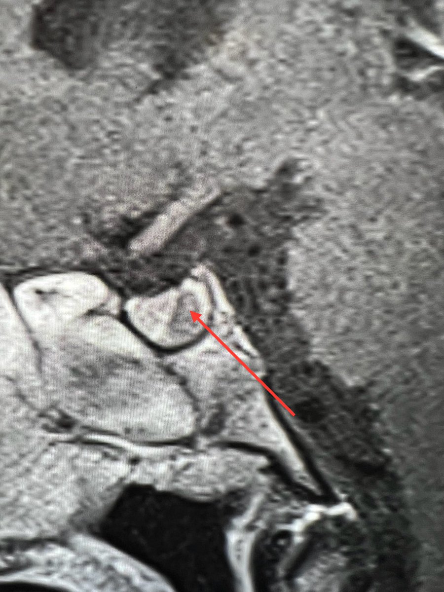
“Can we take the patient off the magnet?!”
Scan check for kids under GA comes with great responsibility!
Don’t rush.
Keep an eye on the scan from the get go.
Know your patients ahead of time.
Based on what you see, ask yourself if you need any additional sequences to clarify
The hardest post yet! Are you up for the challenge?
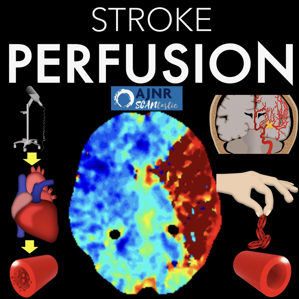
How stroke perfusion imaging works!
Ever wonder why it’s Tmax & not Tmin?
Here’s what to know from @theAJNR SCANtastic!
https://t.co/SuQyawSP8V
Clinically, there are 2 main perfusion parameters used:
(1)Cerebral blood flow (CBF) = how FAST blood gets to the tissue
(2) Tmax or time to max residue function. Everyone knows Tmax is for penumbra, but does anyone know what it really is??? You will now
Pretend we want to know how fast a kitchen prepares food—Restaurant Continental Breakfast Flow or rCBF. If we know when ingredients arrive & we know when food gets on our table, we can back calculate kitchen speed--& that’s what we do for the real CBF
When the ingredients arrive is the arterial input function. We measure over a cerebral artery to see when blood first arrives. It’s equal to how long it takes the restaurant to get ingredients from the supplier—how long it takes the artery to get blood after injection
How fast food is building up on our table is tissue concentration. We measure in brain
parenchyma to detect the buildup of contrast. How long it takes for blood to get from injection to tissue is equal to how long it takes ingredients to be turned into food on our table
Time for the kitchen to turn ingredients to food for the table is CBF. We want to find CBF by dropping contrast right in a brain artery & seeing how fast it washes out to tissue. This is kitchen time--how long for a blood drop to wash out from artery (kitchen) to tissue (table)
If we know the time for blood to get from injector to artery & time to get from injector to tissue, we can back calculate how long it takes to get from artery to tissue. So we use the arterial input function & tissue concentration to back calculate the artery to tissue time
In this month’s @AJNR, Proner et al. looked at factors affecting quality in CT perfusion and found arrhythmias could cause non diagnostic imaging. This is understandable, as it affects the delivery of contrast. These pts need longer scan times.
Now you know how perfusion imaging works!
Hopefully this Tmax-ed out your knowledge!!
But this just scratches the surface. Follow @theAJNR and check it out for yourself:
https://t.co/SuQyawSP8V
It’s an alphabet soup!!
Do you know you Tmax from your MTT from your CBF??
Here’s what to know from @theAJNR SCANtastic!
https://t.co/SuQyawShjn
In a stroke, remember for the core:
—FLOW is LOW (cerebral blood flow or cbf is low)
—Tmax is maxed (Tmax is very high)
—V is for vague (cerebral blood volume or cbV is usually low, but not as low as cbf, so it’s vague or harder to see!
But this just scratches the surface. Follow @theAJNR and check it out for yourself:
https://t.co/SuQyawShjn
1/The hardest thread yet! Are you up for the challenge?
How stroke perfusion imaging works!
Ever wonder why it’s Tmax & not Tmin?
Here’s what to know from @theAJNR SCANtastic!
https://t.co/2o9S3rVp6X
Are you agile on the sagittals??
Sagittal T1 images are for anatomy, but only if you know how to use them!
Here is how I remember the sagittal anatomy
1. Start lateral
2. Find the gyrus that looks like a triangle, that is the pars triangularis of the inferior frontal gyrus
3. In front of it is the pars orbitalis—easy it’s over the orbit
4. Behind it is the pars opercularis. Remember oper sounds like OVER so its OVER the sylvian fissure
5. Behind the pars opercularis is the precentral gyrus
6. Right behind the precentral is the post central—duh post follows pre!
7. Behind the post central is the supramarginal gyrus. Remember supra means above, so it’s above the sylvian fissure. Margin = at the posterior margin of the sylvian fissure
8. Behind the supramarginal gyrus is the inferior parietal lobule. Remember inferior is below supra, so inferior parietal lobule is below the supramarginal
9. More medial is the lateral orbital gyrus. It’s easy, it above the orbit!
10. Even more medial is the gyrus rectus. Rectus means straight, so if you see a gyrus that is straight like a line, that is the gyrus rectus!
Hopefully this gives you a handle on the sagittal!
Stay tuned for the midline sagittal anatomy!!
The most dangerous, common, and overlooked bad habit in the world:
Your phone.
It's why you're stressed, depressed, and your mind can't stop procrastinating.
Here're 5 ways it destroys your brain and ONE app to break free (from a Harvard-trained psychiatrist):
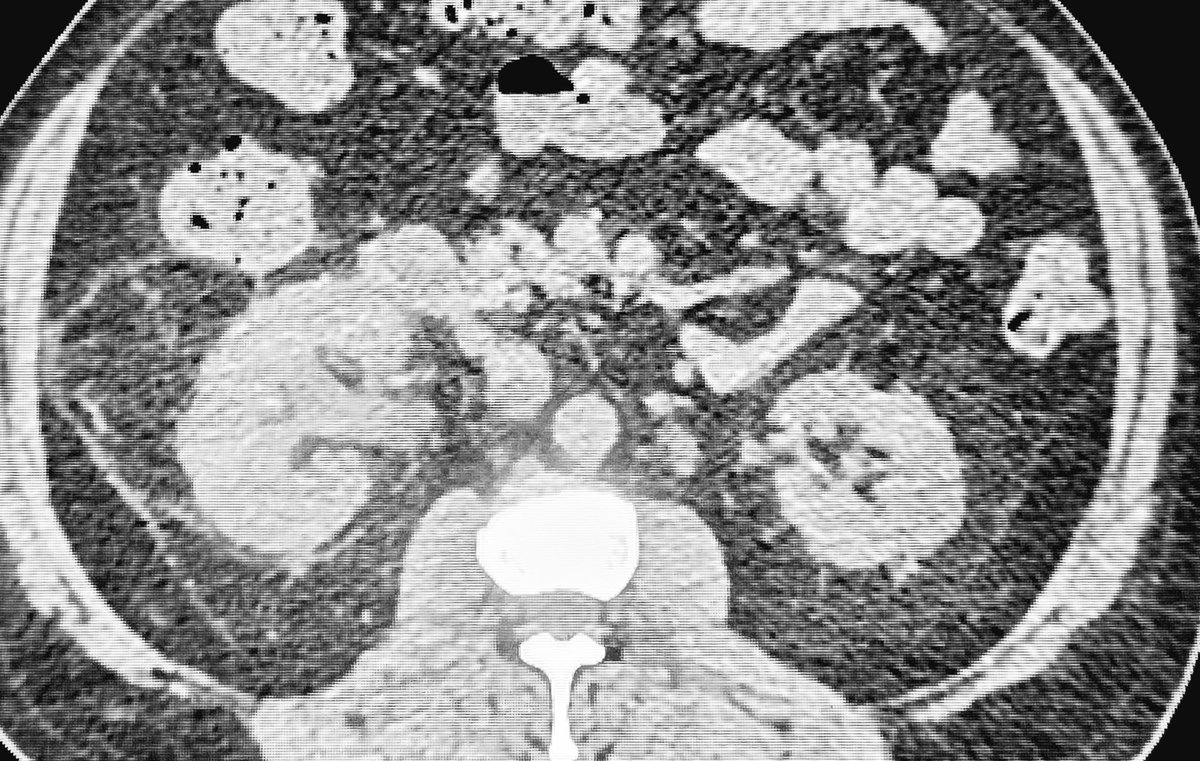
Poorly controlled - DM
Hydro-ureteronephrosis
No ureteric cal
Diagnosis high on the list:
"Urteric obstruction due to necrosed papilla"
You have to prompt it to your urologist..or it might be treated as usual pyelonephritis.
#MedTwitter#FOAMed#Radiology@AJR_Radiology
If you are only looking at the optic nerve on Orbital MRIs, you need to open your EYES!
Don’t be blind to all the other nerves in the orbit!
Branches of trigeminal V1 &V2 and the oculomotor nerve all can cause pathology in the orbit
But if you don’t look, you won’t see!
Let this video show you some of the nerves you can see in the orbits (CN6 is only seen proximally bc its orbital course is so short & CN4 is too small to reliably see)
Hopefully, now you will have an eagle eye when it comes to orbital pathology!
Wish you had a sixth sense to localize that sixth cranial nerve palsy?
Feeling six feet under when you see a sixth nerve deficit?
Here’s a cheat sheet to help you locate the lesion in a sixth cranial nerve palsy!
Just remember the Six Syndromes of the Sixth Cranial Nerve!
There are six syndromes of CN6—one for each of its five stops along the way to its destination in the orbit—and one that is idiopathic
1. Brainstem
🔸CN6 nucleus is at the facial colliculus—looks like a baby’s butt of the brainstem
🔸Many important structures here
🔸Rarely get an isolated palsy & uniquely can get associated MOTOR deficits
2. Subarachnoid space
🔸CN6 exits under the dad bod belly of the pons & enters Dorello canal
🔸Susceptible to changes in intracranial pressure—can get stretched!
🔸ICP changes can cause BILATERAL palsies here
3. Petrous apex
🔸Exits Dorello canal & travels under the petroclinoid ligament, like under a seat belt
🔸Many other nerves here
🔸Rarely get an isolated palsy & uniquely can get associated HEARING deficit
4. Cavernous sinus
🔸Many nerves in close proximity!
🔸Rare to get an isolated palsy
🔸Many pathologies can affect the cavernous sinus—so many differentials & many deficits!
5. Orbit
🔸Short orbital course—quick lateral turn after the superior orbital fissure to innervate the lateral rectus
🔸Rare to have isolated palsy
🔸Get a SOF syndrome or orbital apex syndrome
6. Idiopathic
🔸No other identifiable etiology
🔸Like transverse myelitis—likely microvascular or post-inflammatory
🔸1/4th of CN6 palsies are idiopathic
But you must exclude the other five first!
Now you know the Six Syndromes of the Sixth Cranial Nerve.
Hopefully now when you see a CN6 palsy, there will never be six degrees of separation between you & the etiology!
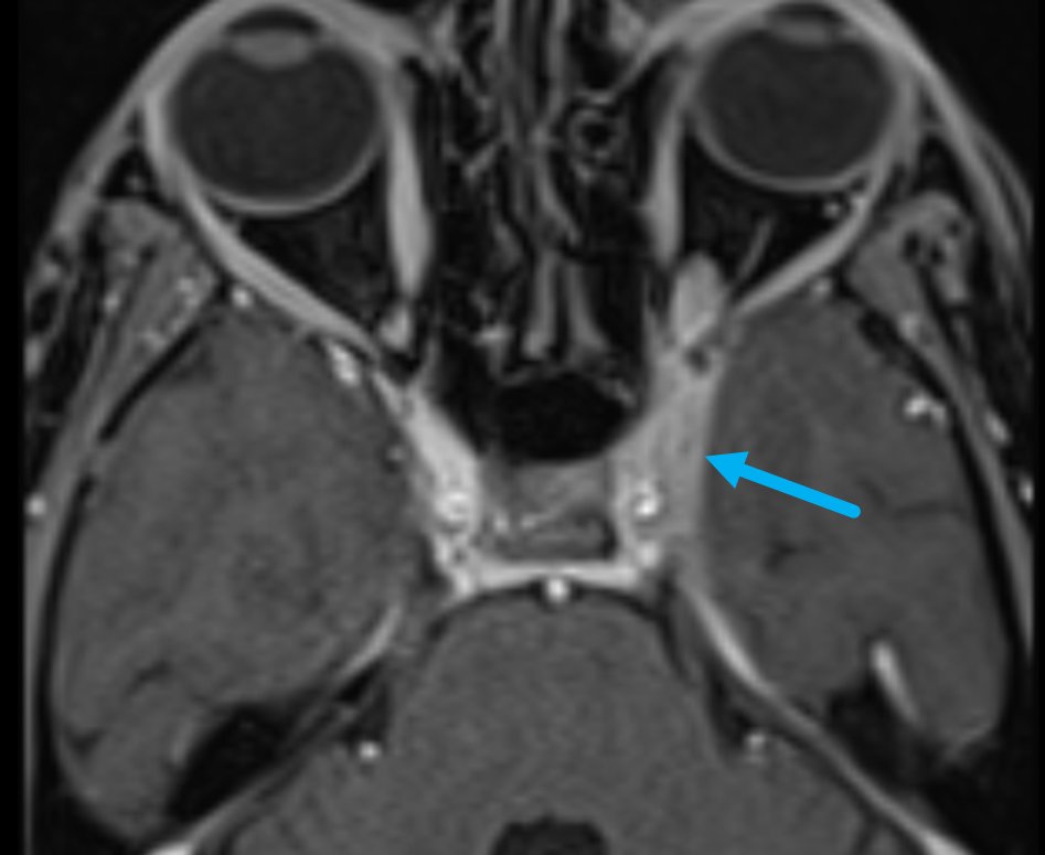
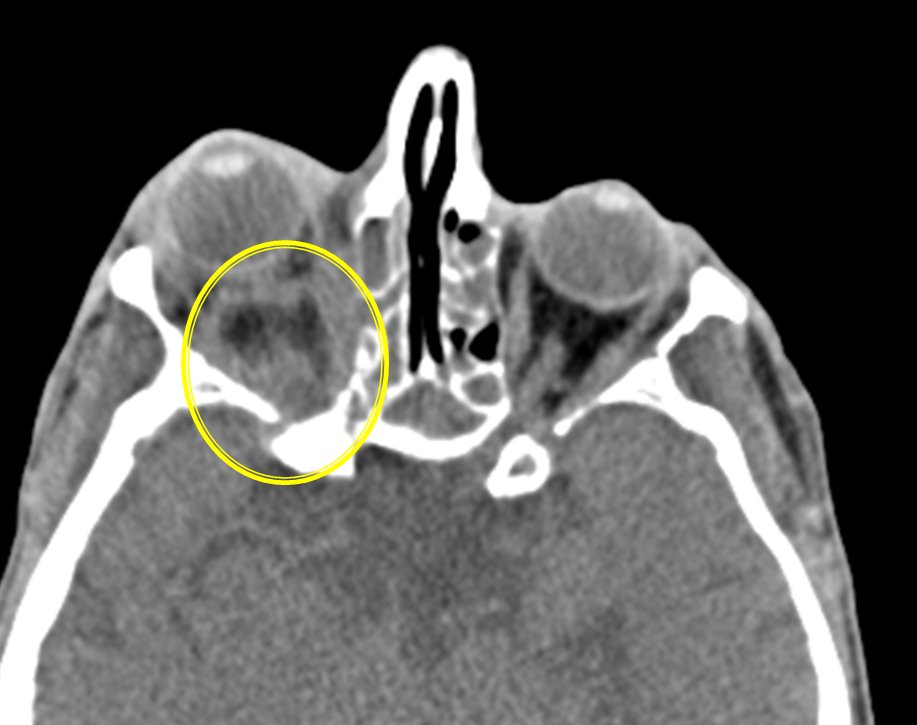
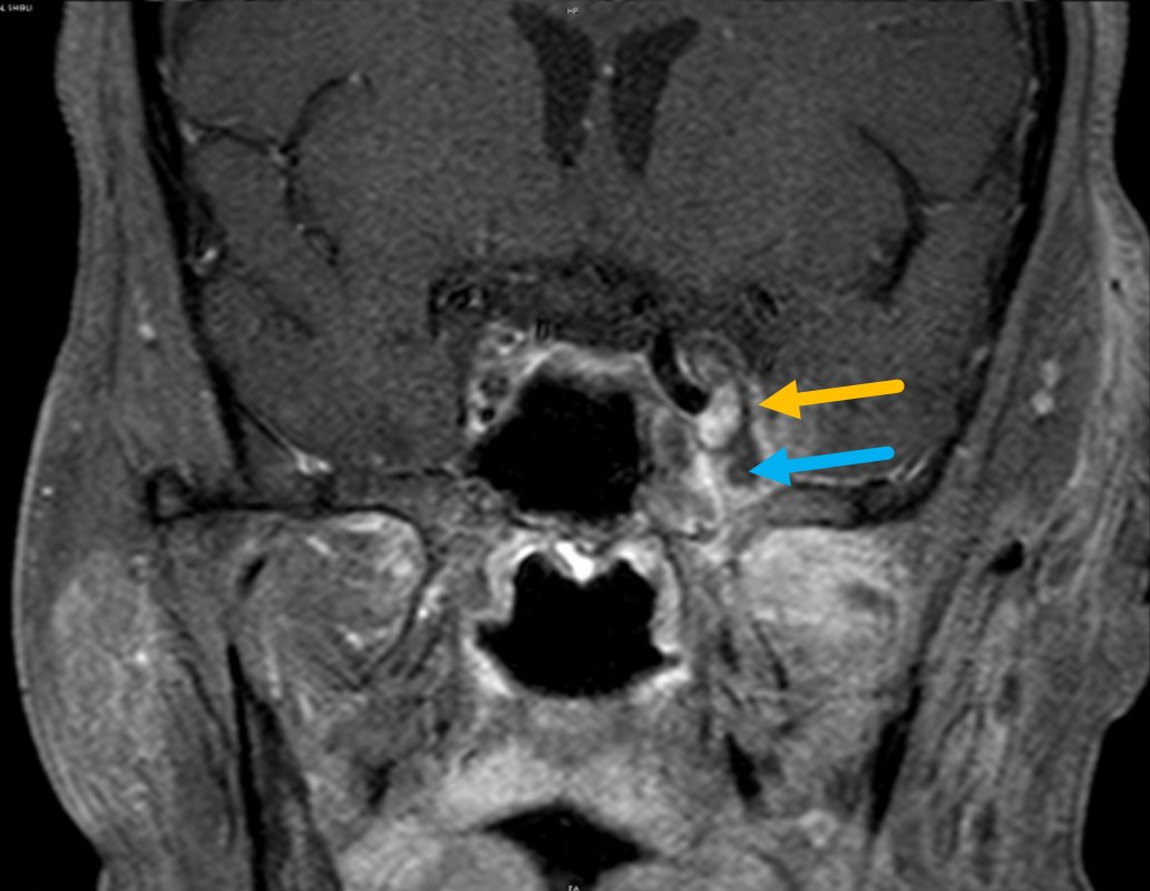
Cavernous sinus thrombosis on imaging:
Direct signs
1. Loss of concavity with lateral bulging (i.e., convexity)—best seen on coronal CECT/CE-MRI (image 1)
2. Heterogeneous filling defects within the
cavernous sinus (image 1)
Indirect signs (when you see this, look for cavernous sinus carefully)
1. Thickened dural enhancement along the cavernous sinus margins (image 2)
2. Orbital apex inflammation (image 3)
3. Exophthalmos (image 3)