Welcome to our OCTOBER 16 issue of #JACCCaseReports@JACCJournals on simultaneous publications from the 37th #GreatWall#GW-ICC/AHS2026
https://t.co/HEW4oV37b4.
https://t.co/5Lekmbd8H1
Thank you @Kentso987 and @MiChen__ on representing the Journal moderating our session at the meeting!
@minnowalsh
@hmkyale
An Adrenal Adenoma as the Culprit Behind 30 Years of Recurrent Syncope
https://t.co/G00ovxKtHF
Progressive Dilated Cardiomyopathy in a 24-Year-Old Male: A Rare Manifestation of Propionic Acidemia
https://t.co/IyKRPNbZhI
Exercise-Induced Ventricular Arrhythmia: Initial Presentation of Occult Barlow Syndrome Unmasked by Stress Testing
https://t.co/p2daE6I6t2
A Fish Bone Penetrating the Oesophagus Into the Left Atrium
https://t.co/E8UtCQBRl4
Optical Coherence Tomography–Guided Calcium Modification for a Complex Calcified Nodule With Circumferential Calcium: From Debulking to Fracturing
https://t.co/1UhKbeJQLF
Intraoperative ST-Segment Elevation Myocardial Infarction Complicating Ruptured Abdominal Aortic Aneurysm Repair
https://t.co/EK4AxtUWR2
Pregnancy-Related Right Atrial Appendage Tachycardia Requiring ECMO and Hybrid Ablation
https://t.co/EQHaHxGwiF
Low-Frequency Whole-Body Vibration and Isometric Exercise in Cardiac Rehabilitation: Improved Capacity and Triglyceride-Glucose Index
https://t.co/IeTtHr8kMY
Perioperative B-Cell–Targeted Therapy for High-Risk Valve Surgery in Active Primary Sjögren's Disease
https://t.co/aQ6W23aRPQ
Recurrent In-Stent Restenosis and Accelerated Neoatherosclerosis in a Middle-Aged Female With High Inflammatory Burden
https://t.co/61wvsyk6Fv
First-in-Human Axillary Vein Implantation of a Leadless Pacemaker: Breaking the Access Barrier
https://t.co/JW0SYxLVwS
Ablation in Dextrocardia With Interrupted Inferior Vena Cava and Azygos Continuation to Coronary Sinus
https://t.co/v4wdx16oVp
Vascular Plug Closure of Delayed Leaflet Perforation Post-TEER in Patient With Multiple Implantable Devices
https://t.co/DTi5HBNSLt
Tachycardia Initiation by Dual-Mode PVC on Ambulatory ECG: Pattern Discovery and Invasive Electrophysiological Characteristics
https://t.co/UPw3DTYE1t
Novel SCN5A Variant in a Patient With Dilated Cardiomyopathy Complicated by Conduction Disorders and Recurrent Ventricular Arrhythmias
https://t.co/miThXofpBR
Laparoscopic Renal Denervation and Perirenal Adipose Tissue Debulking for Refractory Hypertension in End-Stage Renal Disease
https://t.co/PhKN1WX3q5
Multimodality Imaging of Blood Supply in Intramyocardial RCC Metastasis With Double Primary Malignancies
https://t.co/csbTCKToUS
Perioperative and Surgical Management of a Ruptured Mycotic Coronary Artery Aneurysm Secondary to MRSA
https://t.co/bG5IbXglH8
Coronary Visual Intelligence System (CorVIS) Guided Complex Percutaneous Coronary Intervention
https://t.co/B6dwtdFx3O
Catecholamine-Induced Cardiomyopathy Masquerading as Acute Myocardial Infarction
https://t.co/fxx43Szwpc
Successful Guidewire-Guided Ethanol Ablation for PVCs Originating From Epicardial Anterior Interventricular Groove Adjacent to LAD
https://t.co/SYBe8H019g
Transcatheter Adrenal Arterial Embolization for Life-Threatening Pheochromocytoma Crisis
https://t.co/yJQOUF6mym
@aroniskn
@CraigBasman@JACabreraCardio@MiChen__@aabha_divya@MarcEngels7@sarano_maurice@Miho_Fukui_@CardioOncCO@MMarinCuartas@Nishaki1@ChrissyMD2000@ACHDocTMoe@PanjrathG@jsaef1@Kentso987@TagliariPaula@m_taramasso@SyedZaidMD
Author’s Handles:
@LydiaMessele@chu_jiapeng@Dingyu1989
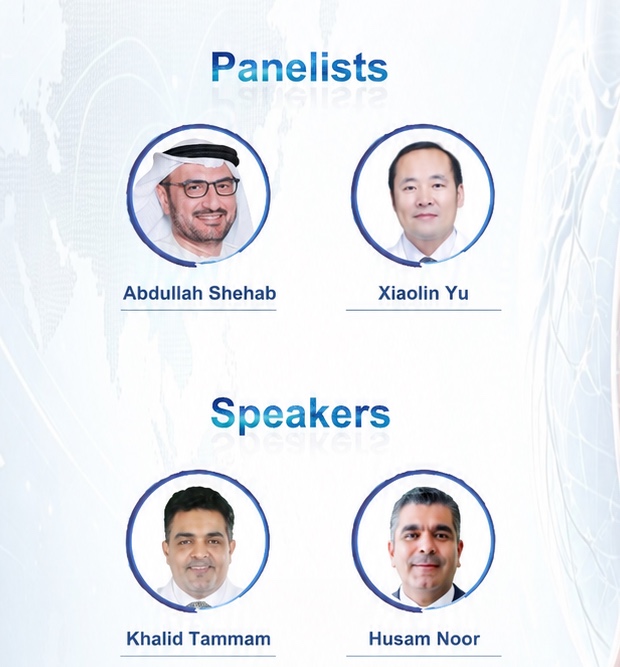
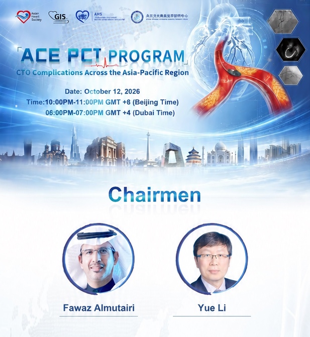
🌏 Complex PCI Across Asia | AHS Jointly with Gulf Intervention society Webinar
Join our expert chairs and panellists from the Asian Heart Society (AHS) for an engaging discussion on Complex PCI Across Asia,
sharing clinical insights and perspectives from across the region.
📅 Monday, 12 October 2026
🕒 10:00 PM Beijing (GMT+8) | 6:00 PM UAE | 5:00 PM KSA
Connect with the experts and be part of the discussion!
Register now:
https://t.co/s8i3MI5OTC
#AHS #AsianHeartSociety #ComplexPCI #InterventionalCardiology #CardiologyWebinar #GIS
@Falmutairi2018@HusamNoor1@KhalidKtammam
OSTIAL CORONARY LESIONS: SMALL DISTANCE, BIG TECHNICAL CHALLENGE
An ostial lesion is generally defined as disease within about 3 mm of a vessel origin.
The location matters because precise stent placement is critical.
1. Aorto-ostial lesions
Involve the origins of the RCA, left main stem, or a bypass graft from the aorta.
2. Non-aorto-ostial lesions
Involve the origins of major coronary branches arising from another coronary artery, such as the LAD or LCx.
3. Branch-ostial lesions
Involve branch origins, such as diagonal, obtuse marginal, PDA or posterolateral branches.
Why are ostial lesions difficult?
Ostial plaques can be fibrotic, calcified and resistant to balloon expansion. Aorto-ostial lesions can also be challenging because guide-catheter engagement and stent positioning are less forgiving.
The key PCI principles:
🟣Confirm the lesion.
Angiography can be limited by foreshortening, overlap and guide-catheter engagement. Physiology and intravascular imaging can help when severity is uncertain.
🟢Get the right angiographic view.
A small change in projection can completely change your perception of whether the stent actually covers the ostium.
🔵Cover the ostium completely.
Too distal = geographic miss.
Too proximal = unnecessary extension into the parent vessel or aorta.
🟡Optimize the stent.
Underexpansion is a major mechanism of PCI failure. IVUS is particularly valuable for left main and complex lesions, helping with vessel sizing, lesion assessment and post-stent optimization.
⚫Think beyond the angiographic picture.
The goal is not simply a beautiful angiogram. It is complete lesion coverage, adequate expansion, good apposition and preservation of adjacent branches.
The 2024 ESC Chronic Coronary Syndromes guideline has strengthened the role of IVUS-guided PCI in anatomically complex disease, while a 2026 EAPCI/EBC consensus specifically addresses intracoronary imaging for left-main PCI.
For ostial PCI, millimeters matter.
Reference: Jokhi P, Curzen N. Percutaneous coronary intervention of ostial lesions. EuroIntervention. Updated with contemporary ESC/EAPCI guidance.
https://t.co/UjCRhos5HI
🫀 Rare fluoroscopy: a Starr–Edwards ball-in-cage valve, first implanted in 1960. ⚙️ A landmark in valve surgery, with extraordinary durability—some functioning for 50+ years. ❤️ A remarkable chapter in prosthetic valve history.
ECG From Scratch #43: Myocardial Ischemia
Before learning ischemic ECG changes, understand the physiology.
Myocardial ischemia occurs when myocardial oxygen supply cannot meet oxygen demand.
Three major determinants of LV oxygen demand are:
◻️Heart rate
◼️Contractility
◻️Ventricular wall stress, strongly influenced by systolic pressure
Exercise increases these demands, which explains why stable angina may appear with exertion.
Ischemia is not the same as infarction.
Transient or reversible ischemia may occur without cell death. Prolonged, severe ischemia can cause myocardial necrosis, producing myocardial infarction (MI).
Modern definitions also recognize that MI can result from acute coronary thrombosis or from an oxygen supply-demand imbalance without an acute coronary thrombus, as in type 2 MI.
ACS encompasses acute coronary presentations including unstable angina and MI (STEMI or NSTEMI). Diagnosis integrates symptoms, ECG findings and cardiac troponin, with alternative causes considered.
#ECG takeaway:
Ischemia can produce dynamic ST-T changes, but a normal or nondiagnostic ECG does not exclude myocardial ischemia or MI.
This is the foundation for understanding the ischemic ECG patterns coming next.
A 23-year-old athlete collapses during a football match and cannot be revived.
He had no previous cardiac symptoms. At autopsy, the heart shows marked thickening of the interventricular septum with a narrowed left ventricular cavity.
What is the most likely diagnosis, and what is the mechanism of sudden cardiac death?
Este corazón continúa latiendo fuera del cuerpo mientras espera ser trasplantado.
Un sistema de perfusión lo mantiene caliente y recibiendo sangre oxigenada, permitiendo conservar el órgano en condiciones adecuadas hasta el trasplante.
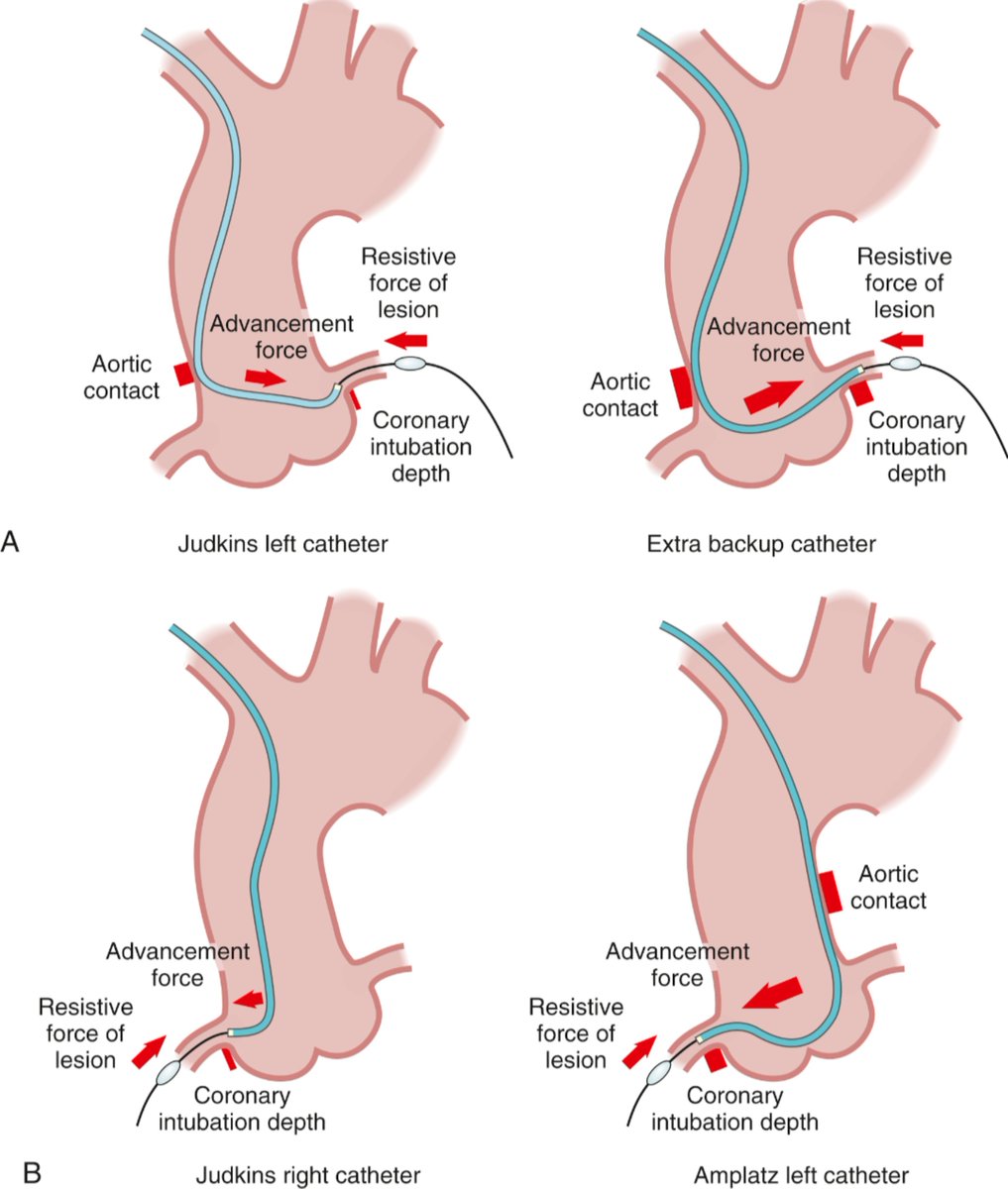
Choosing the Guide Catheter
A good guide catheter does more than deliver devices.
It creates the stable platform needed to cross difficult lesions, deliver balloons and stents, and perform complex PCI safely.
The key principle is:
Guide support = shape + size + anatomy + access site.
A larger French size generally provides greater passive support, but bigger is not automatically better. The guide must match the patient's artery and the planned intervention. Larger radial sheaths and guides increase the risk of radial artery occlusion.
⭕ Left coronary artery
For difficult or calcified lesions, EBU, XB and CLS guide shapes can provide strong backup support because their secondary curve contacts the aortic wall while the tip engages the coronary ostium.
⭕ Right coronary artery
A Judkins Right (JR) guide is often sufficient for straightforward RCA PCI.
More challenging anatomy may require an Amplatz Left (AL) or another supportive shape, depending on the RCA takeoff and aortic root anatomy.
The guide should be coaxial with the coronary ostium. Poor alignment can reduce support and increase the risk of ostial injury or dissection.
What about guide size?
6 Fr remains extremely versatile and can accommodate most PCI techniques.
Larger guides may be needed when the procedure requires greater lumen capacity, such as certain complex bifurcation techniques or simultaneous device delivery.
But the goal is not to choose the largest guide possible.
Choose the smallest guide that safely provides the support and device compatibility the procedure requires.
And if the guide still lacks support, options include guide extensions, buddy wires, anchoring techniques or carefully selected deep intubation.
Guide selection is part of PCI strategy, not just catheter selection.
Source: Textbook of Interventional Cardiology, 8th ed., Eric J. Topol & Paul S. Teirstein.
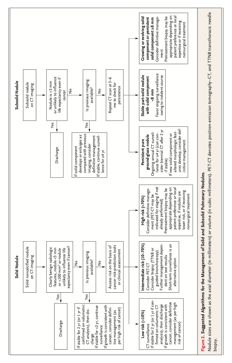
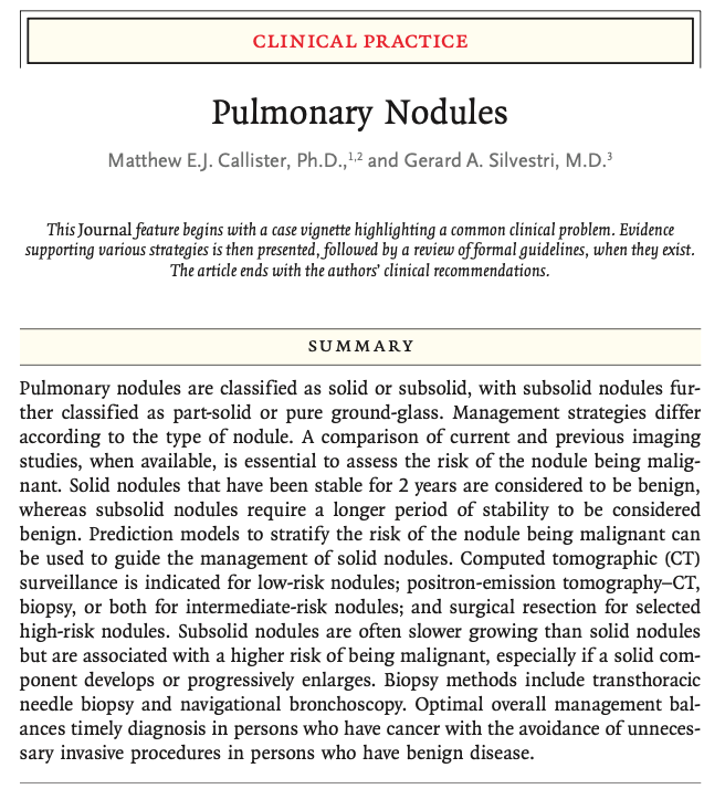
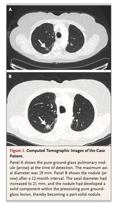
Nódulo pulmonar, la última revisión de NEJM (2026). Puntos clave:
🟢 Un nódulo sólido estable a 2 años es benigno y se da de alta (1 año basta si se confirmó por volumetría). Los subsólidos crecen más lento: requieren ≥4 años de estabilidad antes de llamarlos benignos.
🟢 Importante estratificar con un modelo (Brock si es tamizaje o subsólido; Mayo/Herder si es incidental sólido): <10% → vigilancia por CT; 10–70% → PET-CT y/o biopsia; >70% → manejo definitivo, con cirugía posible sin biopsia previa. Umbrales para PET: ≥8 mm o ≥300 mm³ (abajo de eso es inútil)
🟢 Hasta 60% de los subsólidos desaparecen, así que repite CT a 3–6 meses para confirmar persistencia antes de hacer cualquier cosa.
🟢 La biopsia por broncoscopia navegada puede ser tan buena como la transtorácica (rendimiento 74% (aguja transtorácica) vs 79% (broncoscopía navegada), lo malo: neumotórax 28% vs 3%.
🟢 Solo 3–4% de los nódulos detectados son cáncer; pero 50% de los pacientes sufre distrés durante el proceso.
So far the most accurate multiclass segmentation model on brainchop is available for testing (at brainchop-test). 0.9 Dice on HCP, and 0.86 on a wide selection of data. Pull a sample from openneuro to test or drag&drop your favorite problem sample.
ECG Patterns of Occlusion Myocardial Infarction (OMI)
STEMI criteria alone can miss up to a quarter of true acute coronary occlusions. Here’s a complete guide to subtle but high-risk OMI