More on rare presentations of APL➡️
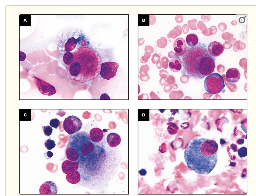
APL with circulating myeloblasts, and "aleukemic" promyelocytes confined to the marrow may cause diagnostic challenges:
If diagnosis relied only on peripheral blood findings, the promyelocytic component hidden in the marrow may be missed, particulalry that the typical myeloblastic morphology identified in the blood (as seen in the top left figure below) and phenotype (bottom left figure) may not alerting to perform STAT FISH for PML::RARA, delaying appropriate treatment.
The top right figure shows promyelocytes in the bone marrow aspirate smear, with a phenotype classic of APL by flow cytometry analysis (negative CD34 and bright MPO expression).
For more on this + figure credits➡️
https://t.co/4YLV7CTlfH
#Hemepath #leusm #PathX #PathTwitter #Surgpath #Molpath #SoMe #MedEd
Acute promyelocytic leukemia (APL) in the setting of primary or secondary MPO deficiency can create diagnostic challenges, as bright MPO expression by flow cytometry analysis is a key feature of APL.
Six cases of APL with reduced MPO expression (2 cases) and absent MPO expression (4 cases) have been described so far: Four of the microgranular variant and two classic APL cases.
One of the MPO negative microgranular APL has been extensively analyzed and found to have a germline heterozygous MPO loss of function mutation (MPO c.2031 -2A > C) with somatic uniparental disomy of 17q, resulting in homozygous MPO deficiency and subsequent MPO negativity in the myeloblasts.
Three different flow cytometric patterns of immunoreactivity with the MPO protein have been described in the setting of MPO deficiency:
(a) Dim/absent MPO expression, characteristic of patients with primary complete MPO deficiency.
(b) Moderate MPO expression, typical of patients with primary partial MPO deficiency.
(c) Bright MPO expression, typical of patients with secondary MPO deficiency (a functionally inactive form of the enzyme is present at normal levels).
MPO deficiency was previously thought to be extremely rare; However, recent studies have suggested that the incidence might be much higher, with a reported frequency of 1 per 200 to 4,000 in the US. The prevalence of heterozygotes for MPO c.2031-2A > C may close to 1% in the population.
In the setting of an acute leukemia with morphologic and immunophenotypic features typical of APL, with dim to absent MPO expression, evaluating MPO expression in background granulocytes is very helpful to highlight the possibility of a primary (hereditary) form of MPO deficiency (partial or complete):
Figure 1 below illustrates a case of primary MPO deficiency with dim/negative MPO expression in granulocytes, while Figure 2 illustrates bright MPO expression in granulocytes in normal control.
For more on this + figure credits, check this article by @Kritika_Krish@ChoudhuriJui@YanhuaWang3
➡️ https://t.co/V2DZqtF5Vj
#Leusm #hemepath #molpath #pathX #pathTwitter #SoMe #MedEd #BMTsm
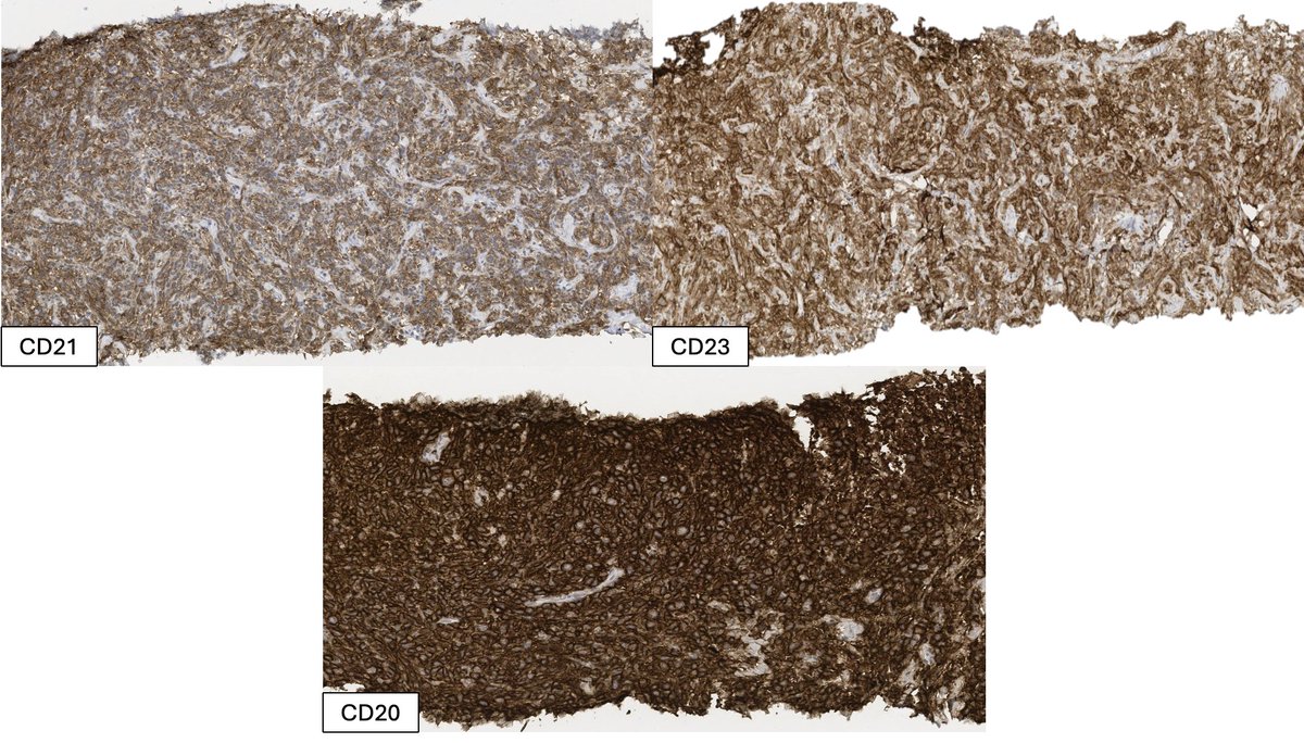
A new fear unlocked: Leukemic non-nodal mantle cell lymphoma may involve the bone marrow in a very subtle sinusoidal way, as illustrated in figure 1 below with H&E and CCND1 stain highlighting a sinusoidal neoplastic lymphocytic infiltrate.
In addition to this sneaky pattern of infiltration, leukemic non-nodal mantle cell lymphoma may be CD5-negative occasionally, which may create a great diagnostic pitfall, as other CD5-negative B-cell lymphomas, classically known for their sinusoidal pattern of infiltration (such as splenic marginal zone lymphoma, splenic diffuse red pulp small B-cell lymphoma and splenic B-cell lymphoma/leukemia with prominent nucleoli), may be thought of in this scenario long before a CD5-negative Leukemic non-nodal mantle cell lymphoma.
Figures credit and more on mantle cell lymphoma with a sinusoidal pattern of infiltration of the bone marrow
➡️https://t.co/7iXz7VRDx6
#Hemepath #Lymsm #PathX #PathTwitter #MedEd #SoMe
4 the young #pathologists here, who may not know: angiomyofibroblastoma, retiform hemangioendothelioma, spindle cell liposarcoma, cellular angiofibroma, soft tissue angiofibroma, etc were among many entities recognized & described by Dr. Fletcher (& trainees)
Give him a shoutout the next time you diagnose one of these 🔬May he RIP🤍
The wait is over 🎊
The @WHO heme 5th is now available in PRINT
https://t.co/S6Podveh8c
Can’t wait 4 mine 🤩 and yes.. I had to kill a few trees, but I really wanted to smell and flip the pages 📑 🙈 a pdf just won’t do…
#hemepath#leusm#mpnsm#mdssm
📄Analysis of 222 CCUS cases confirmed the comparable mutational spectrum between #CCUS and #MDS, reflecting the biological continuum. However, significant variations were observed in both the frequency and VAF of specific gene mutations: https://t.co/IM7i0wbKUz
Follicular lymphoma with abundant extracellular eosinophilic material, mimicking amyloid deposition: An unsual variant to be aware of #hemepath#lymsm#pathtwitter#MedEd#SoMe
Check a similar but more prominent case by @sanamloghavi @BloodJournal➡️ https://t.co/FgGDXEZwZw
we have come such a long way in our understanding of IgG4-related disease. This is a volume of seminars in diagnostic pathology devoted to Ig4-related disease. Covers virtually every organ. written by Folks that have spent their careers writing about this disease. The focus is not on how one makes the diagnosis, but how one avoids making the diagnosis😄
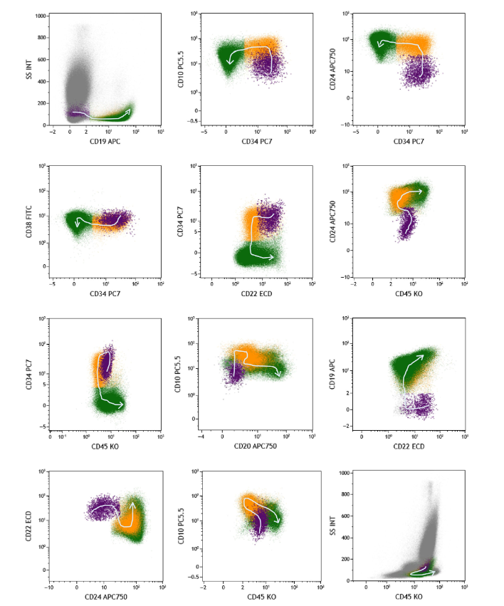
We know about Stage 1, 2 & 3 hematogones, but Stage 0 hematogones? Read all about this CD19-negative population and the implications for MRD testing in patients receiving anti-CD19 therapies, in this pathbreaking paper just published in Clinical Cytometry!
https://t.co/1T7JEFbsRd
#Hemepath pitfall alert! Thrombopoietin-mimetic agents such as eltrombopag (Promacta) & romiplostim (Nplate) cause changes in megakaryocytes that exactly mimic MDS/MPNs. The paper below describes some abnormal forms, but I've seen the whole gamut. Be wary!
https://t.co/C9Jjq1gvMo
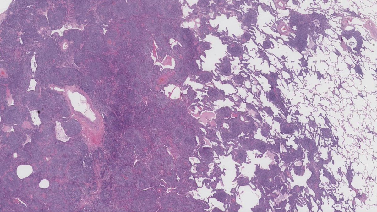
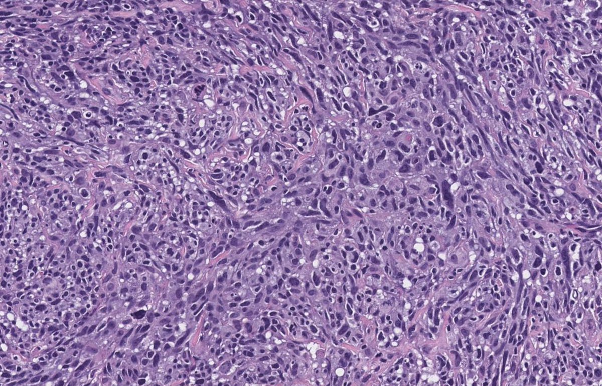
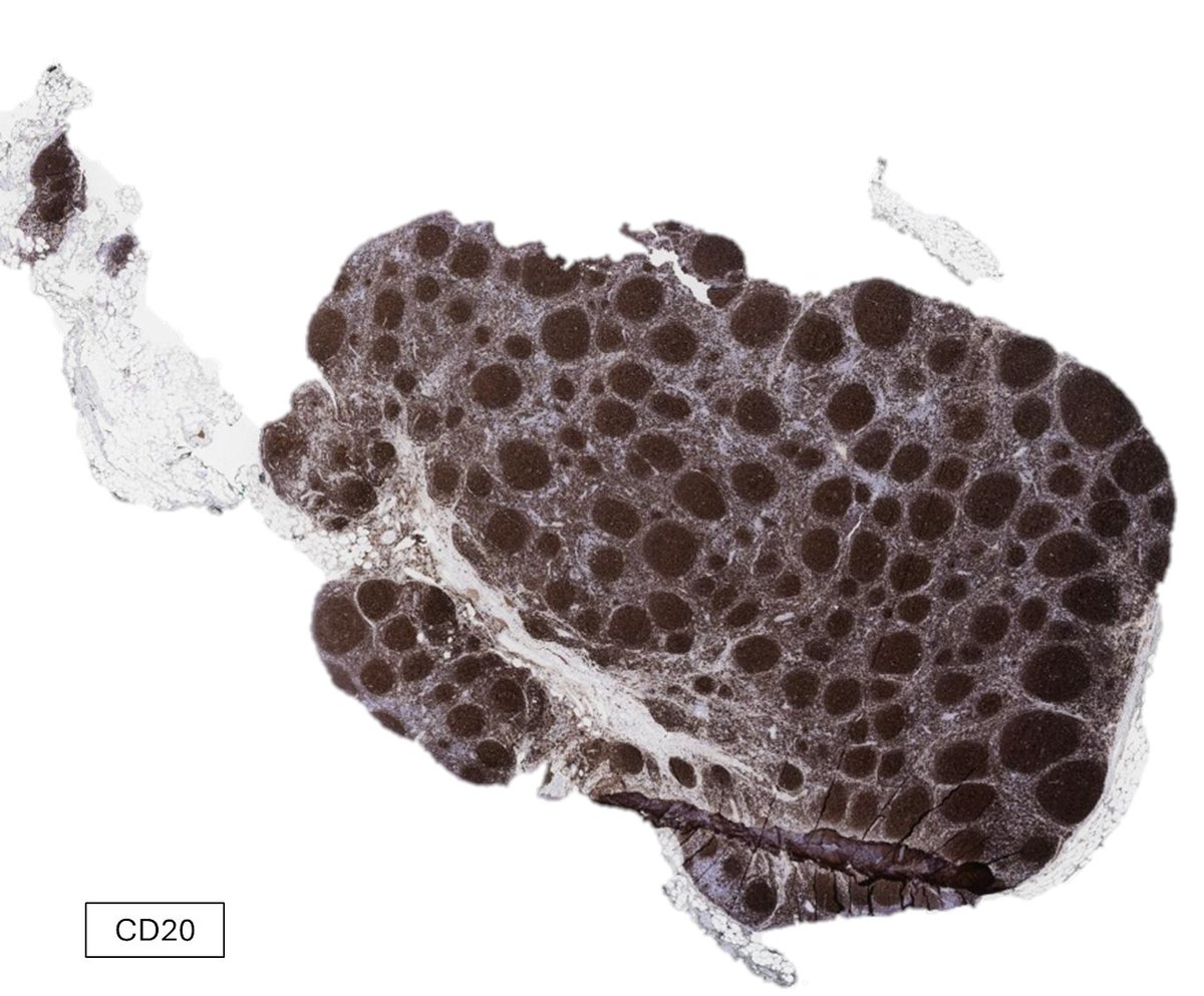
An example of EBV-negative extra-nodal marginal zone lymphoma (ENMZL), involving the lung, 6 years post transplant
PTLDs occurring later after the first year of transplantation, are more frequently EBV-negative. The late PTLDs are also more commonly monomorphic
Mutations described in EBV-negative ENMZL in PTLD setting include mutations seen in ENMZL in general: namely, TNFAIP3 , TNFRSF14 , LRP1B, FAS , and NOTCH2
As recipients live longer, the burden of EBV-negative processses in the post transplant setting seems to be increasing, and more cases of EBV-negative PTLD may be encountered in the future
Although most cases of EBV-negative PTLD are clinically aggressive, posttransplant EBV-negative MZLs seem to be clinically indolent and can be managed conservatively
More on EBV-negative marginal zone lymphoma in the post transplant setting ➡️https://t.co/8fsZ2UHbRH
#hemepath #bmtsm #lymsm #PathTwitter #pathX #MedTwitter #medX #SoMe #surgpath #thoracipath
To be or not to be… is there such a thing as type G LyP?
Am J Surg Pathol, 2024
Lymphomatoid Papulosis With T-cell Receptor-Gamma Delta Expression: A Clinicopathologic Case-series of 26 Patients of an Underrecognized Immunophenotypic Variant of Lymphomatoid Papulosis
The WHO Classification of Haematolymphoid Tumours, 5th edition, is now finalized 👏😊 and is available on-line at
https://t.co/c1bweKYbJV
It will soon be available as a two volume printed book. #hemepath#lymphoma#WHO
Are you a pathology trainee interested in a #hemepath fellowship? Big news: most hemepath fellowship programs will be moving to a match process for positions that begin in 2026! For more information: https://t.co/2WFaouRS8t