Rare diagnostic pitfall
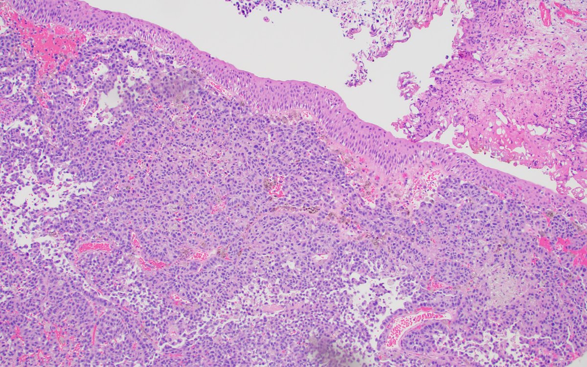
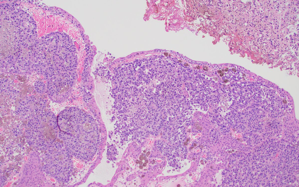
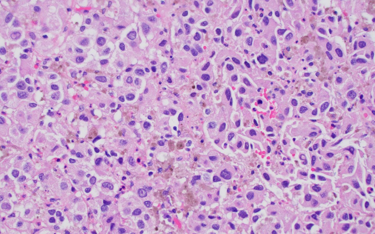
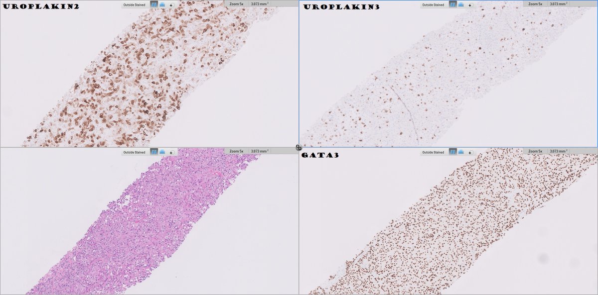
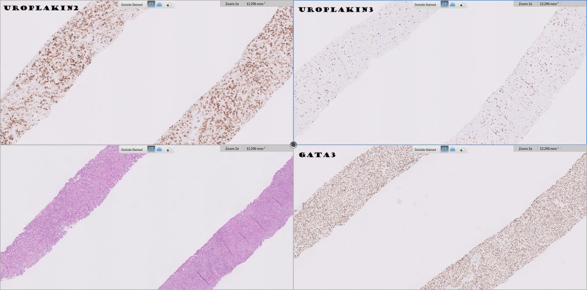
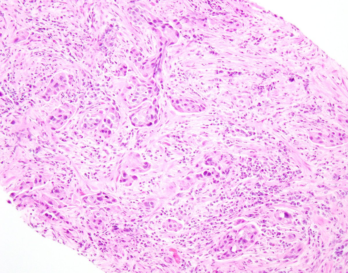
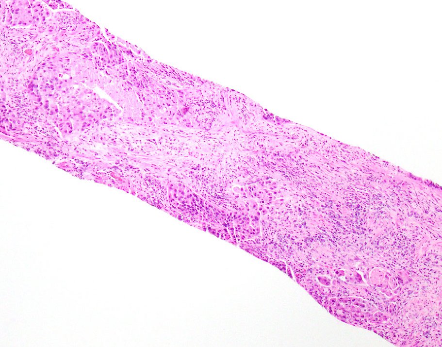
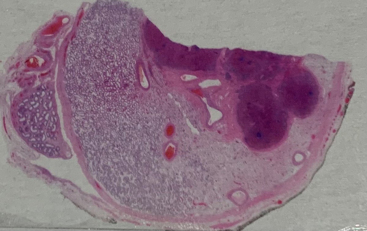
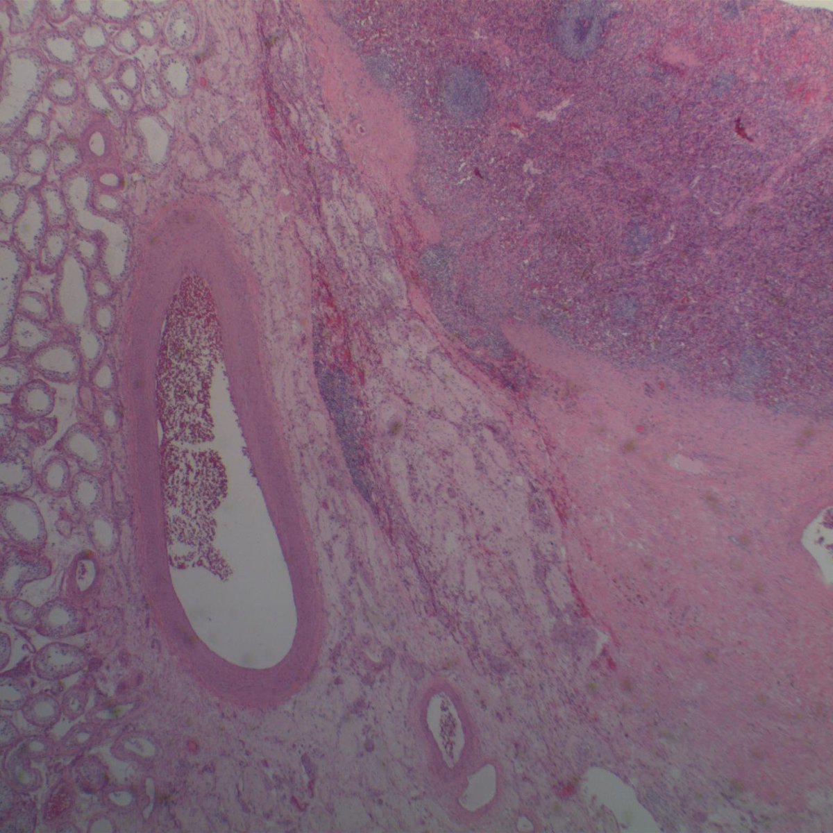
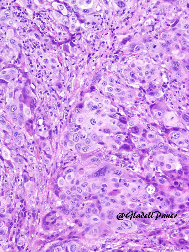
Bladder mass in a 70yo male.
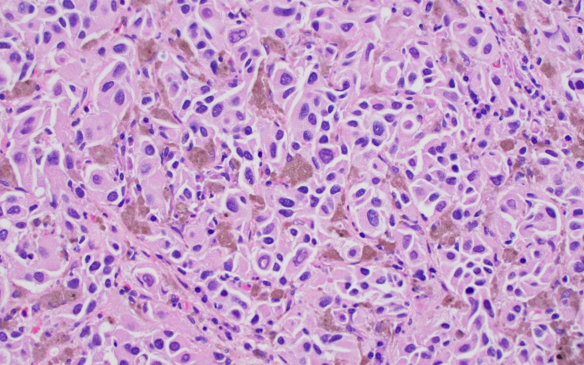
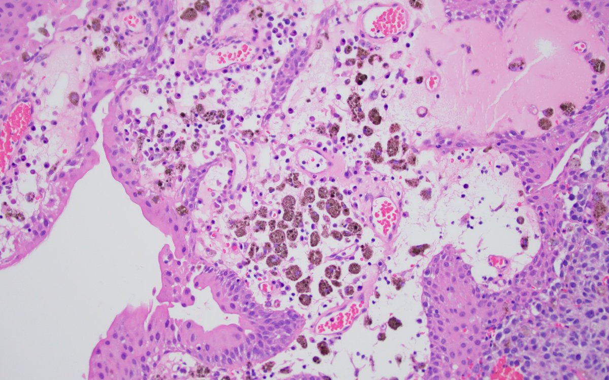
Epithelioid cells with focal spindle cell morphology and atypical mitoses, marked variation in nuclear size and shape, hyperchromatic, occasional intranuclear inclusion.
Note the absence of in situ carcinoma.
see next tweet.
This is a case of metastatic melanoma to bladder. This mass was black in cystoscopy. Less than a 30 are reported in the literature, but the true prevalence is higher, autopsy series on melanoma patients reported 3-18% prevalence.
M : F = 15 : 8.
#gupath#pathology#bladdercancer
Histology flash card
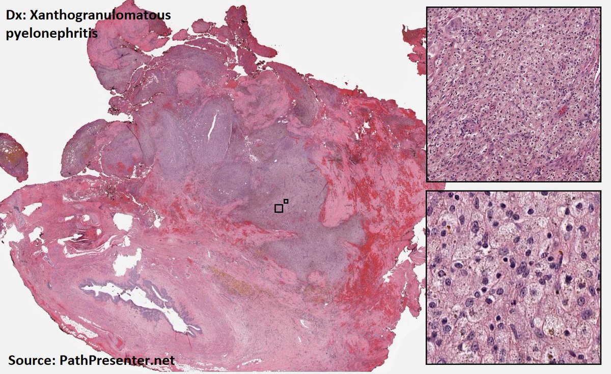
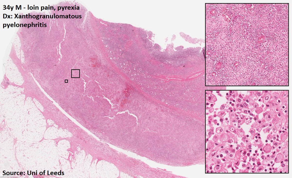
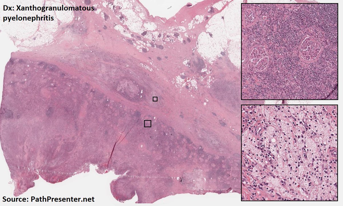
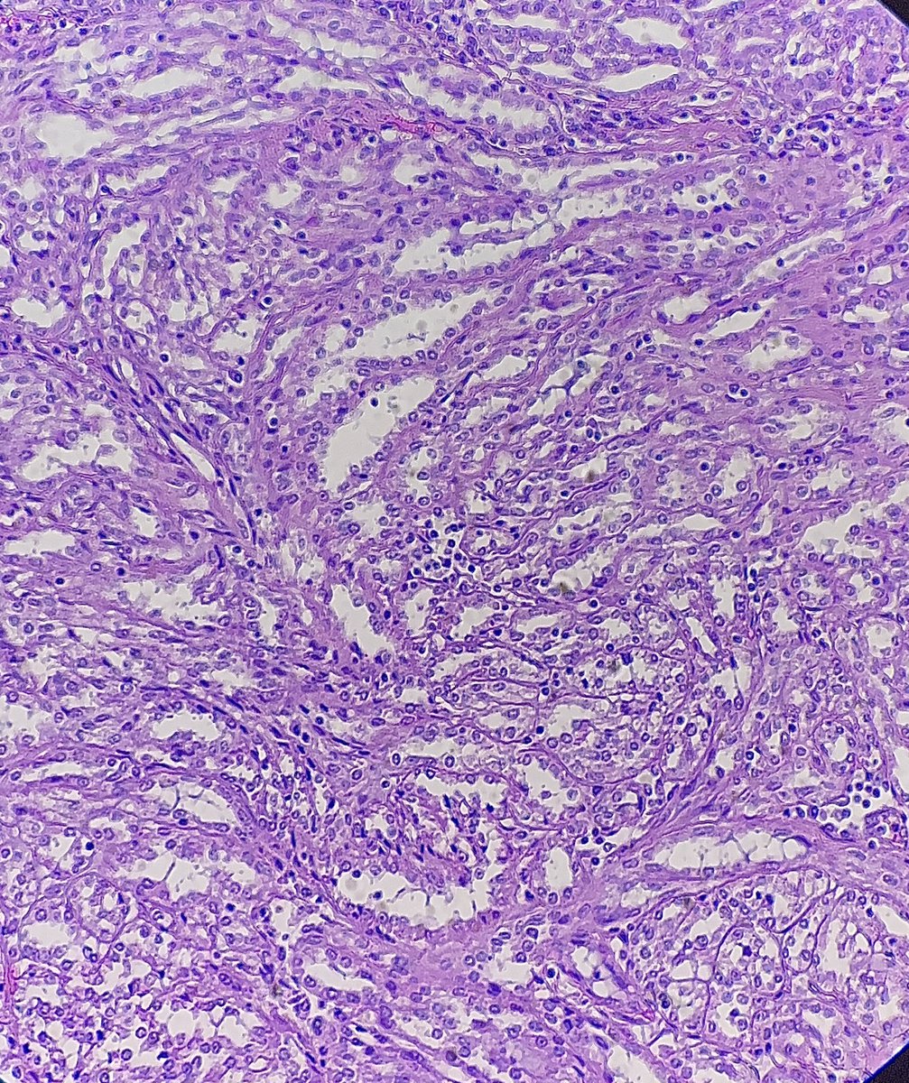
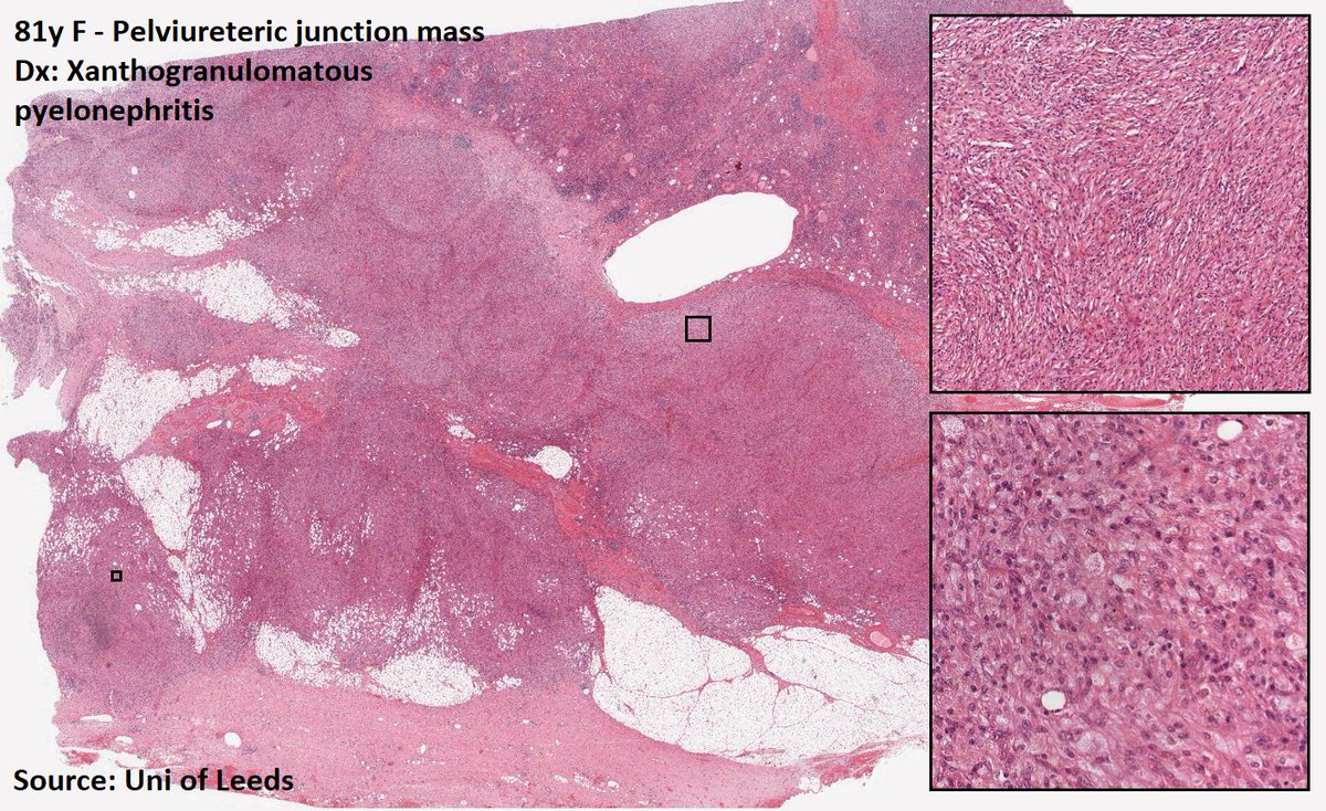
Xanthogranulomatous pyelonephritis
Uncommon/rare
Destructive inflam process that may resemble RCC
Oft assoc with chronic pyelonephritis due to E. coli or Proteus
Histo
Extensive foamy histiocytes
#MedTwitter#PathTwitter
Just published! Great review by @SAMBITKMohanty1@LoboAnandi and Sourav
Precision Medicine in #bladdercancer: Present Challenges and Future Directions https://t.co/yGY2AKuMgK
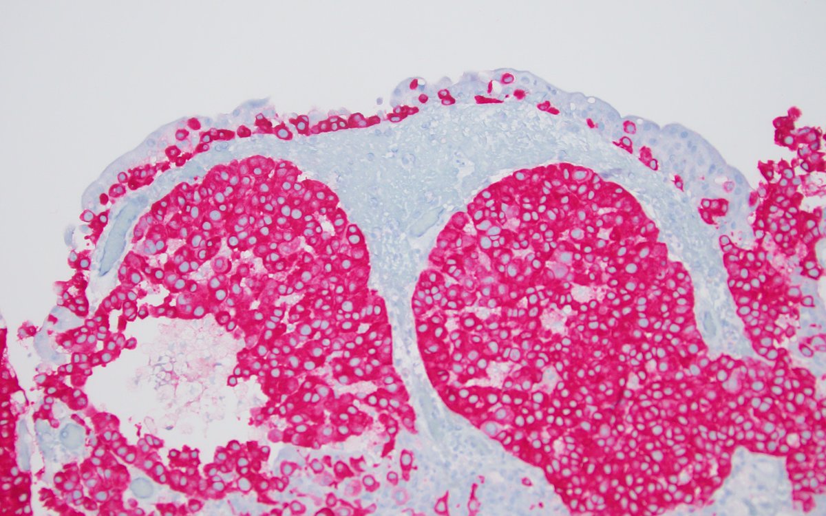
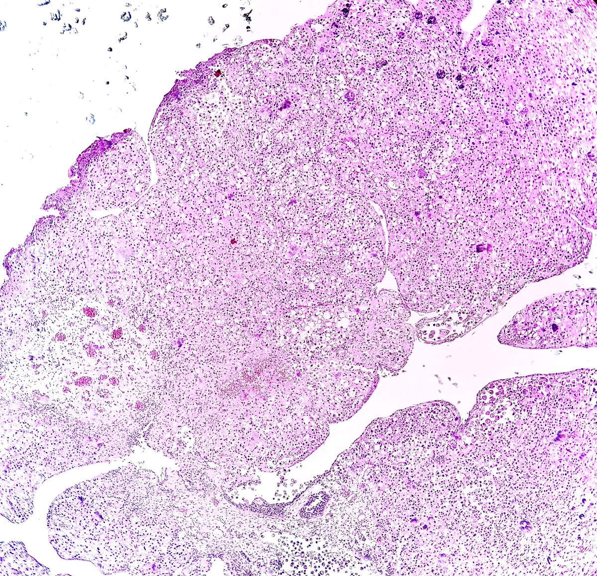
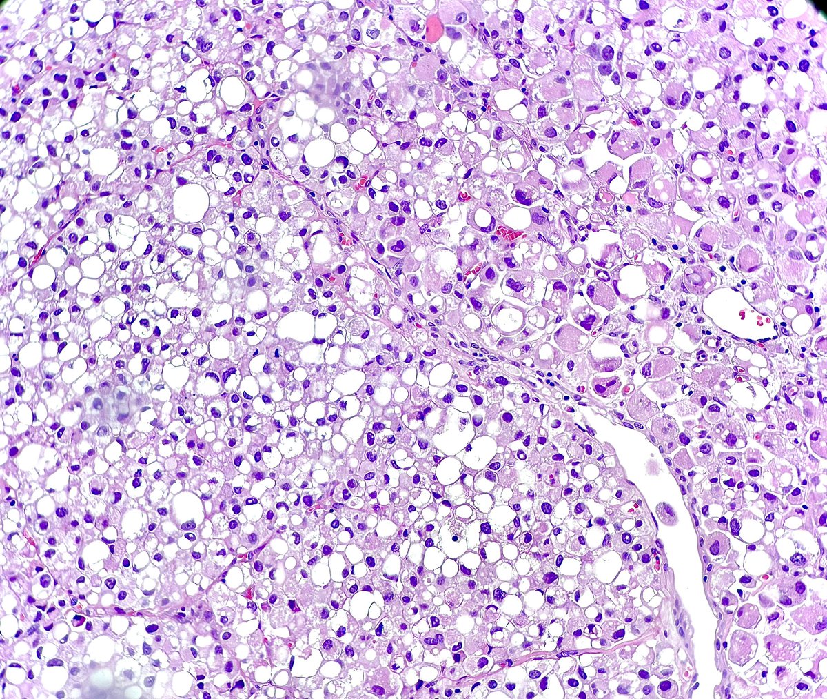
Lipid-rich urothelial carcinoma
✔️frequently associated with conventional urothelial carcinoma
✔️ lipid nature of the vacuolar content has been confirmed by EM
✔️ poor prognosis
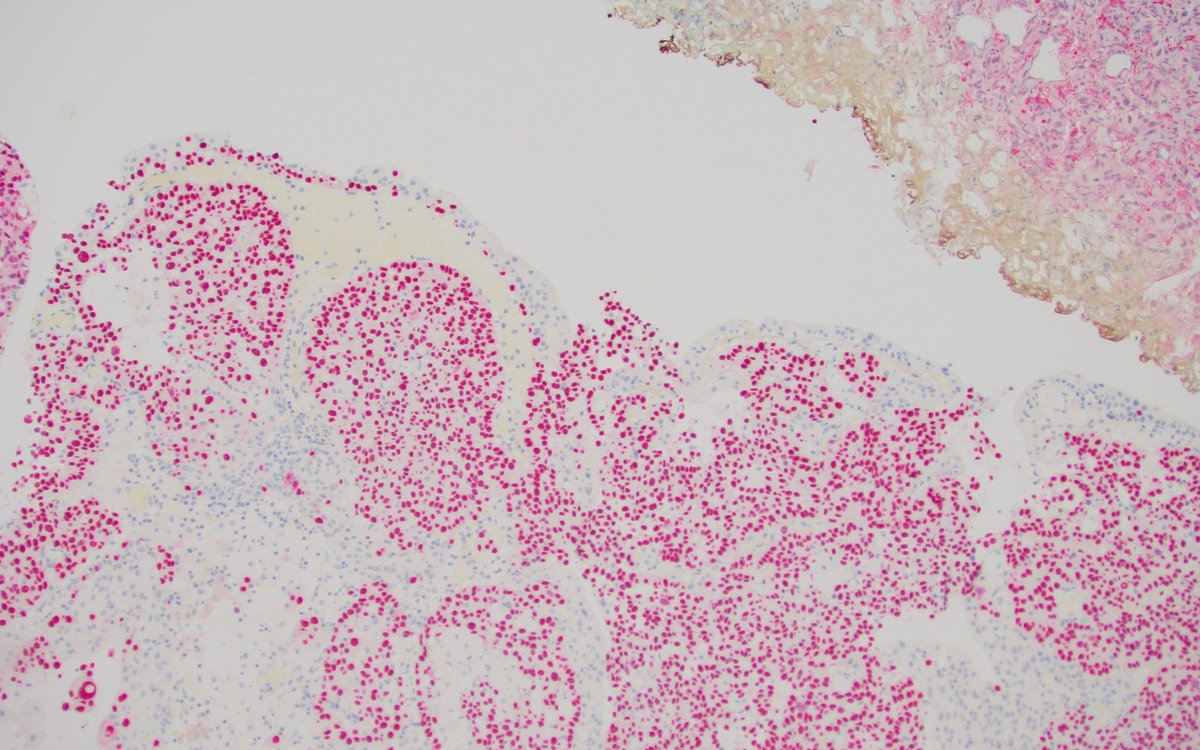
✔️ + CK7, CK20 (photo), 34betaE12
#gupath#pathology#uropathology
Histology flash card
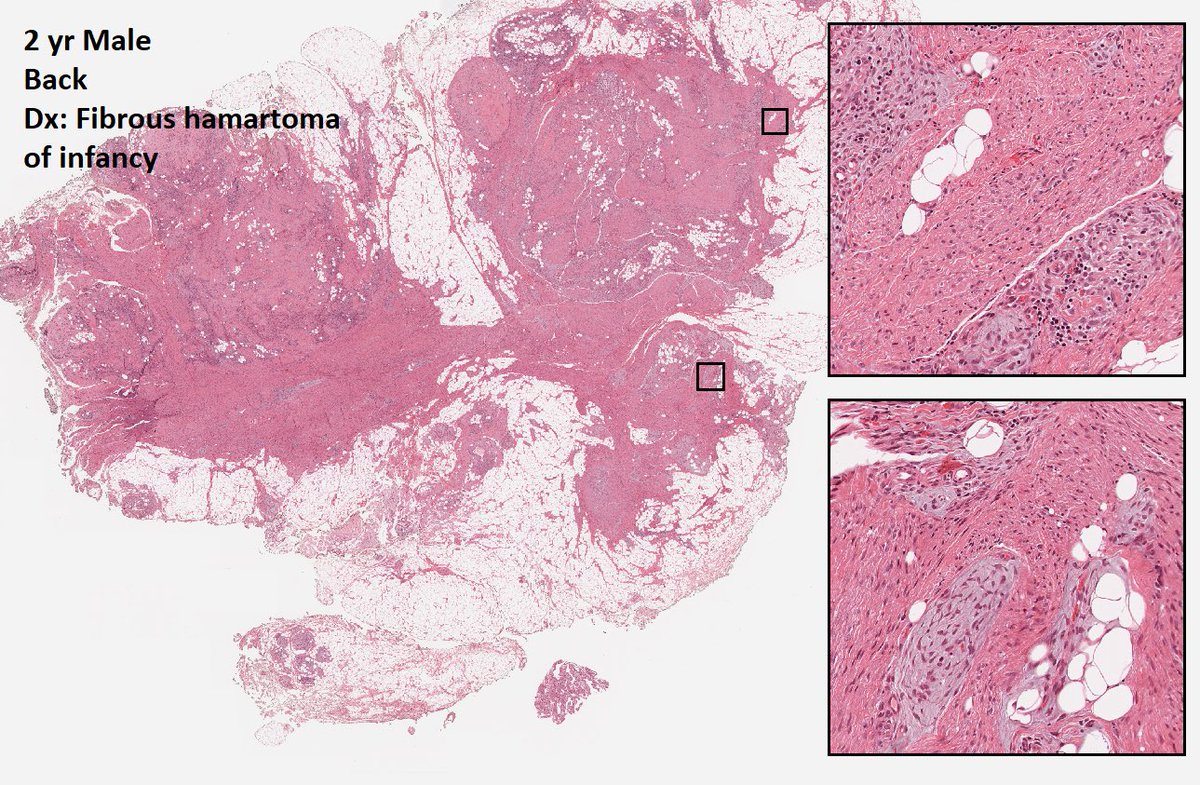
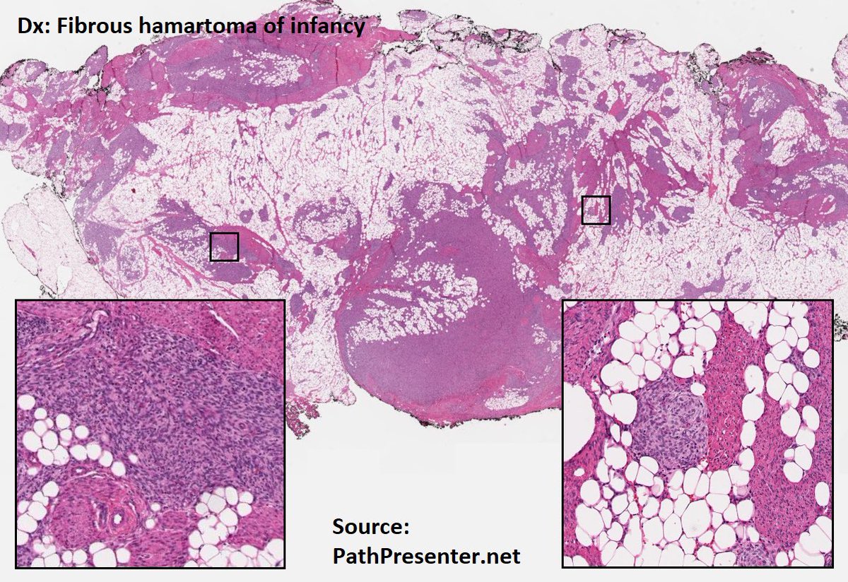
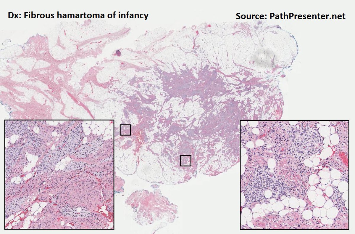
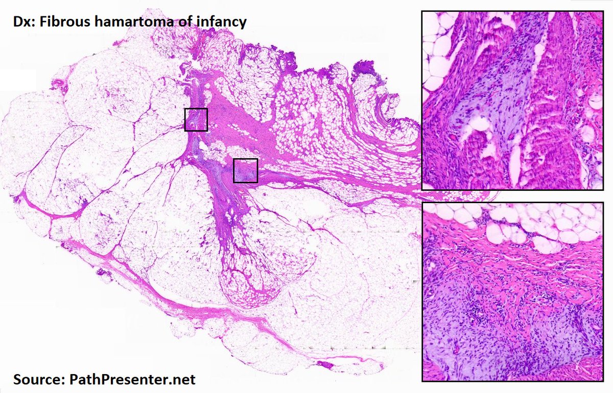
Fibrous hamartoma of infancy (FHI)
Rare
Usually <2yrs (90% <1yr)
Oft solitary
Location: subcut or reticular dermis
Benign
Rarely recurs (16% and may be explained by incomplete excision)
Histo
Mature adipose tissue
Fibrous tissue
Myxoid & basophilic areas
May is #bladdercancerawarenessmonth - important to recognize lesions which mimic #bladdercancer
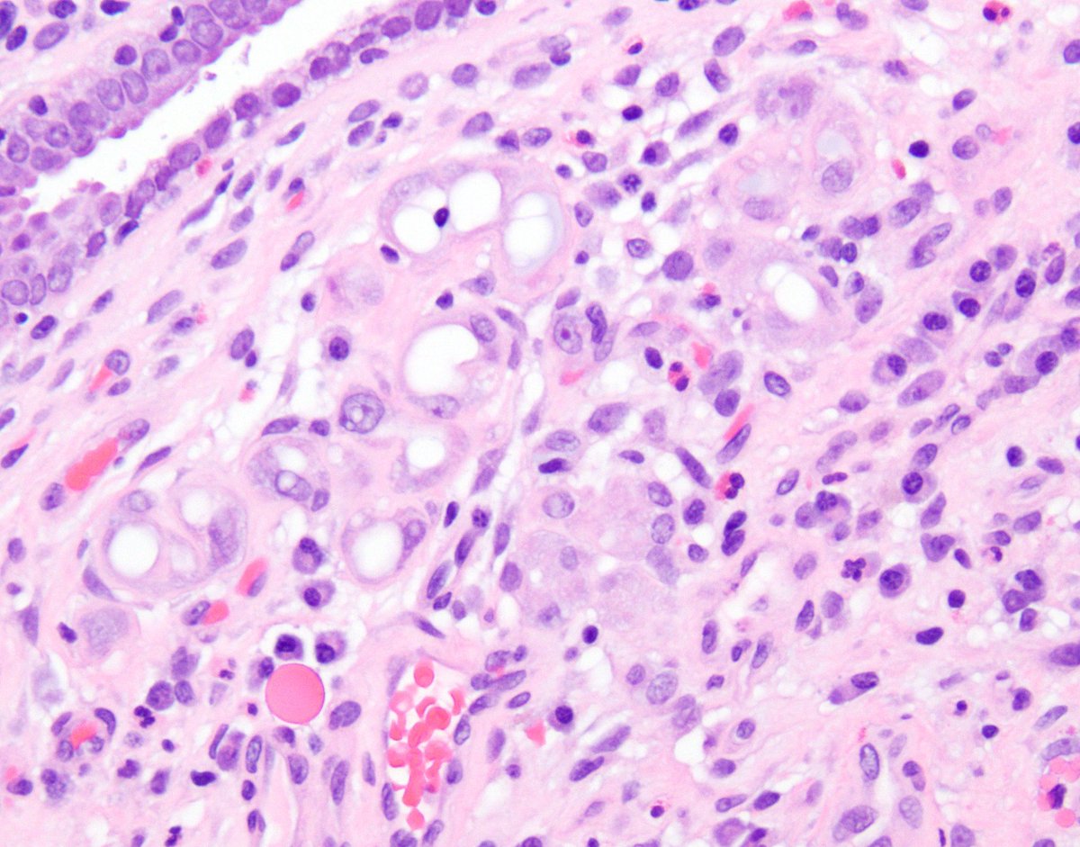
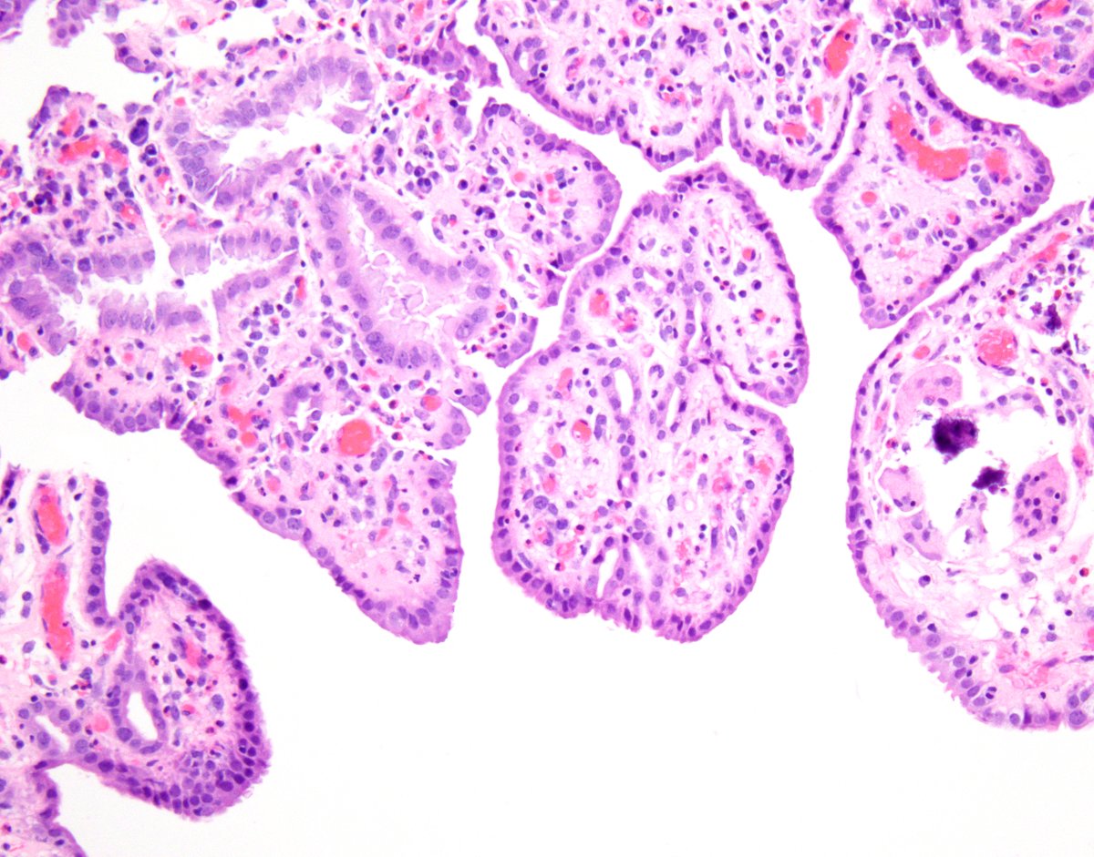
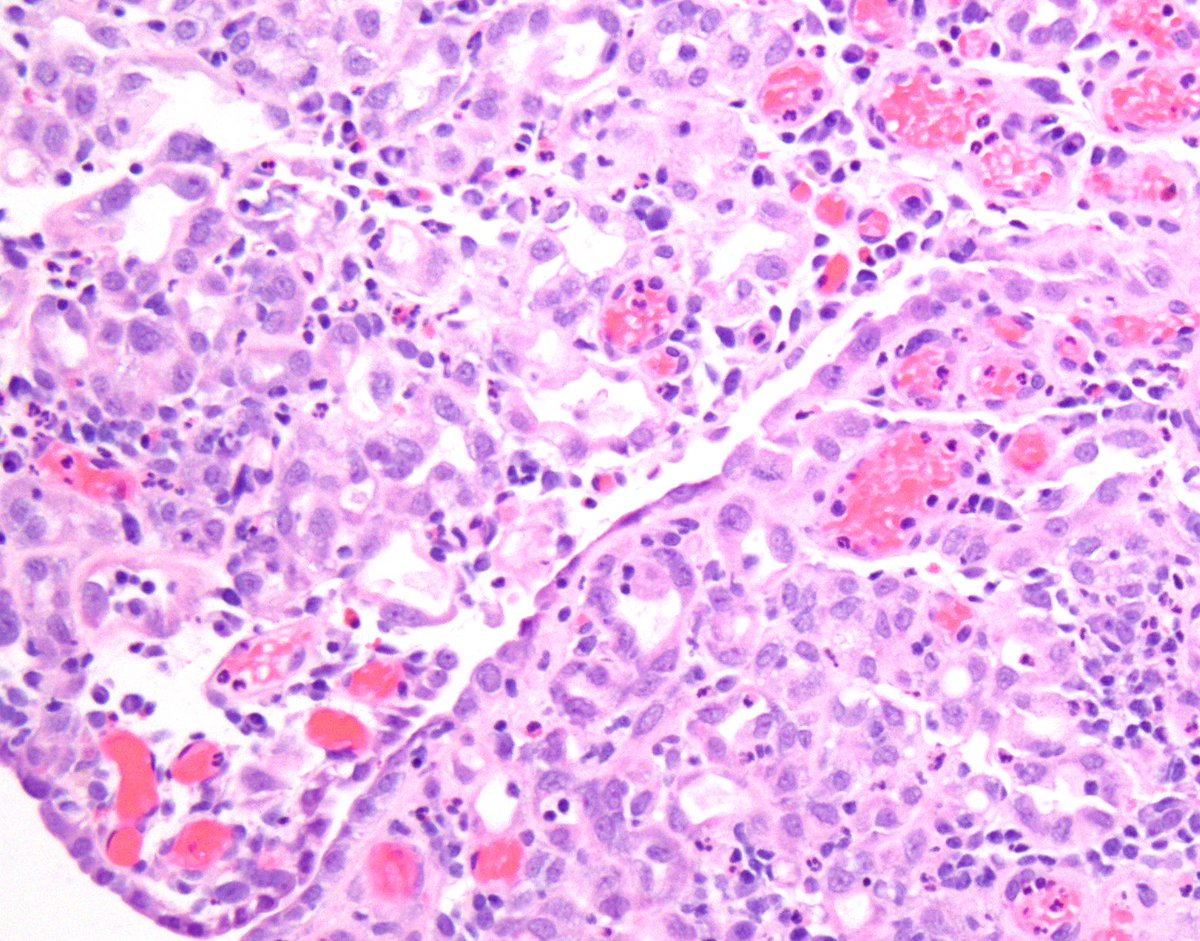
Nephrogenic metaplasia/adenoma:
- various architectures (papillary, tubular/glandular, vascular-like...)
- cuboidal to hobnail nuclear features
- prominence of basement membrane
Their presence can be subtle 🔬
Urothelial carcinoma with trophoblastic differentiation.
Note the syncytiotrophoblasts occasionally surround (cap) the urothelial cells and are thinned out.
See IHC in comment. #GUPath
Case of the month May 2023
47F with bladder wall mass
➕ GATA3, SYN, CG, INSM1
➖ CK AE1/AE3
S-100 protein highlights sustentacular cells
Authors: Katrina Collins (@SueEPig), Jessica L. Davis, Andres Acosta (@Andres_M_Acosta)
@IndianaPath#IUpath
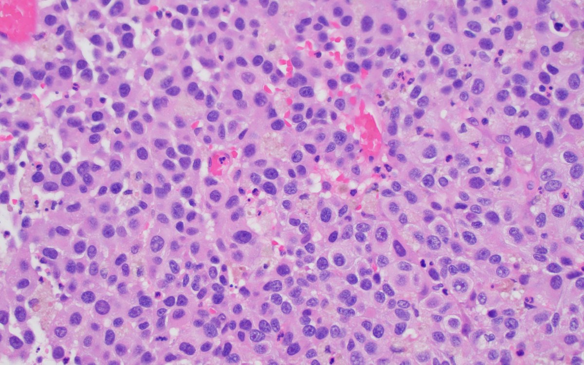
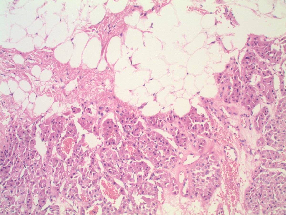
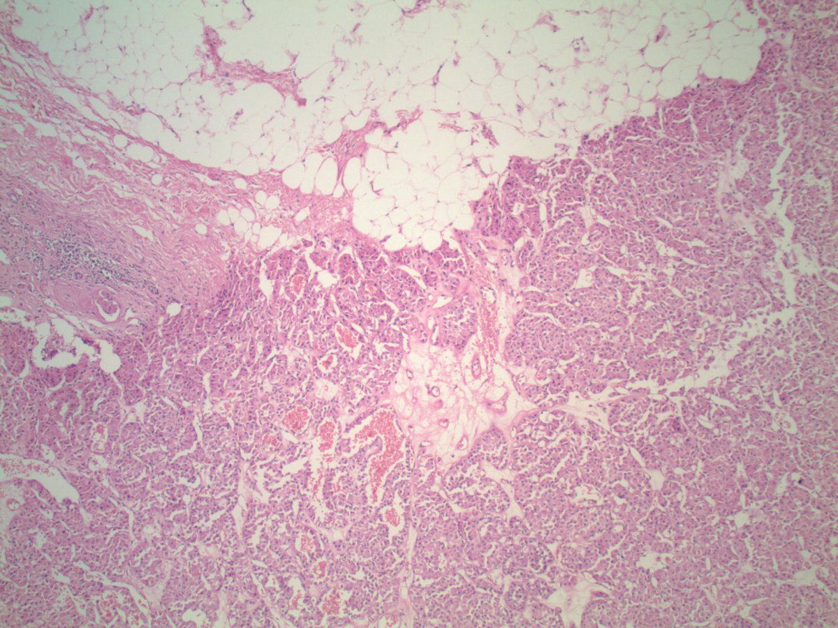
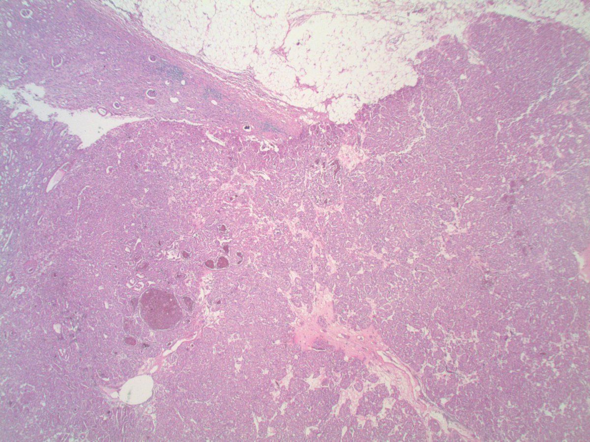
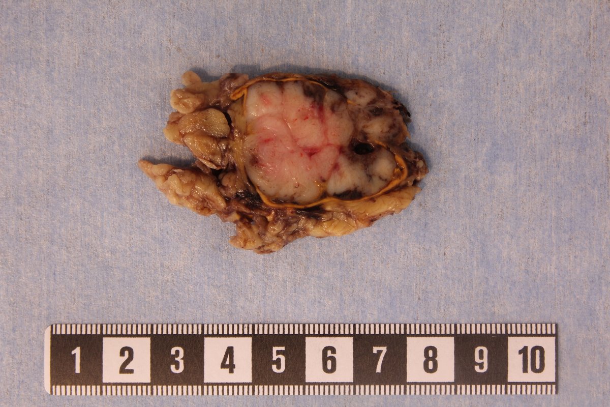
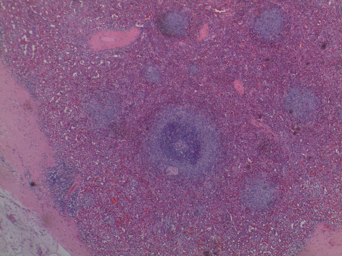
Left-sided adrenalectomy in an adult. Localized mass, non-producing. White cut surface, firm, lobulated, with extraadrenal extension.
Histological images in the thread below!