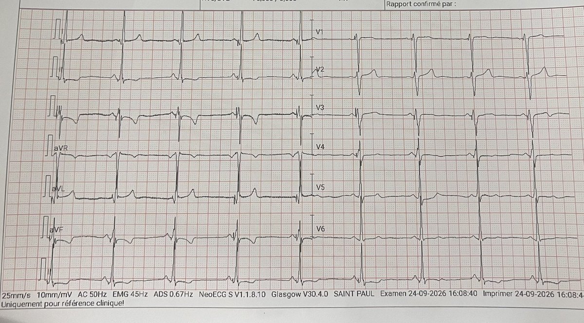
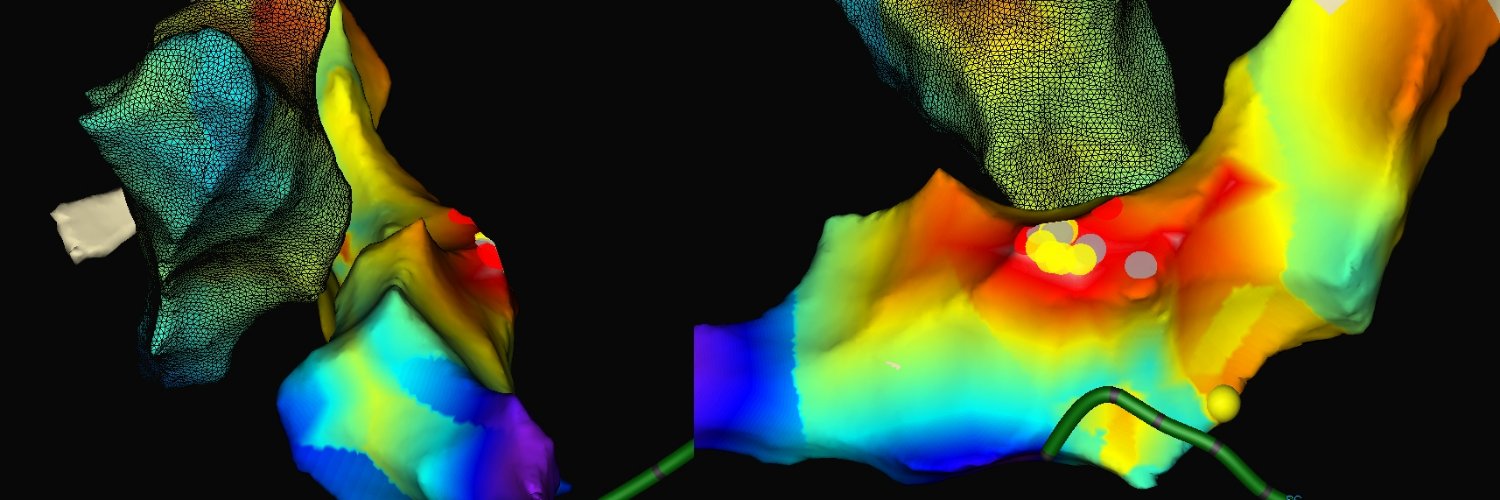
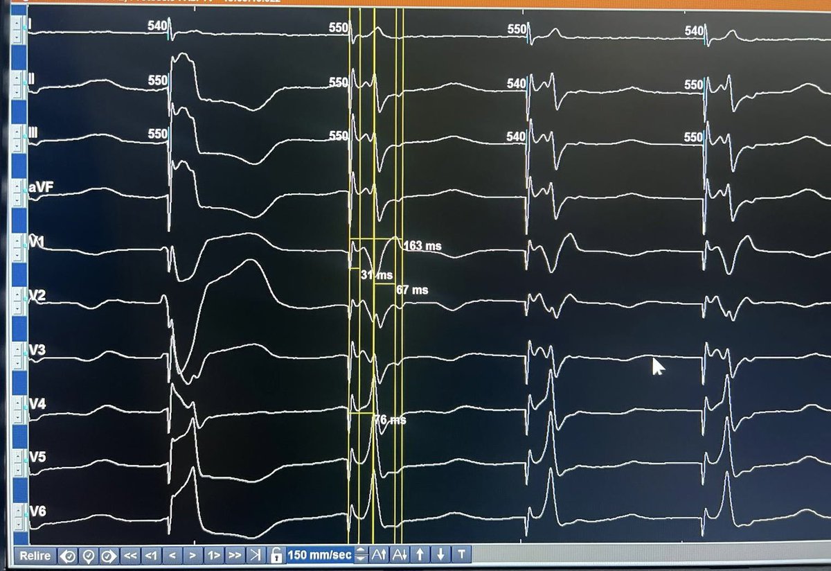
@syamkumarmd Look at this tracing. There was some retrograde VA conduction. Sometimes 3/2 sometimes 2/1. I started w SP ablation. But the pat felt pain even though I was not inside CS, no junctional beats. So after 2 RF pulses I went to NCC. Do you have an explanation for different QRS??
PVC on His (5th beat) and late APC without reset. PPI after VOP variable. No constant coupling interval AVV ➡️ JET. During tachy, earliest His posterior to AVN. Did not dare to ablate there (20% risk of AVB). ➡️ 20-25 W RF NCC. Firing ➡️ No sustained tachy inducible 😅
@DrAJRestrepo Agreed. EPS should be done more often, especially in young patients. Perhaps EPS and AVNRT ablation before the left atrial procedure could have even avoided PVI in a 52 yo patient?
@jeffrey_vinocur And she did have syncopes with the AP „intact“. Ablating it or not may be a more academic thought. We discussed it in the team, there were different opinions. I was for ablation but I wouldn’t have pushed it in case of a difficult ablation. A pacemaker had to be implanted anyway
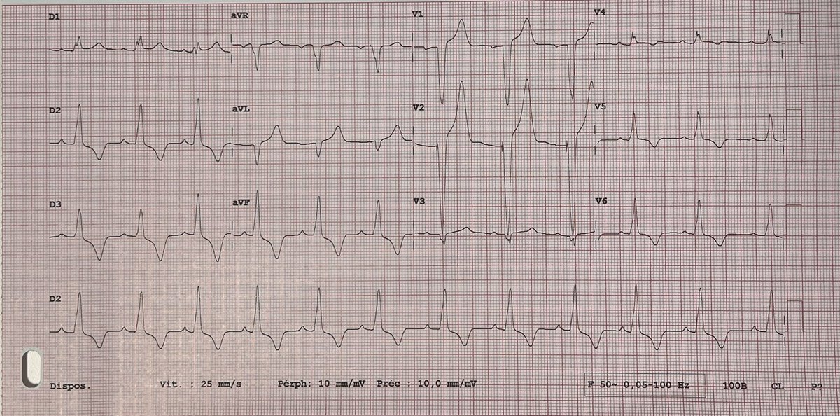
Would you have ablated this AP, #EPeeps? 68 y/o lady + syncopes + WPW, but a very slowly conducting AP. AP block at 600 ms during IAP. Ablation unmasked LBBB, AV conduction disease with AH 100 ms and paroxysmal complete AV block. AP ablated + Micra AV implanted.
@cclang It’s possible that she’ll be paced >40%. Main argument was to find a solution during the same procedure. What would you have done? LBBP? This is the paced ECG
@cclang Thx for your answer! Anterograde ERP was 580, retrograde 420 ms. Unfortunately I didn’t test w isoproterenol. I ablated the AP as 1) AP conduction is not reliable 2) to prevent tachycardia in the future. The pat needed a pacemaker but the decision to ablate is surely debatable.
Thank you, Dr Warren “Sonny” Jackman for sharing your extensive experience in another great webinar on accessory pathways! It’s a true delight to listen to someone who still practices the beautiful art of electrophysiology!
#Epeeps Sandwich 🥪 ablation of symptomatic LV summit PVC: earliest signal -30 ms before QRS in both, LVOT and GCV. LV ablation (40 W) supressed PVC, subsequent recurrence. Sandwich ablation from GCV eliminated PVC w 1st pulse (20 W). 1 min fluoro. Happy patient
@StellEkaterina In this case of loss of LBB capture after a previous procedure, we tried CS first but didn’t succeed. Then, this position of LBBP was the 1st attempt. I implant often a bit more midseptal and sometimes more inferior (LIFP) than described by Huang ➡️ less TR, easier implant
66 yo lady w #DCM, NYHA 3+, LVEF 12%, LBBB, QRS 170 ms. 3 months ago, I upgraded her #CRT-D w #LBBP. Today: NYHA 1-2, LVEF 35%, QRS 130 ms. But the most important result was not on the echo or ECG. She told me: „Thank you, doc! This operation has changed my life.”
@StellEkaterina Thank you. “Fortune favors the prepared mind” 🤓 (Pasteur). In this case, fusion of LBBP and intrinsic AV conduction to the RV first helped to create this slim QRS. This is the QRS with VVI pacing
@Emilioamigo_ The QRS is beautiful isn’t it? It could be HBP but it isn’t. You can see the pacing lead in the middle of the septum. The QRS is so narrow because of fusion of LBBP avd intrinsic AV conduction to the RV. Therefore you don’t see a Qr in V1 as in VVI pacing during the implant