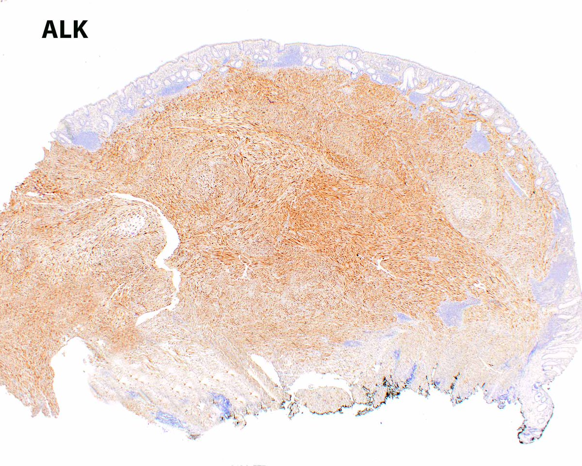
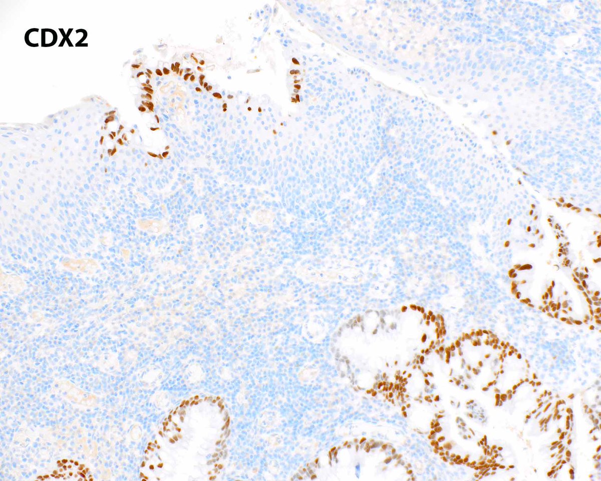
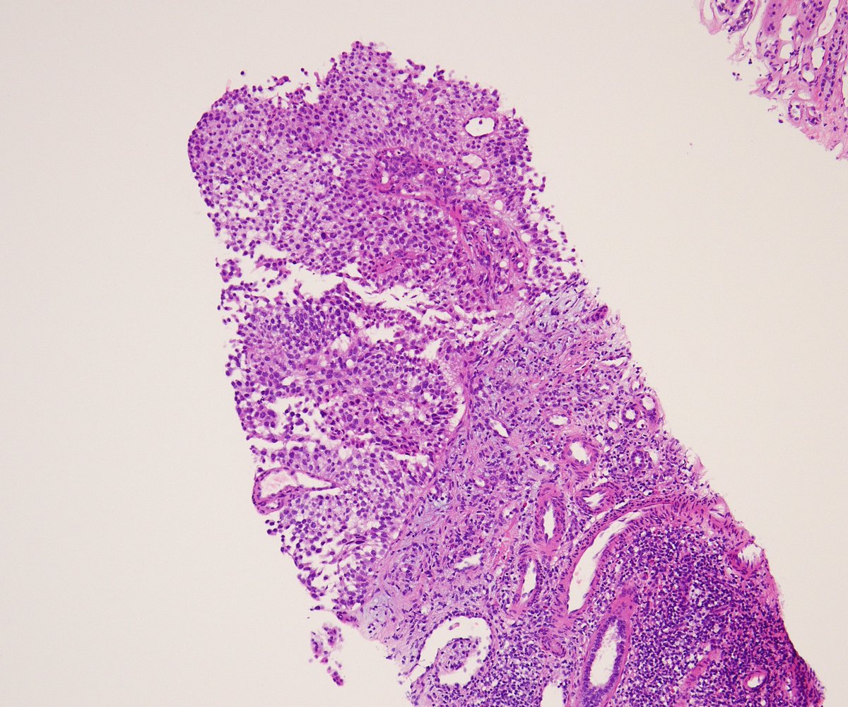
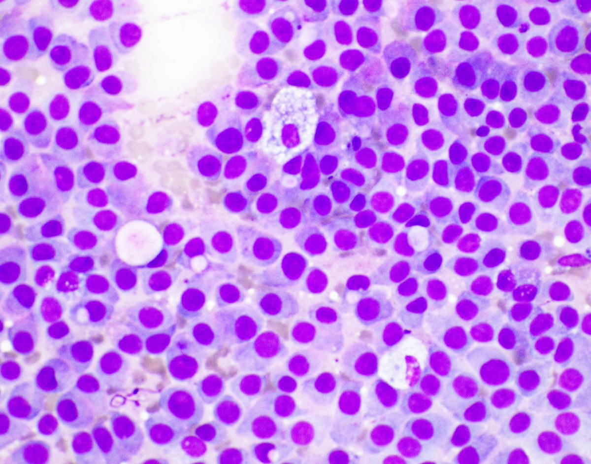
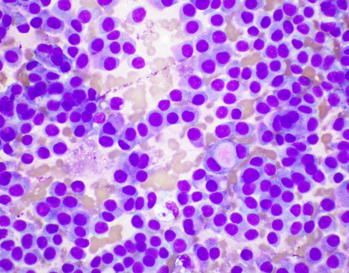
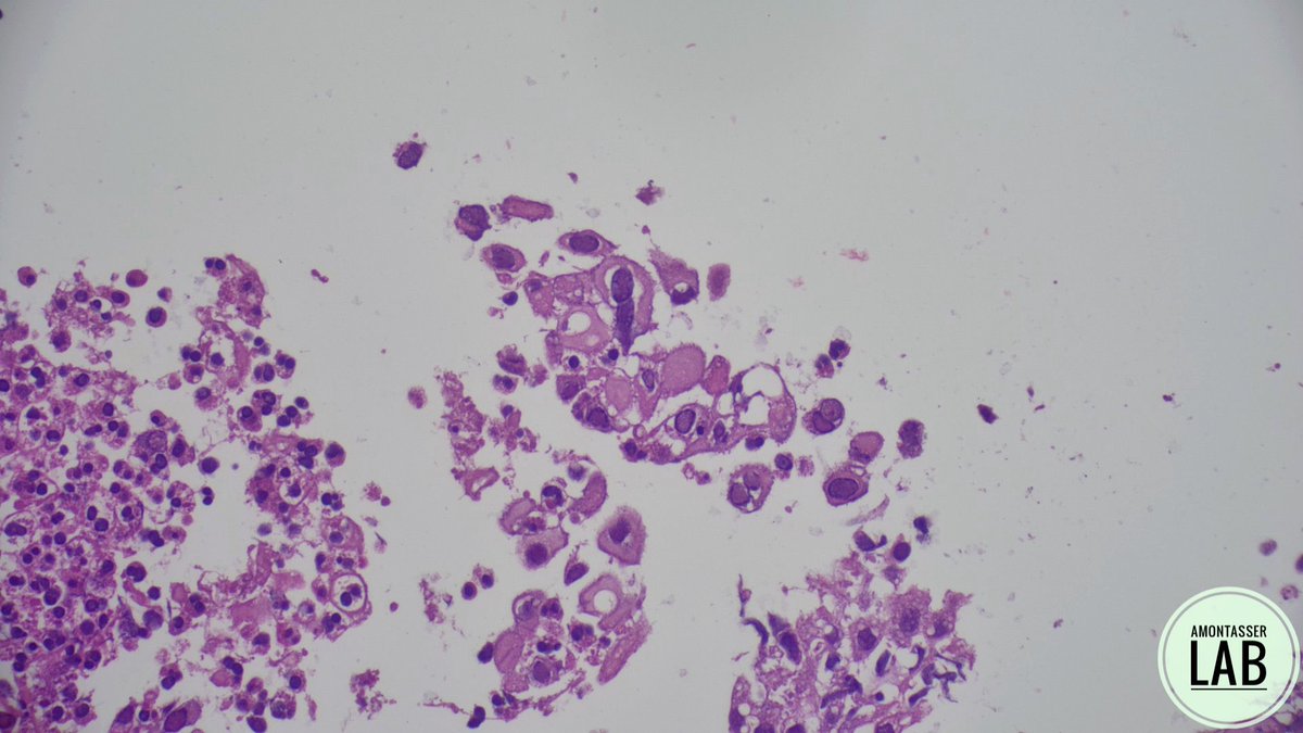
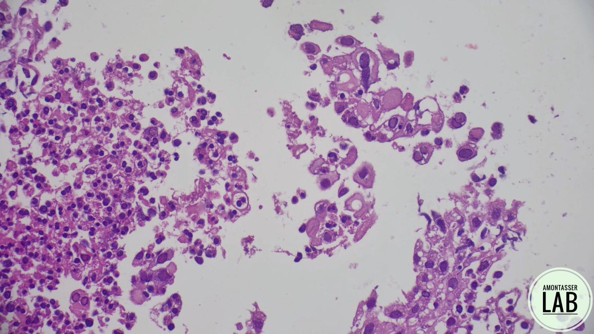
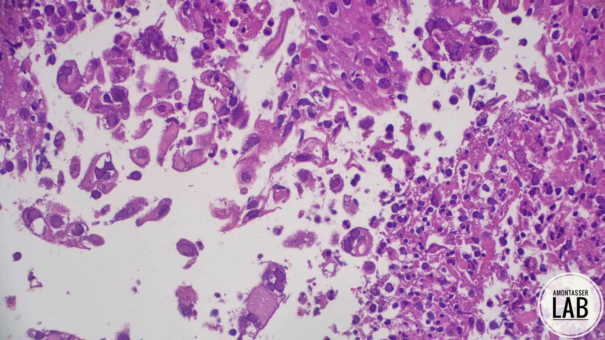
When inflammatory myofibroblastic tumor (IMT) extends into the submucosa and mucosa, it can mimic a gastrointestinal tract schwannoma because of its cuff of lymphoid aggregates. Generally, IMTs are located in the mesentery and schwannomas in the muscularis propria. #UMiamiPath
High-grade DCIS associated with metaplastic carcinoma.
Even if the entire tumor is keratin-negative, the presence of DCIS supports the diagnosis of metaplastic carcinoma (see pic)
When other features are lacking, sample extensively. Go back to the specimen and take more sections from the periphery—leaf-like architecture (phyllodes) and DCIS are often found there
Dr. Wen. Interactive microscopy, Practical Updates from the International Society of Breast Pathology (ISBP) #USCAP #PathX #pathology
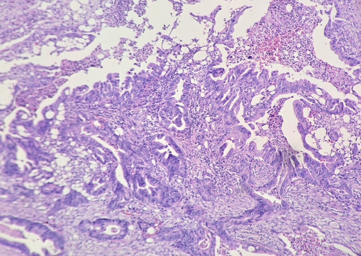
Omental cake biopsy in a case of metastatic adenocarcinoma of colonic primary. Note luminal dirty necrosis within the malignant glands.
#pathology#GIPath#CRC
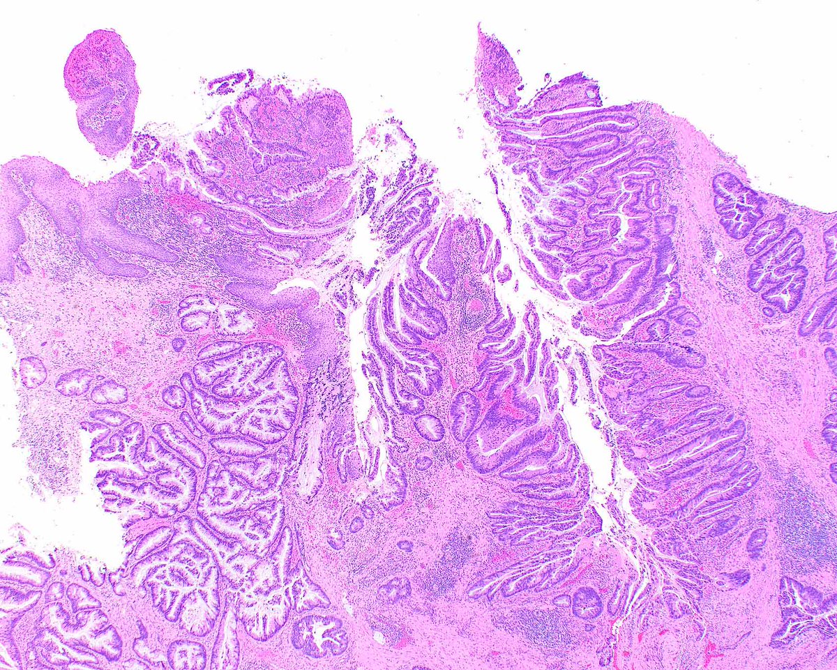
Primary perianal adenocarcinoma with pagetoid intra-epithelial extension - these look a bit like HPV-associated lesions, lack HPV transcripts, and have TP53, ERBB2, and MYC abnormalities, without typical rectal adenocarcinoma mutations (APC, KRAS, BRAF). https://t.co/ajb2kPFkBI
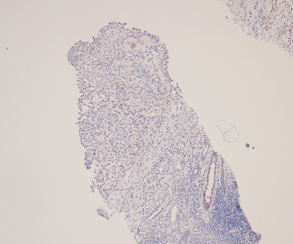
Renal biopsy in a middle-aged woman with a known history of NSCLC. Clinical query: primary renal tumor vs. metastasis?
PAX8 shows weak nuclear expression 🧐
So, renal cell carcinoma or is there another diagnosis here? 🔬
#GUPath#PathTwitter
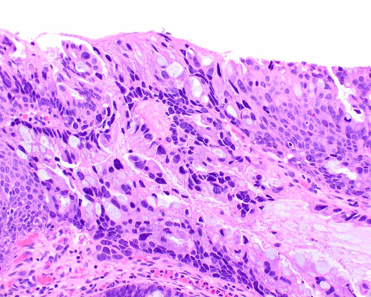
This pattern of esophageal injury is often associated with medications (immunotherapy frequently) or various other immunologic conditions, but this patient had a history of esophageal lichen planus. Apoptotic bodies are indicated.
https://t.co/KctUX85qSk
https://t.co/GKkBB7p0aa
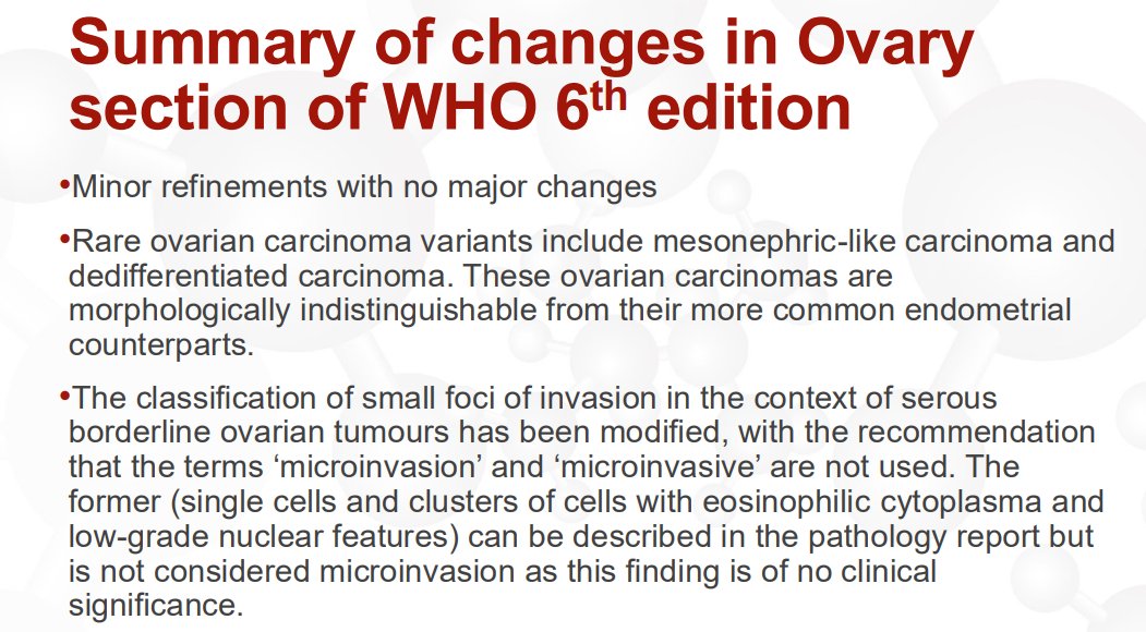
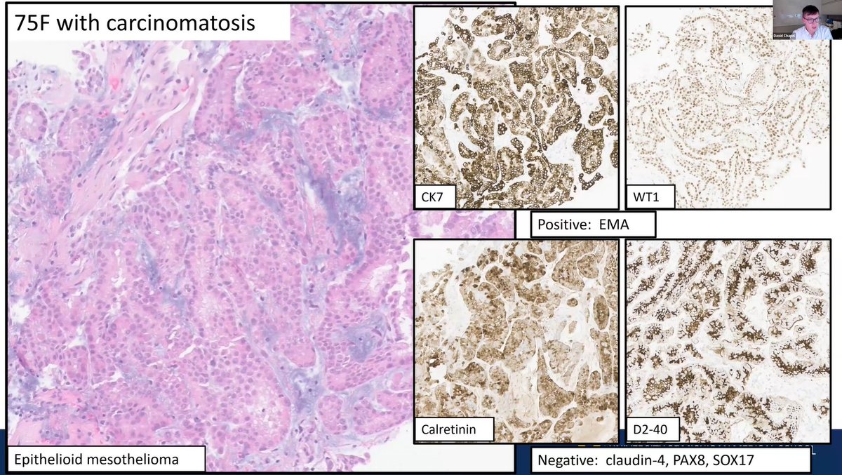
Ovary – Changes to WHO Classification (6th edition, 2026)
Serous borderline ovarian tumors:
The terms “microinvasion” and “microinvasive” are no longer recommended
What was previously called microinvasion (single cells and clusters of cells with eosinophilic cytoplasm and low-grade nuclear features) has no clinical significance (see pic 2). It can be mentioned in a comment if desired; Dr. Gilks previously reported it but no longer does
What was previously called microinvasive carcinoma represents small foci of low-grade serous carcinoma (see pic 3). Report the size and extent of these foci and diagnose them as a small focus of low-grade serous carcinoma arising in a serous borderline tumor
Dr. Gilks. Gynecologic Pathology Masterclass 2026
#USCAP #pathology #pathX
Very happy to share newest online publication in JAMA Oncology @JAMAOnc, "FGFR Alterations in Bladder Cancer- From Molecular Pathogenesis to Precision Oncology," written by Dr. Katrina Collins @katcollmd
In this review, we examine how FGFR biology is reshaping the molecular classification and precision management of patients with bladder cancer. We highlight the central role of FGFR3 alterations and the fast-growing range of molecular biomarker testing at every stage of disease. We discuss comprehensive genomic profiling, liquid biopsy (including urine-based molecular testing), and emerging molecular residual disease (MRD) assessment. We also cover how each is used in diagnosis, treatment selection, surveillance, and disease monitoring.
On the therapeutic front, erdafitinib has established FGFR-directed precision therapy in advanced urothelial carcinoma. Combination approaches, intravesical FGFR-directed treatments, and next-generation FGFR3-selective inhibitors are extending this strategy into new clinical settings. Emerging technologies, including AI-assisted histopathology and integrated multi-omic biomarkers, may further refine patient selection.
Read the full article: https://t.co/FXLvelufYS
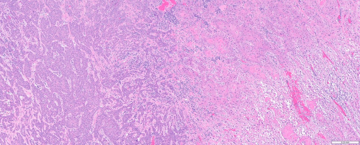
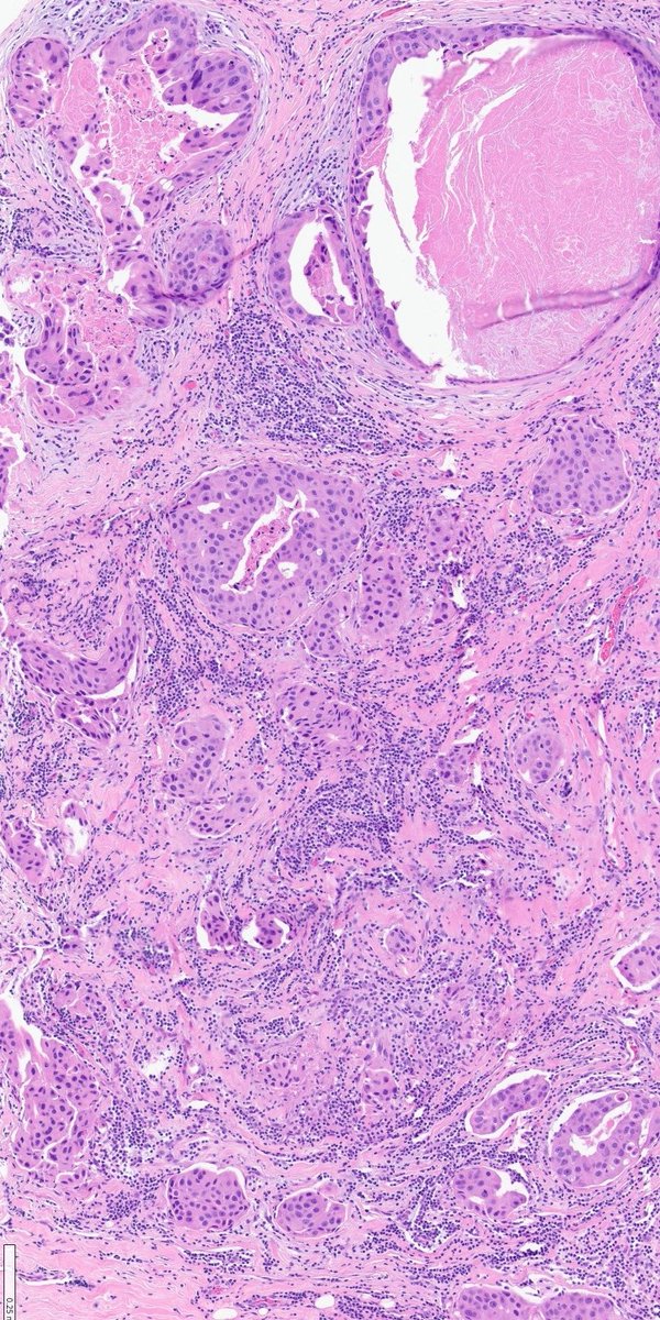
#GUpath TURBT:
invasive urothelial carcinoma (left half) with squamous differentiation (right half)
in the absence of background traditional urothelial ca elements,
DDx includes primary bladder SCC, direct invasion, met (#IHCpath typically not helpful!)