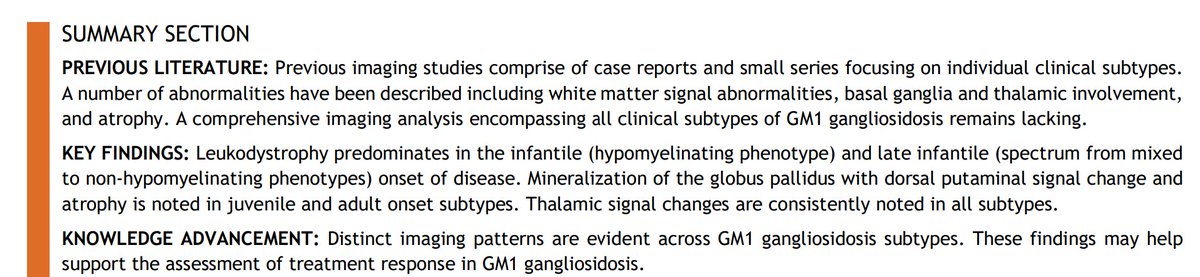
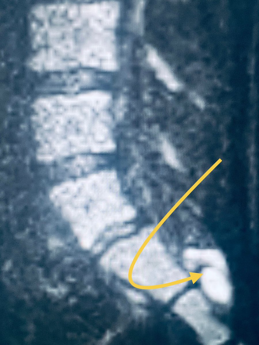
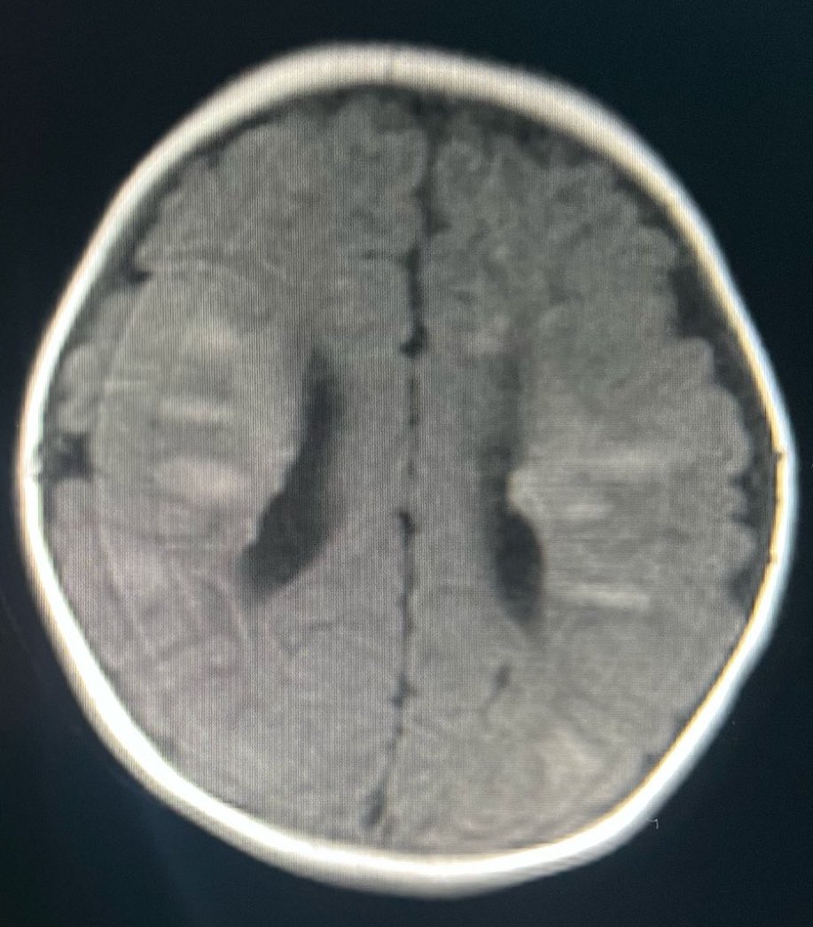
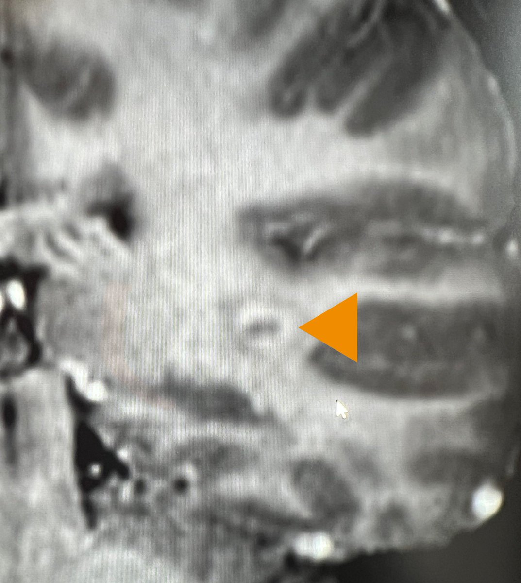
Absolutely chuffed to share our work- Neuroimaging spectrum of GM1 gangliosidosis published in @TheAJNR . Grateful to my colleagues in London, Turin, Philadelphia, Riyadh, Sao Paolo and Porto Alegre for their incredible contributions! https://t.co/Z6sc00OpwF
🧠 This week’s SPIN-POV: Multifocal lesions in the cerebrum, cerebellum, brainstem, and deep gray nuclei can be the initial presenting feature of Hemophagocytic Lymphohistiocytosis.
Don’t miss it, as early testing, systemic therapy, and HSCT can improve outcomes.
Great Ormond Street Hospital for Children NHS Foundation Trust is offering a
Locum Consultant Paediatric Neuroradiologist post !
https://t.co/XKh3XUqWul
🔥TOMORROW🔥
Still time to register ⏱️
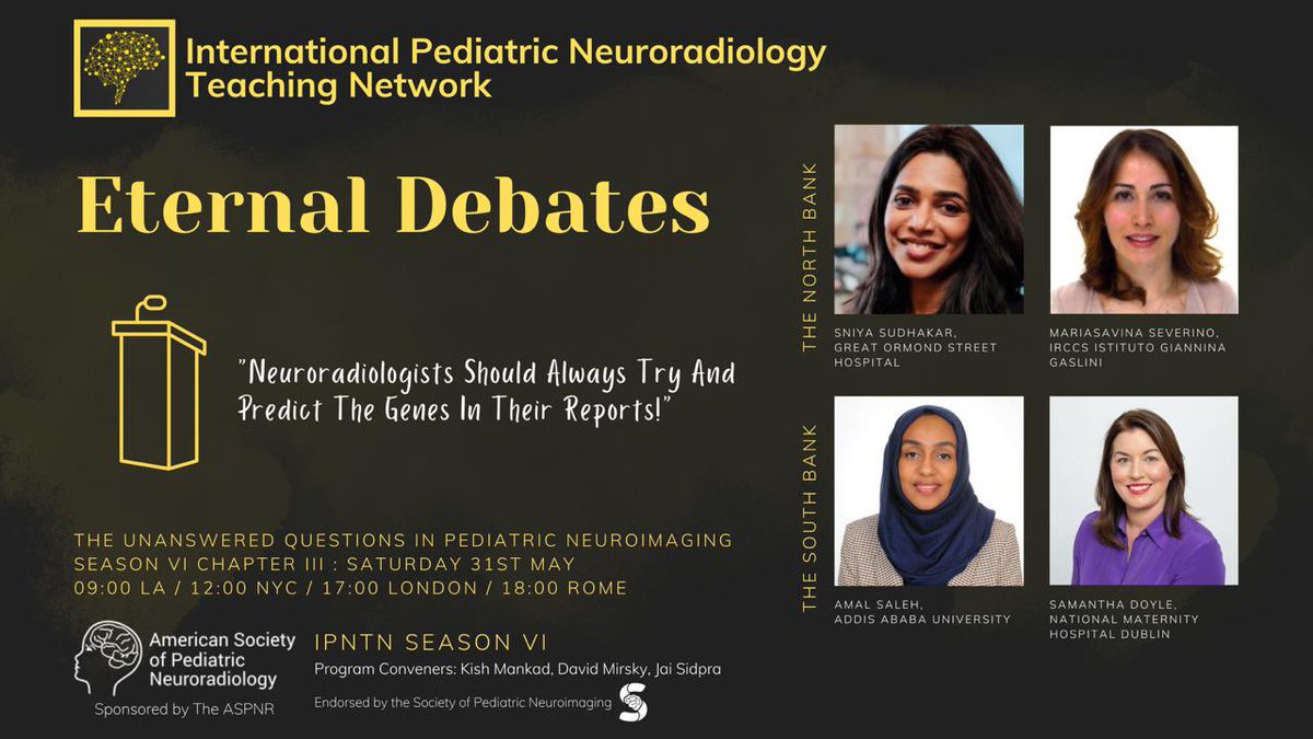
International Pediatric Neuroradiology Teaching Network
Season VI: Eternal Debates
Chapter III: The unanswered questions in pediatric neuroimaging “Neuroradiologists should always try and predict the genes in their reports!”
May 31, 2025
Register here: https://t.co/gayxpzyGkz…
This week’s #SPIN-#POV: Have you ever spotted “dark white matter” on FLAIR images in a patient with seizures? That subtle FLAIR hypointensity can help identify the seizure focus. It’s not pathognomonic and can be seen in a variety of conditions (this was MOGAD).
Read on, here’s the weekly #SPIN-#POV. If you encounter a cystic lesion where a previous myelomeningocele was repaired, proceed to perform diffusion imaging. An epithelial inclusion cyst will light up like here. These can grow and surgeons are keen to get them out.
#SPIN-#POV: This week, a very instructive case from a dynamic friend of #SPIN@aisfagan. The point of view made is that in a neonate, T1 trumps over T2 to show the dysplastic lesions of Tuberous Sclerosis #TS. Thank you Dr Fagan!
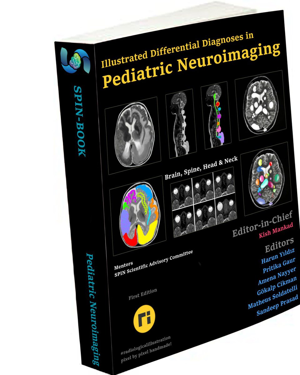
The #SPINBOOK is releasing soon….Your illustrated guide to differential diagnoses in Pediatric Neuroimaging. Our editors are working hard to get this one completed. Thank you in advance to all our wonderful contributors. As we say, when it’s innovating, then it’s spinning!
This week’s SPIN-POV here with @drmankad. It may look like multiple sclerosis and satisfy McDonalds criteria for DIT & DIS with that enhancing lesion! Yet it remains a “Radiologically Isolated Syndrome” until the child has a clinical event and or OG bands- both negative here.
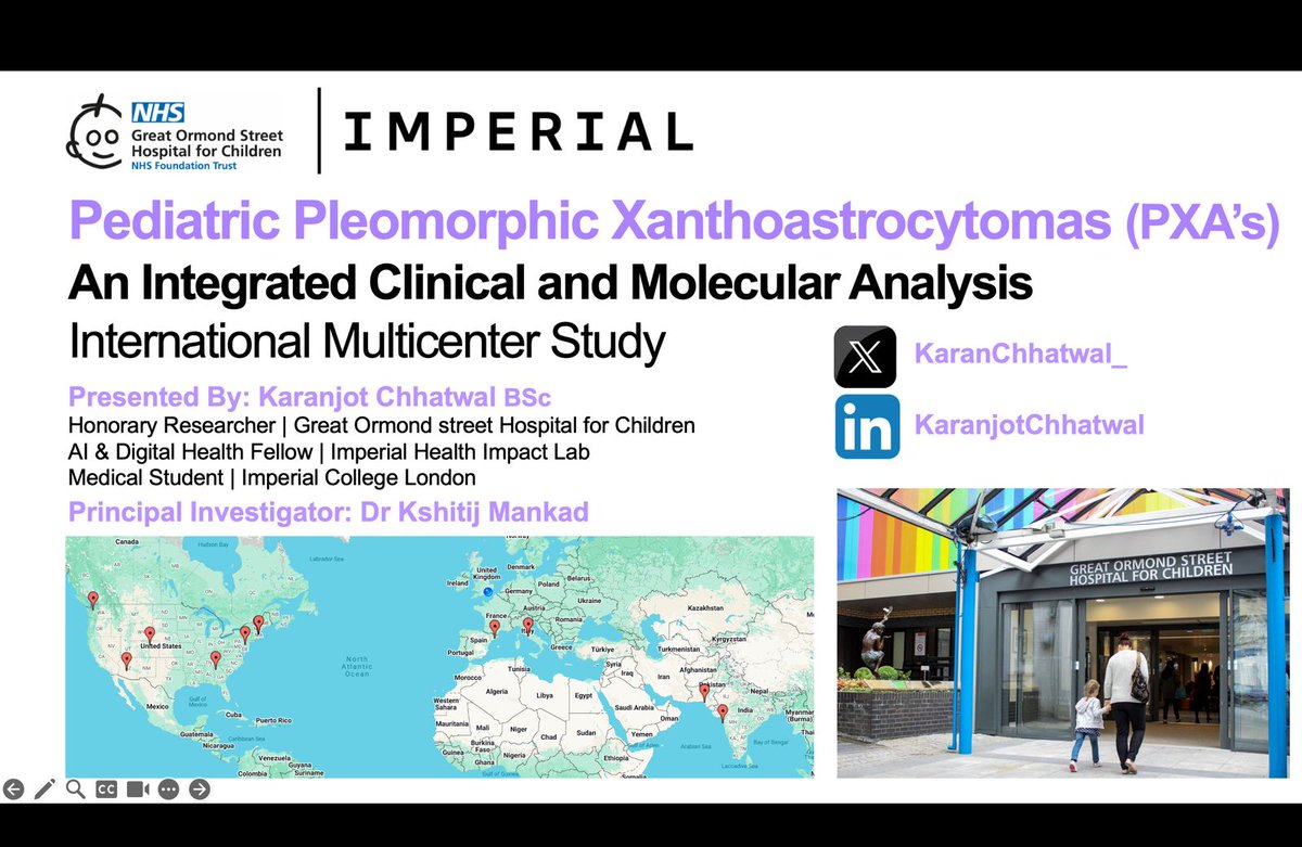
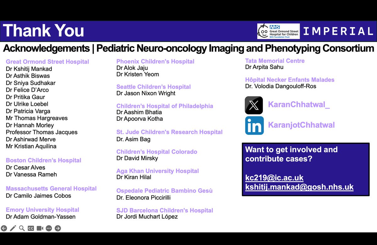
Can’t wait to present our project on Pediatric Pleomorphic Xanthoastrocytomas at @The_ASPNR in Austin, Texas!
Huge thanks to the team at @GOSHneurorad and our amazing collaborators worldwide. Excited to share key findings on neuroimaging and survival predictors!#ASPNR#Radiology
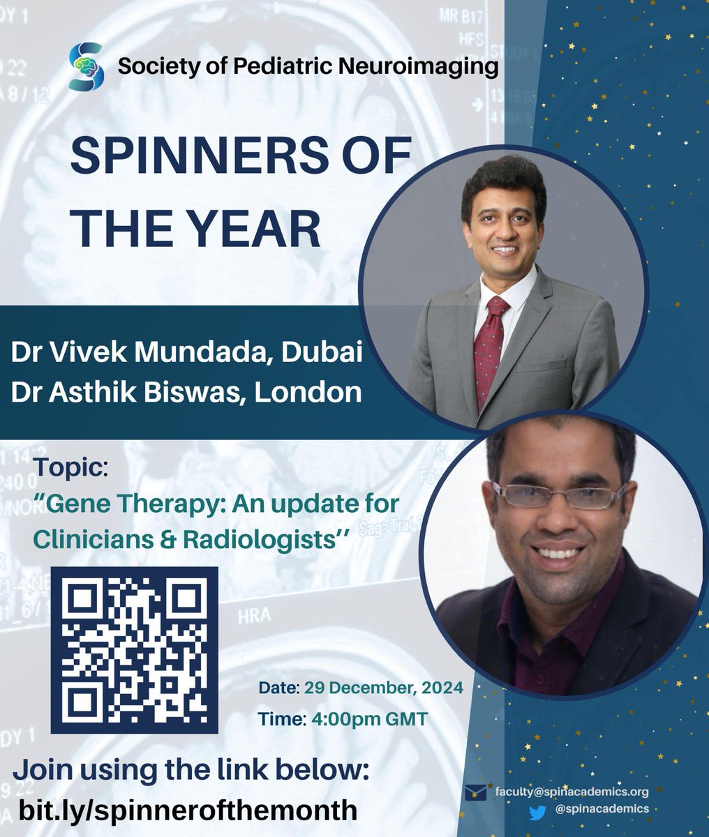
The spinner of the month series wants to end the year with a bang, as we set the stage for greater collaborative pursuits in the next. Join the #SPIN movement to enhance your academics. Welcome two incredible speakers this month, sharing their updates on #genetherapy.
Thrilled with this one, very ably led by @stikkman11 @GOSHneurorad: Imaging readiness in the gene therapy era‐exploring standardized protocols for response assessment - Biswas - Journal of Inherited Metabolic Disease - Wiley Online Library https://t.co/KOngtLrwRm
'Tumour mimics in paediatric neuroimaging' https://t.co/KwoC3aPJoH : Useful article highlighting the importance of early identification of tumor mimics, as well as #radiologists guiding early intervention where needed. Thank you @FiorenzaCoppola for leading on this paper.
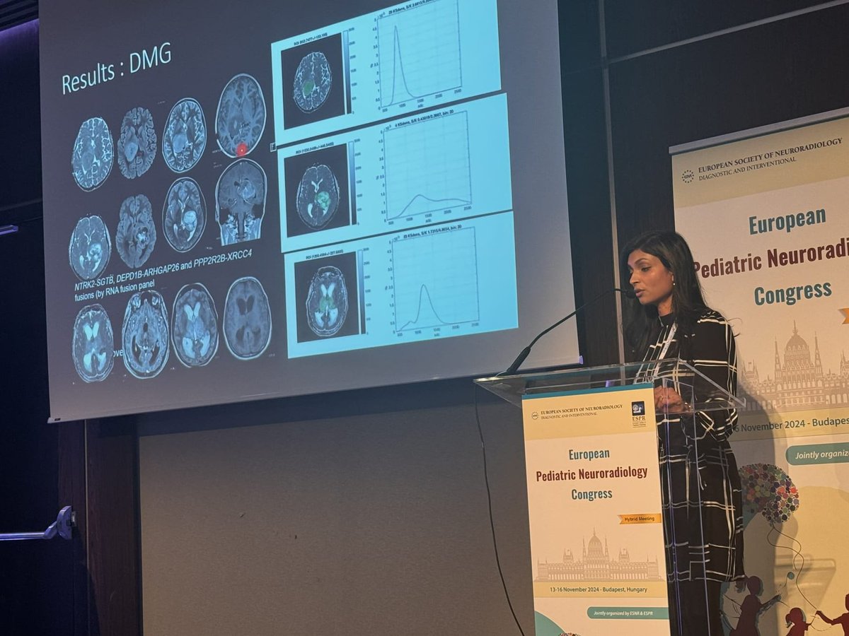
I am grateful for the opportunity to honour my mentor Zoltán Patay with this lecture. Such a beautiful and moving session. We miss him, his ideas and advise in our daily work and at our meetings.
Sincere thanks to Maria Agyropoulou & @AndreaRossi_NRX for the invitation.