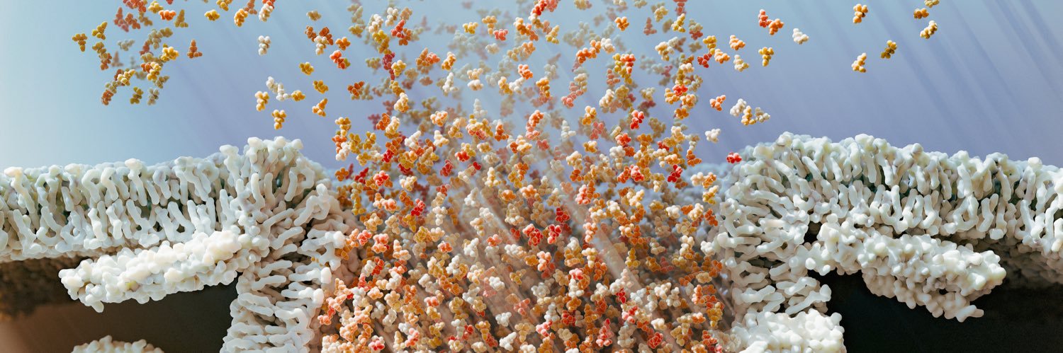
Dear fellow scientific animators, here's a tutorial on my approach to 3D vesicle fusion animations in Houdini: https://t.co/HZ8GRM3JSl
This tutorial addresses the problem of lipid persistence/flickering that occurs in many approaches by using a particle solver in Houdini.
I created this image of the PhD process as part of a job interview earlier this year.
I didn’t get the job in the end, but I had a lot of fun making it, and it makes me smile when I look at it.
Hopefully it makes you smile too!
The less accurate part is that the transmembrane domains are actually missing, so in reality they would stick out of the membrane a bit further than shown - a future Blender challenge to solve!
Here’s a short clip I made a few months ago while enrolled on a medical animation course! I had a lot of fun improving my understanding of materials and lighting (amongst many other things). Also this scene was infinitely easier to set up thanks to #MolecularNodes
The Delta ligand is shown in red, and the Notch receptor is shown in blue, and the final binding state is the accurate part (using the PDB structure: 4XLW).
My first 3D print - a frame from my animation of neural progenitor cells in the developing spinal cord.
It was so cool to see it gradually emerge from the printer and then even cooler to be able to actually hold it!
@vizcie This looks great, I love that you even have the rotation of the particles included! First thing that springs to mind that I would use this for is particles inside a vesicle and their release, or maybe morphogens diffusing across a developing tissue…
Turns out synaptic vesicles are absolutely packed with glutamate (around 8000 molecules on average). This is what that would look like in an average 40nm diameter vesicle!
Also shown are some of the main vesicle proteins present in their reported average abundance.
Thread (1/4)
@JeffYoshimi The other surprise came from my ignorance of this research area and then seeing electron microscope images where you can actually see the massive ATPases present on the surface of the vesicles in the images! The image I attached here shows a VATPase at the bottom of a vesicle
@JeffYoshimi Thank you! I basically didn’t have an intuition for what the reported concentrations in the literature looked like in an average sized vesicle. I’ve only ever seen cartoon representations of it in figures where a few neurotransmitters are drawn.