👨🏻⚕️ استلقِ لمدة ١٠-١٥ دقيقة فقط، وستشعر باسترخاء رقبتك التي كانت متوترة بإذن الله ❤️
💡أنصح به بشدة لمن يقضون وقتًا طويلًا أمام الكمبيوتر المحمول أو ينحنيون على هواتفهم 👍
👨🏻⚕️A. Acromioclavical Shear Test
اختبار القص الأخرمي الترقوي (AST) هو إجراء فحص سريري يستخدمه أخصائيو العلاج الطبيعي وجراحو العظام لاكتشاف أمراض وإصابات مفصل الكتف. يستهدف الاختبار على وجه التحديد تقييم الألم أو الخلل الوظيفي أو الانفصال في المفصل الذي تلتقي فيه عظمة الترقوة بعظمة لوح الكتف
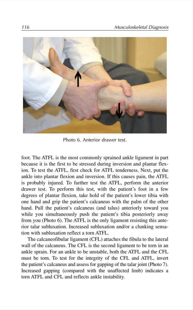
كتاب ممتاز في تشخيص مشاكل الـMusculoskeletal
يشرح كيف تفرق بين أنواع الألم، وتربط بين الـHistory والفحص السريري والـSpecial Tests للوصول للتشخيص الصحيح، مع تبسيط واضح لفهم الـRadiculopathy وتقييم مفاصل الجسم بشكل دقيق
رابط الكتاب📚
https://t.co/hyI1pdDcBN
A) العصب المتوسط او الناصف "Median Nerve"✅
📚 العصب المتوسط أحد الأعصاب الرئيسية الخمسة التي تنشأ من الضفيرة العضدية. يُعتبر العصب الوحيد الذي يمرُ عبر النفق الرسغي، وإذا حدث ضغط للعصب داخل النفق، فتحدث متلازمة النفق الرسغي
📍ينشأ العصب المتوسط من الجذور البطنية C5,C6,C7,C8,T1
أفضل مساعد شخصي لشغل الـPower BI 📊
- استخدم Copilot .. أداة رسمية من مايكروسوفت
تكتب له طلبك بلغة بسيطة، وه�� يجهز لك :
- تقارير
- أكواد DAX
- مخططات بدون ما تتعب
https://t.co/xAMDMBHs2a
بنفس فكرة الموقع السابق 👇
هذا الموقع يعطيك خريطة جسم تفاعلية تختار منها العضلة ويعرض لك التمارين مباشرة
تمارين تناسب جميع المستويات (مبتدئ / متوسط / متقدم) مع شرح وفيديو لكل تمرين
يدعم اللغة العربية 👌
مفيد لشرح التمارين للمر��جعين ولأي شخص يتمرن
https://t.co/JLQR5MDGoJ
The knee: the largest and most commonly injured joint in the body.
This animation shows the femur, tibia, and patella in motion with the ACL, PCL, MCL, LCL, and menisci all visible. #KneeAnatomy
All animations created using the SciePro 3D anatomy model.
من الصور الرائعة التي تصف مواقع مشاكل الكتف 😍⭐️
( يمكنك ترجمتها للعربي )
A great image that illustrates the most common shoulder problems in different regions, including tendon inflammation, injuries, and joint-related conditions.
1- Anterior join line pain - localised and tender, often aching and dull in nature - consider capsulitis (early frozen shoulder) - x-ray and MRI scan
2- Biceps groove pain - localised and tender, worse with lifting and carrying - considerbiceps tendonitis - ultrasound scan
3- Anterolateral pain - diffuse and radiates down arm - aching, but sharp pain with overhead activities (eg. brushing hair) - consider rotator cuff pathology (impingement, tendon tear, calcific tendonitis) - ultrasound scan
4- AC Joint pain - very specific and localised - dull ache, but sharp with extreme overhead activities - consider ACJ Arthritis in older, osteolysis in younger - x-ray and MRI scan
5- Pain over trapezius - persistant, dull ache - can radiate down arm and unaffected by shoulder movements - consider cervical radiculopathy - MRI cervical spine
6- Coracoid pain - rare - localised tenderness over coracoid and sharp pain with shoulder adduction - consider coracoid impingement, but capsulitis may be more likely; also pec minor tightness secondary to other shoulder pathology (eg. impingement, instability) - MRI scan
7- Sternoclavicular joint pain - localised and tender, increased with overhead and adduction - consider SCJ arthritis in older, osteitis in young - MRI scan
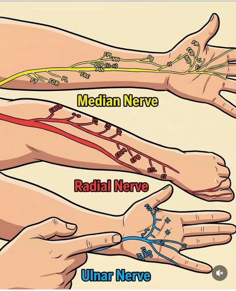
📚 تتلخص الأعصاب الرئيسية الثلاثة في الذراع واليد فيما يلي:
1. العصب المتوسط (Median Nerve)
•المسار: يمر أسفل الجزء الداخلي للذراع ويعبر من مقدمة المرفق وصولاً إلى الرسغ عبر "النفق الرسغي".
•الوظيفة الحركية: يتحكم في العضلات التي تساعد على ثني الرسغ والأصابع.
•الوظيفة الحسية: يوفر الإحساس في الإبهام، السبابة، الإصبع الأوسط، ونصف إصبع البنصر. 
•NewYork-Presbyterian
2. العصب الكعبري (Radial Nerve)
•المسار: يمتد على طول الجزء الخلفي من عظمة العضد وصولاً إلى اليد.
•الوظيفة الحركية: يتحكم في العضلات الباسطة (التي تساعد على فرد المرفق والرسغ والأصابع).
•الإصابة الشائعة: قد يؤدي تلفه إلى حالة تُعرف بـ "سقوط الرسغ" (Wrist Drop). 
3. العصب الزندي (Ulnar Nerve)
•المسار: يمر خلف المرفق (فيما يعرف بالعظمة المضحكة) ويمتد على طول جانب الساعد حتى اليد.
•الوظيفة: يتحكم في معظم العضلات الصغيرة في اليد التي تسمح بالحركات الدقيقة، ويوفر الإحساس في إصبع الخنصر ونصف إصبع البنصر.
•الإصابة الشائعة: قد يؤدي تلفه إلى حالة تُعرف بـ "اليد المخلبية" (Claw Hand).
What happens in the neck in cervical myelopathy?
As we get older, the spine can slowly change. The discs lose height, bones can form extra edges, and ligaments may thicken or even turn into bone. Over time, these changes can make the space around the spinal cord smaller.
When that space narrows, the spinal cord can become compressed. This may lead to symptoms like pain, weakness, numbness, or trouble with balance and walking.
These changes are often described as “degeneration” on a scan. But for patients, the effects can be very real and can worsen if not recognized early.