@PNASNews: Myosin VI oligomerises on cardiolipin membranes and transports vesicles processively, linking it to mitophagy!
Using dynamic #MassPhotometry, we track Myo6 assembly states directly on lipid bilayers.
https://t.co/jcqSzBWrmc
@LMU_Muenchen@JohannesDietri7@AronVenczel
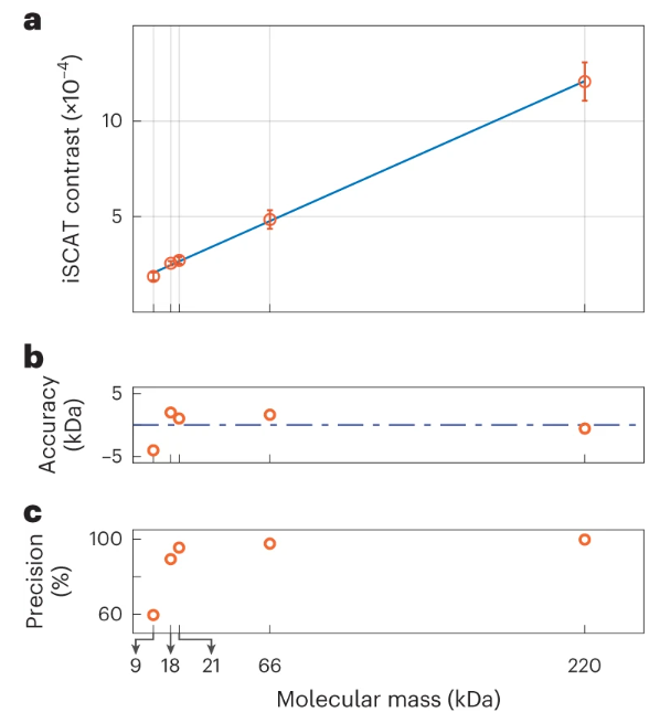
From the @SandoghdarLab An unsupervised machine learning approach for anomaly detection improves the mass sensitivity of iSCAT (mass photometry) by a factor of 4 to below 10 kDa. https://t.co/Hvy3SmVw4k
In this article, we speak with Dr. Nikolas Hundt, a Ludwig Maximilian University of Munich researcher, to discover the benefits of a #massphotometry approach to shed new light on fundamental biological processes: https://t.co/1tw2sEZfnQ
@refeynit @HundtNikolas@LMU_Muenchen
@PhilippKukura Thanks @PhilippKukura for all the years of great mentoring. Your support made me stick to it. I hope the work pays out and our conclusions and approaches will help researchers investigating actin and other polymers in the future.
Delighted to present confocal iSCAT microscopy Friday morning at #ELMI2022, developed in @SandoghdarLab.
Preprint out: https://t.co/bcVeGc0JER
Video: label-free imaging of nanoparticle action in a live COS7 cell. Recognize anything? Size 15x15µm.
Check out our protocol for mass-sensitive particle tracking on lipid membranes with #iSCAT in @JoVEJournal:
https://t.co/7goLuEwJFb
It comes with a nice little video and analysis code to download.
#massphotometry@MPI_Biochem@LMU_Muenchen
@naturemethods Also very happy that we could publish this breakthrough in #massphotometry back-to-back with the @PhilippKukura lab. Check out their article, too: https://t.co/hQVubLb8Nw
Very excited about our new @naturemethods article. It was an amazingly productive and fun collaboration with the Schwille lab. Now we can do #massphotometry on membrane-associated proteins.
My first review about #massphotometry:
Label-free, mass-sensitive single-molecule imaging using interferometric scattering microscopy https://t.co/sOsSZAChm6