The view of a large brain arteriovenous malformation (AVM) prior to a successful removal. Note the complex network of dilated, abnormal vessels. This is just the surface, this particular AVM was also 5 cm deep.
AVMs are congenital tangles of abnormal blood vessels in the brain. Because of the high blood flow through them, they carry a risk of rupture that can lead to life-threatening brain hemorrhage. Depending on their location, AVMs may also cause seizures, headaches, or other neurological deficits.
Treatment options may include observation, microsurgical resection, endovascular embolization, radiosurgery, or a combination of these approaches. The best treatment plan is individualized and depends on several factors, including the AVM’s size, location, vascular architecture, and the patient’s overall condition.
#AVM #Neurosurgery #BrainHealth #AVMTreatment #Neurology #BrainAVM #CerebralAVM #Surgery #VascularNeurology #AVMSurgery #Brain
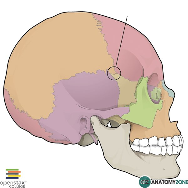
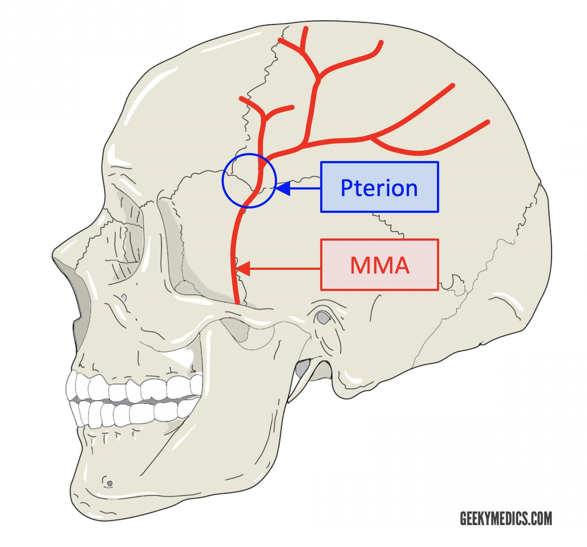
الـ pterion هي منطقة في الجمجمة وهي مو عظمة بحد ذاتها بل هي نقطة التقاء أربع عظام في نفس المكان وهي الـ frontal bone والـ parietal bone والـ temporal bone والـ greater wing of the sphenoid bone، وتقع على الجانبين من الجمجمة في المنطقة الصدغية يعني فوق الأذن وللأمام قليلاً، وشكلها عند الالتقاء يشبه حرف H أو أحياناً يشبه حرف K.
اللي يخلي هذي المنطقة مهمة جداً من الناحية الطبية إنها تُعتبر أضعف نقطة في الجمجمة كلها، والسبب في ضعفها إن العظم في هذي المنطقة رفيع جداً مقارنةً بباقي مناطق الجمجمة وأحياناً سماكته ما تتجاوز ملليمترات قليلة، ف لو جاء أي ضربة أو صدمة على هذي المنطقة حتى لو كانت خفيفة نسبياً ممكن تكسر العظم بسهولة، واللي يزيد الأمر خطورة إن تحت هذي المنطقة مباشرةً يمر شريان مهم جداً اسمه الـ middle meningeal artery وهو فرع من الـ maxillary artery، وهذا الشريان يسير في أخدود أو قناة في السطح الداخلي للجمجمة في نفس منطقة الـ pterion، ف لما تصير كسر في الـ pterion هذا الكسر ممكن يمزق الـ middle meningeal artery، ولما الشريان يتمزق يبدأ الدم يتجمع بين العظم والـ dura mater في المساحة اللي نسميها الـ epidural space أو فوق الـ dura، وهذا التجمع الدموي يُسمى الـ extradural hematoma أو الـ epidural hematoma.
هنا تجي أخطر علامة سريرية لازم كل طالب طب يحفظها وهي الـ lucid interval، يعني المريض بعد الإصابة يفقد وعيه لفترة قصيرة ثم يرجع وعيه ويبدو بخير تماماً ويتكلم ويمشي وكأن ما صار شي، وهذه الفترة تسمى الـ lucid interval وهي خادعة جداً لأن الدم في هذا الوقت لا يزال يتجمع ببطء خلف العظم، وبعدين فجأة الضغط داخل الجمجمة يرتفع بشكل حاد ويضغط على الدماغ ويبدأ المريض يتدهور بسرعة ويفقد وعيه مرة ثانية وهذه المرة بشكل أخطر، وإذا ما تدخلنا بسرعة الضغط المتزايد يسبب الـ transtentorial herniation يعني الدماغ يبدأ ينزل من فتحة الـ tentorium cerebelli ويضغط على الـ brainstem وعلى العصب الثالث اللي هو الـ oculomotor nerve فتتسع الحدقة في نفس جهة الإصابة وهذي علامة خطيرة جداً اسمها الـ blown pupil.
التشخيص يتم بسرعة عن طريق الـ CT scan اللي يظهر فيه تجمع دموي عدسي الشكل أو biconvex لأن الدم يتجمع بين العظم والـ dura اللي هي ملتصقة بالعظم من الجوانب فيعطي شكل العدسة المحدبة من الجهتين، والعلاج هو جراحة طارئة عاجلة عن طريق عمل ثقب في الجمجمة اسمه الـ burr hole أو craniotomy لتفريغ الدم المتجمع وإيقاف النزيف قبل ما يصير ضرر دائم في الدماغ، وعشان كذا في الطوارئ أي مريض جاء بضربة على جانب الرأس حتى لو كان بخير وواعي لازم ما نطمن وننتبه للـ lucid interval ونراقبه بجدية.
Suppa et al. explore rigidity in Parkinson’s disease, highlighting inconsistencies in its definition and interpretation. They propose new approaches to better characterise its mechanisms and clinical impact. https://t.co/IX61OXzRY8
What happens in the neck in cervical myelopathy?
As we get older, the spine can slowly change. The discs lose height, bones can form extra edges, and ligaments may thicken or even turn into bone. Over time, these changes can make the space around the spinal cord smaller.
When that space narrows, the spinal cord can become compressed. This may lead to symptoms like pain, weakness, numbness, or trouble with balance and walking.
These changes are often described as “degeneration” on a scan. But for patients, the effects can be very real and can worsen if not recognized early.
When you’re asked to localize the lesion in a patient with aphasia, do you suddenly feel speechless?
At a loss for words to categorize the type of aphasia?
Never fear—here is the decision tree for patients w/aphasia and the associated anatomic correlates
Three main questions:
1. Fluency? Nonfluency indicates damage to the FRONTAL language regions anterior to the fissure of Rolando
2. Comprehension? Impaired comprehension indicates damage to the TEMPOROPARIETAL language regions posterior to the fissure of Rolando
3. Repetition? Impaired repetition indicates damage within the core PERISYLVIAN language zone
The answers will lead you both to the type of aphasia and the location of the lesion.
Keep this figure with you for quick reference—and then when you’re asked about a patient with aphasia, you will have a lot to say!