#Thiamine in the ICU: 10 high-yield pearls
1. Unexplained lactate? Think thiamine.
Thiamine is required for pyruvate dehydrogenase. Deficiency blocks conversion of pyruvate to acetyl-CoA, shunting pyruvate toward lactate and impairing mitochondrial aerobic metabolism. Thus, thiamine deficiency belongs in the differential diagnosis for otherwise unexplained lactic acidosis.
2. Thiamine deficiency in the ICU is not rare.
~30-70% of critically ill patients are thiamine-deficient. Many arrive deficient; others develop deficiency during critical illness because of increased metabolic demand, inadequate intake, or losses.
3. The bedside phenotype is more important than waiting for a thiamine level.
High-risk situations include malnutrition, alcohol-related liver disease, bariatric surgery, renal replacement therapy, polyuria/DKA, chronic diuretic therapy, unexplained encephalopathy, unexplained lactate elevation, and sepsis.
4. Starting nutrition can unmask the deficiency.
Glucose administration increases carbohydrate metabolism and thiamine utilization. A malnourished patient may therefore develop rising lactate after initiation of enteral/parenteral nutrition. Refeeding syndrome is not simply a phosphorus/potassium problem; thiamine deficiency can be central to its pathophysiology.
5. Dialysis and polyuria are major clues.
Thiamine can be lost during continuous RRT, hemodialysis, and peritoneal dialysis. Renal losses also rise with polyuria, including the diuretic phase of AKI, post-transplant polyuria, DKA, and chronic diuretic therapy in heart failure.
6. Do not wait for the classic Wernicke's triad.
Altered mental status, ocular abnormalities, and gait ataxia are rarely all present together. I think I have seen it only a few times. In an at-risk ICU patient with otherwise unexplained encephalopathy, absence of the complete triad should not reassure you.
7. A plasma thiamine concentration is not a great “rule-out” test.
Testing is often unavailable and expensive, and blood concentration does not necessarily reflect tissue thiamine status. I don't even remember when I last ordered the test (if ever...). Erythrocyte transketolase activity is physiologically more informative, but its cutoffs are poorly defined. Just recognize the risk population and consider a therapeutic trial rather than wait for laboratory confirmation.
8. The sepsis data are more nuanced than “thiamine works” or “thiamine doesn't work.”
Thiamine is not expected to improve hard outcomes in the overall septic-shock population. Some subgroup analyses and meta-analyses suggest signals for improved lactate clearance and organ function, particularly renal outcomes. I don't consider it a magic drug...
9. Thiamine alone ≠ HAT (hydrocortisone + ascorbic acid +thiamine) therapy.
Do not interpret negative trials of HAT as proof that correcting thiamine deficiency is useless. Large HAT RCTs failed to demonstrate the dramatic clinical benefit initially claimed, and a VICTAS secondary analysis raised concerns about longer-term outcomes. That is a different question from replacing thiamine in a patient likely to be deficient.
10. The kidney may be an important target organ.
Renal tubular cells are highly energy dependent. Across secondary analyses, observational data, and meta-analytic evidence, thiamine supplementation -particularly in deficient patients- shows a signal toward less AKI progression, better renal recovery, and less need for RRT, although individual RCTs have not consistently demonstrated significant benefit in unselected populations.
Rule of thumb:
“⬆️ lactate + risk factor + no good explanation" → think B1 deficiency”
Especially think about it when you see malnutrition/refeeding, alcoholism/cirrhosis, dialysis, polyuria/DKA, chronic diuretics, unexplained encephalopathy, sepsis, or otherwise unexplained persistent hyperlactatemia.
I usually include it in the admission orders for most of my ICU patients along with DVT and GI prophylaxis...!
Thiamine deficiency behaves less like an obscure vitamin deficiency and more like a potentially reversible form of mitochondrial energy failure. Or that's how I like to think about it.
#foamed #foamcc #meded
Baran DA, Naidu SS, et al. 2026 SCAI SHOCK Classification Expert Consensus Update: Refined Staging and Incorporation of Clinical Phenotypes and Patient Populations. J Soc Cardiovasc Angiogr Interv 2026. DOI 10.1016/j.jscai.2026.105736 (Article in press)
#ICU Evidence #008: 2026 SCAI SHOCK Classification Update: Summary and Comments
@SCAI just updated its cardiogenic shock classification (Baran and Naidu, co-chairs), endorsed by ACC, EACTS, ISHLT, SCCM, STS and others. It’s still “article in press” in JSCAI. The method is a narrative literature review plus consensus voting (75% agreement). The pyramid stays A to E. The definitions and guidance around them have changed.
Summary:
Philosophy: stage comes from the exam, labs and hemodynamics, not simply from what we did. Pressors, inotropes, MCS and ECLS don’t define the stage by themselves, and the stage should be reassessed serially.
What changed:
A (at risk) is better defined. It now focuses on meaningful risk from the combination of baseline vulnerability and an acute stressor, disease or procedure: large ACS, high-risk PCI, VT ablation, structural interventions, advanced HF.
B vs C depends on HYPOPERFUSION, not hypotension. Low BP with good perfusion is B. Hypoperfusion with a normal BP is C.
D means the initial strategy is failing: worsening or non-improving hypoperfusion or lactate despite treatment. They propose reassessing at 2–3 hours for clear worsening and up to 4–6 hours to judge failure to respond, and admit the best interval is uncertain.
E is actual or impending collapse: features include lactate ≥8, pH ≤7.2, bolus pressors or profound hypotension despite maximal support.
Recovery: a patient who normalizes perfusion on ECMO or MCS can be restaged as C while still on support.
“Arrest” modifier: reserved for cardiac arrest when neurologic recovery remains uncertain. Cardiac arrest itself does not determine the SCAI stage.
Phenotypes: AMI, HF, RV-predominant, mixed (about 20% of CICU shock), postcardiotomy and pediatric shock each get specific discussion. In HF, preserved BP and even a normal lactate do not necessarily mean adequate perfusion.
Implementation: staging across EMS, ED, cath lab and ICU, serial nursing assessment, point-of-care lactate/POCUS, EMR tools and shock teams. The authors explicitly favor sensitivity over specificity for early recognition.
Some personal comments:
1. “Therapy doesn’t define stage” isn’t completely clean. Table 2 (attached) uses escalating pressors or added MCS as evidence for D and bolus pressors as a feature of E. That makes sense because D is fundamentally about response to treatment. The real problem is that treatment varies between hospitals, operators and available resources. I’d call that confounding more than subjectivity.
2. The fix may be more subjective than the problem. Table 2 is full of “typically includes” and “may include,” with no minimum number of criteria and no inter-rater data. Try this one: warm patient on epinephrine, lactate 2.3, CI 2.4, normal mentation, good urine output. What stage? Two good intensivists may disagree.
3. This document is expert consensus, not high-level evidence. A narrative review plus a vote isn’t a systematic review. The decision to keep normotensive hypoperfusion in C is a consensus choice, and the paper itself discusses CSWG data that classified it differently. Stage A remains the least validated part of the framework.
4. Sensitivity over specificity widens the net, especially with Stage A, and could dilute what each stage means.
5. Many authors disclose relationships with device companies. Members with relevant significant conflicts did not vote, an important safeguard, but this remains worth knowing when reading a consensus document in an MCS-heavy field.
6. It’s a classification, not a treatment algorithm. Moving from C to D tells you the initial strategy is failing. It doesn’t tell you what to do next, and Table 4 essentially lists the available options.
Useful common language and a reasonable consensus, in my view, but not a validated decision tool.
How do you stage cardiogenic shock where you work? And do you restage after ECMO goes in?
#CardiogenicShock #SCAI #FOAMcc #Foamed
#ICU / #ED Evidence #007: What if we bring ECMO to the patient instead of bringing the patient to ECMO?
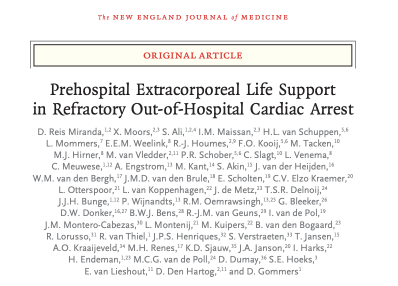
We know ECPR is a race against time, but in most systems we still transport a patient receiving CPR to the hospital before ECMO can even start. The OnScene study, published today in NEJM, asked whether bringing the ECMO team to the patient could change that.
The investigators studied 231 patients ≤ 50 years old with refractory out-of-hospital cardiac arrest in a nationwide Dutch helicopter EMS system. Patients attended by HEMS with prehospital ECPR capability were compared with those receiving conventional care.
The results are hard to ignore:
Survival to hospital discharge: 40% vs 20%
Favorable neurologic outcome at 6 months: 36% vs 13%
And here is probably the number that matters most: median time to ECMO blood flow was 45 minutes with prehospital ECPR (83 patients) vs 77 minutes among the 19 patients who eventually received in-hospital ECPR in the conventional-care group.
That is 32 minutes. In refractory cardiac arrest, 32 minutes is a very long time. The comparator group is tiny and the comparison is nonrandomized, so I would read this as a very interesting signal, not proof that the location of cannulation made the difference.
Before we start putting ECMO machines in every helicopter, though, this was not a randomized trial. The patients were highly selected: only 231 ultimately entered the analysis from 3,362 study-related HEMS dispatches for suspected cardiac arrest. Shockable rhythms were more common in the intervention group (89% vs 75%), neurologic outcome was assessed unblinded, residual confounding is very possible, and cannulation failed in 15% of prehospital attempts (vs 17% of hospital attempts).
So I don’t think OnScene proves that prehospital ECPR is superior to in-hospital ECPR. I think it raises a more interesting question: are we focusing too much on where ECPR happens and not enough on how quickly we establish effective extracorporeal blood flow?
The accompanying editorial by Dr. Demetris Yannopoulos (@DYannopoulos), one of the pioneers in the field, makes another important point. Instead of sequential care (EMS resuscitates, transports, then the hospital prepares for ECPR), perhaps we should think in parallel: identify potential candidates early, mobilize the ECPR team while conventional resuscitation continues, and have the receiving hospital preparing at the same time.
Sometimes the fastest route to ECMO will be bringing the patient to the team. Sometimes it may be bringing the team to the patient. Geography, resources and expertise will matter.
The goal isn’t earlier cannulation. It’s earlier successful reperfusion. That, to me, is the real message of OnScene.
If your system could get ECMO flowing within 45 minutes, would you build it? And who would you pick?
And one last thought: if AI is the hottest thing in tech and finance these days, ECMO is surely the hottest thing in Critical Care.
#ECPR #ECMO #CardiacArrest #FOAMcc #Foamed
The study did not show a clear difference in reported infections between prehospital and in-hospital ECPR, but the numbers are too small to conclude the risks are equivalent.
In Table 4, cannula-site infection was reported in 1/89 (1%) prehospital ECPR patients versus 1/19 (5%) in-hospital ECPR patients. Bloodstream infection was reported as 0 in the prehospital group versus 1/50 (2%) in the conventional-care group. Notice that the denominators differ by outcome, and the bloodstream-infection comparison is reported by study group, not simply as a comparison of the two cannulated groups.
#ICU / #ED Evidence #007: What if we bring ECMO to the patient instead of bringing the patient to ECMO?
We know ECPR is a race against time, but in most systems we still transport a patient receiving CPR to the hospital before ECMO can even start. The OnScene study, published today in NEJM, asked whether bringing the ECMO team to the patient could change that.
The investigators studied 231 patients ≤ 50 years old with refractory out-of-hospital cardiac arrest in a nationwide Dutch helicopter EMS system. Patients attended by HEMS with prehospital ECPR capability were compared with those receiving conventional care.
The results are hard to ignore:
Survival to hospital discharge: 40% vs 20%
Favorable neurologic outcome at 6 months: 36% vs 13%
And here is probably the number that matters most: median time to ECMO blood flow was 45 minutes with prehospital ECPR (83 patients) vs 77 minutes among the 19 patients who eventually received in-hospital ECPR in the conventional-care group.
That is 32 minutes. In refractory cardiac arrest, 32 minutes is a very long time. The comparator group is tiny and the comparison is nonrandomized, so I would read this as a very interesting signal, not proof that the location of cannulation made the difference.
Before we start putting ECMO machines in every helicopter, though, this was not a randomized trial. The patients were highly selected: only 231 ultimately entered the analysis from 3,362 study-related HEMS dispatches for suspected cardiac arrest. Shockable rhythms were more common in the intervention group (89% vs 75%), neurologic outcome was assessed unblinded, residual confounding is very possible, and cannulation failed in 15% of prehospital attempts (vs 17% of hospital attempts).
So I don’t think OnScene proves that prehospital ECPR is superior to in-hospital ECPR. I think it raises a more interesting question: are we focusing too much on where ECPR happens and not enough on how quickly we establish effective extracorporeal blood flow?
The accompanying editorial by Dr. Demetris Yannopoulos (@DYannopoulos), one of the pioneers in the field, makes another important point. Instead of sequential care (EMS resuscitates, transports, then the hospital prepares for ECPR), perhaps we should think in parallel: identify potential candidates early, mobilize the ECPR team while conventional resuscitation continues, and have the receiving hospital preparing at the same time.
Sometimes the fastest route to ECMO will be bringing the patient to the team. Sometimes it may be bringing the team to the patient. Geography, resources and expertise will matter.
The goal isn’t earlier cannulation. It’s earlier successful reperfusion. That, to me, is the real message of OnScene.
If your system could get ECMO flowing within 45 minutes, would you build it? And who would you pick?
And one last thought: if AI is the hottest thing in tech and finance these days, ECMO is surely the hottest thing in Critical Care.
#ECPR #ECMO #CardiacArrest #FOAMcc #Foamed
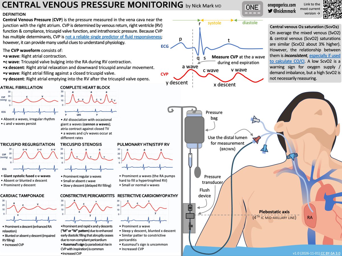
🆕 ICUOnePager on Central Venous Pressure (CVP) monitoring. CVP is not useful for fluid responsiveness but the trend and the waveform can provide many physiological insights! The central oxygen sat (ScvO2) can also provide clues.
1/
#ICU Physiology Secrets: Is vasopressin really “pulmonary-friendly”?
I’ve heard several times that vasopressin is “lung-friendly.” That it may even dilate the pulmonary vasculature. Or that the pulmonary circulation has no vasopressin receptors. Have you heard the same?
It made me wonder how much human evidence actually supports these claims. Does vasopressin constrict the pulmonary circulation less than norepinephrine? Does it lower PVR? Does it improve RV function? Or does it simply raise systemic pressure without raising PA pressure as much? These are related questions, but they are not the same question.
So I went looking for the human evidence. With the caveat that I did not have full-text access to a few potentially relevant studies, this is what I found:
1. Start with the receptor, but don’t stop there
Liu et al. examined human arterial samples and found V1a receptor (AVPR1A) protein was abundant in pulmonary arteries: about 2.2× mesenteric, 2.2× coronary and 1.9× peripheral arteries, and similar to renal arteries. Six samples of each artery type were studied. So the pulmonary circulation clearly does not lack V1a receptors. But expression is not function, and this study didn’t show what a human pulmonary artery does when exposed to vasopressin.
2. Put human pulmonary arteries in an organ bath, and things get interesting
Currigan et al. compared isolated human pulmonary and radial arteries. Vasopressin was a potent radial-artery vasoconstrictor but produced no significant change in pulmonary arterial tone. Norepinephrine constricted both.
Hussain et al. found an even more striking contrast. Maximal contraction of human pulmonary-artery rings was 61% of the KCl response with norepinephrine (12 rings from 4 patients) versus 0.18% with vasopressin (8 rings from 2 patients). That 0.18% gets my attention, but so does the denominator: eight rings from two patients...
These experiments provide a compelling physiologic basis for vasopressin behaving differently in systemic and pulmonary arteries. Still, these are isolated vessels in a bath, not PVR measured in a hypoxemic, ventilated patient with pulmonary hypertension and a failing RV.
3. What happens in actual patients?
Mizota et al. reported a lung-transplant patient with pulmonary hypertension in whom, during pulmonary-artery clamping, a 0.5-U vasopressin bolus raised systemic systolic pressure 35.2% with minimal change in PA pressure. Norepinephrine boluses in the same patient raised systemic systolic pressure 14.8% and PA systolic pressure 6.7%. A beautiful physiologic observation but n = 1, during major surgery, with other vasoactive infusions running.
Tayama et al. studied 9 post-cardiotomy patients with vasodilatory hypotension and pulmonary hypertension. Vasopressin raised mean systemic pressure from 47.3 ± 9.5 to 76.5 ± 12.2 mmHg, while PVR did not change significantly. Cardiac index fell, although it remained in the normal range. The pulmonary/systemic pressure ratio fell from 0.66 to 0.47. Be careful with that: a ratio can fall simply because the systemic denominator rises. A falling ratio is not pulmonary vasodilation.
Adamson et al. studied 28 Fontan patients undergoing catheterization. Vasopressin raised systemic pressure and SVR while PVR fell by 0.4 ± 0.4 Wood units, with no significant change in PA pressure or cardiac index. That is human PVR data, but a small effect from a single study. And Fontan physiology has no subpulmonary ventricle, so it is not a model of the adult ICU patient whose RV is failing against a high pulmonary vascular load.
4. What does the larger comparative evidence show?
Gordon et al. analyzed 779 septic-shock patients from VASST, 241 of them managed with a PA catheter. At similar systemic pressures, vasopressin reduced norepinephrine requirements and heart rate. In the PA-catheter subset, there was no detected difference in cardiac index or stroke-volume index over the first 96 hours. Inotropes were used more often in the vasopressin group.
That is much stronger comparative evidence than an organ bath or a case report. But PVR wasn’t reported, RV function wasn’t measured directly, PA-catheter use wasn’t randomized, and these were septic-shock patients, not patients selected for RV failure.
Geube et al. studied 153 elective cardiac-surgery patients who developed intraoperative hypotension in a single-center, cluster-randomized, multiple-crossover study. They found no statistically significant difference between vasopressin and norepinephrine in the primary time-weighted mPAP/MAP ratio or in RV free-wall strain, a secondary endpoint. That doesn’t support a simple claim of pulmonary selectivity or better RV function in this setting. But a pressure ratio is not PVR and can move with the systemic denominator, and these patients weren’t selected for established RV failure.
5. PA pressure may be the wrong scorecard
Dalla et al. studied 11 septic-shock patients (a small study) in whom norepinephrine was titrated in random sequence to MAP targets of 60, 75 and 90 mmHg. At the highest versus lowest MAP, mean PA pressure rose 19%, which at first glance sounds bad for the RV. But PVRI and effective pulmonary arterial elastance did not increase, while RV function improved: free-wall strain became more negative (−19% to −25%), TAPSE rose 22% and RV stroke-work index rose 15%.
This doesn’t prove norepinephrine is better than vasopressin. It shows why PA pressure alone is a poor scorecard for what the RV is doing.
So where do I land?
Vasopressin’s “pulmonary-friendly” reputation has a legitimate physiologic basis. In small organ-bath studies, human pulmonary arteries show very little direct constriction to vasopressin. In small clinical series, systemic pressure rose without a clear rise in PA pressure. Fontan patients' data show a small fall in PVR, about 0.4 Wood units, in a circulation with no subpulmonary ventricle.
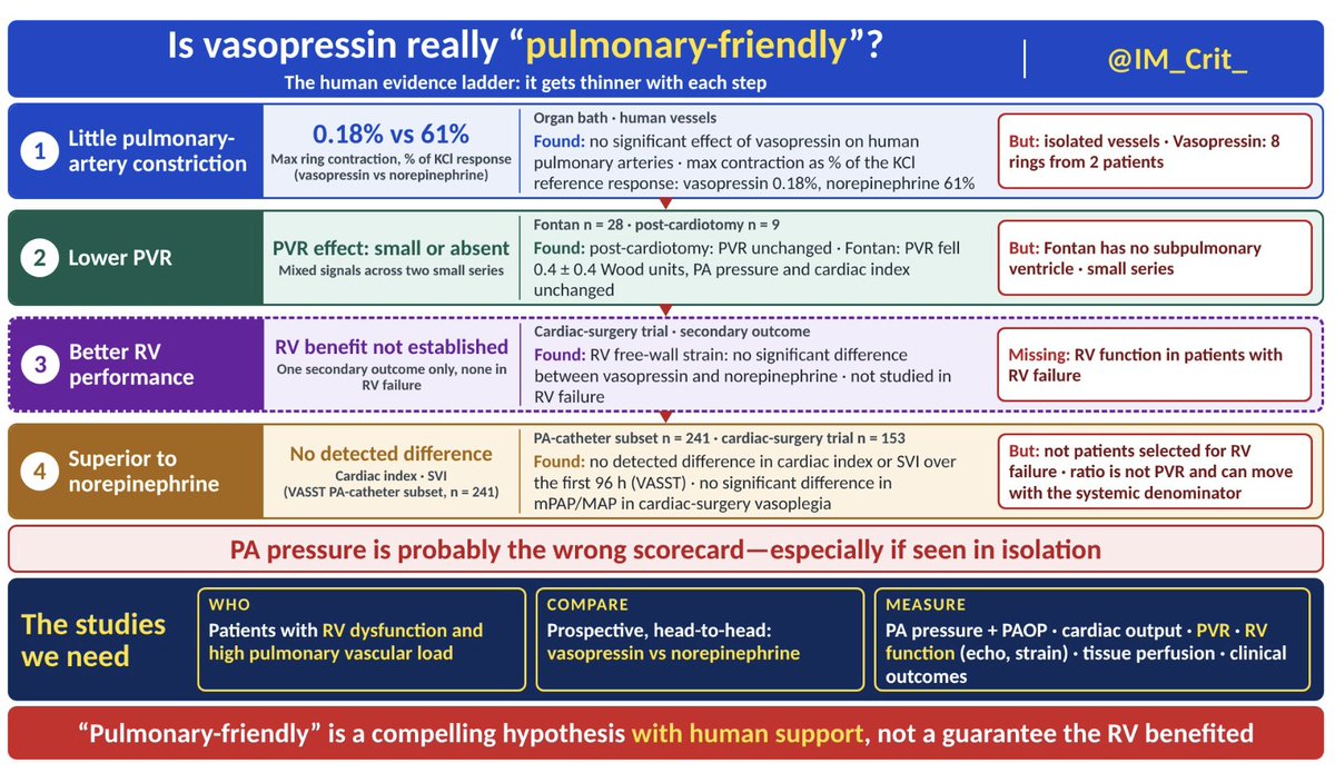
But there’s an evidentiary ladder:
Little pulmonary-artery constriction → lower PVR → better RV performance → clinical superiority over another vasopressor.
Those are four different claims, and the human evidence gets thinner with each step.
What I’d really like to see is a prospective vasopressin-vs-norepinephrine comparison in patients who actually have RV dysfunction with elevated pulmonary vascular load. It should measure systemic and PA pressures, PAOP, cardiac output, PVR, RV function, tissue perfusion and, ultimately, clinical outcomes.
Until then, I’ll still consider vasopressin an attractive pressor when the RV is in trouble. That’s a physiologically informed choice, not a proven RV-outcome advantage. “Pulmonary-friendly” is a compelling hypothesis with human support, not a guarantee that the RV benefited.
When your RV is failing, what pressor do you reach for first and what do you measure to decide whether it helped?
#ICU Physiology Secrets: Is vasopressin really “pulmonary-friendly”?
I’ve heard several times that vasopressin is “lung-friendly.” That it may even dilate the pulmonary vasculature. Or that the pulmonary circulation has no vasopressin receptors. Have you heard the same?
It made me wonder how much human evidence actually supports these claims. Does vasopressin constrict the pulmonary circulation less than norepinephrine? Does it lower PVR? Does it improve RV function? Or does it simply raise systemic pressure without raising PA pressure as much? These are related questions, but they are not the same question.
So I went looking for the human evidence. With the caveat that I did not have full-text access to a few potentially relevant studies, this is what I found:
1. Start with the receptor, but don’t stop there
Liu et al. examined human arterial samples and found V1a receptor (AVPR1A) protein was abundant in pulmonary arteries: about 2.2× mesenteric, 2.2× coronary and 1.9× peripheral arteries, and similar to renal arteries. Six samples of each artery type were studied. So the pulmonary circulation clearly does not lack V1a receptors. But expression is not function, and this study didn’t show what a human pulmonary artery does when exposed to vasopressin.
2. Put human pulmonary arteries in an organ bath, and things get interesting
Currigan et al. compared isolated human pulmonary and radial arteries. Vasopressin was a potent radial-artery vasoconstrictor but produced no significant change in pulmonary arterial tone. Norepinephrine constricted both.
Hussain et al. found an even more striking contrast. Maximal contraction of human pulmonary-artery rings was 61% of the KCl response with norepinephrine (12 rings from 4 patients) versus 0.18% with vasopressin (8 rings from 2 patients). That 0.18% gets my attention, but so does the denominator: eight rings from two patients...
These experiments provide a compelling physiologic basis for vasopressin behaving differently in systemic and pulmonary arteries. Still, these are isolated vessels in a bath, not PVR measured in a hypoxemic, ventilated patient with pulmonary hypertension and a failing RV.
3. What happens in actual patients?
Mizota et al. reported a lung-transplant patient with pulmonary hypertension in whom, during pulmonary-artery clamping, a 0.5-U vasopressin bolus raised systemic systolic pressure 35.2% with minimal change in PA pressure. Norepinephrine boluses in the same patient raised systemic systolic pressure 14.8% and PA systolic pressure 6.7%. A beautiful physiologic observation but n = 1, during major surgery, with other vasoactive infusions running.
Tayama et al. studied 9 post-cardiotomy patients with vasodilatory hypotension and pulmonary hypertension. Vasopressin raised mean systemic pressure from 47.3 ± 9.5 to 76.5 ± 12.2 mmHg, while PVR did not change significantly. Cardiac index fell, although it remained in the normal range. The pulmonary/systemic pressure ratio fell from 0.66 to 0.47. Be careful with that: a ratio can fall simply because the systemic denominator rises. A falling ratio is not pulmonary vasodilation.
Adamson et al. studied 28 Fontan patients undergoing catheterization. Vasopressin raised systemic pressure and SVR while PVR fell by 0.4 ± 0.4 Wood units, with no significant change in PA pressure or cardiac index. That is human PVR data, but a small effect from a single study. And Fontan physiology has no subpulmonary ventricle, so it is not a model of the adult ICU patient whose RV is failing against a high pulmonary vascular load.
4. What does the larger comparative evidence show?
Gordon et al. analyzed 779 septic-shock patients from VASST, 241 of them managed with a PA catheter. At similar systemic pressures, vasopressin reduced norepinephrine requirements and heart rate. In the PA-catheter subset, there was no detected difference in cardiac index or stroke-volume index over the first 96 hours. Inotropes were used more often in the vasopressin group.
That is much stronger comparative evidence than an organ bath or a case report. But PVR wasn’t reported, RV function wasn’t measured directly, PA-catheter use wasn’t randomized, and these were septic-shock patients, not patients selected for RV failure.
Geube et al. studied 153 elective cardiac-surgery patients who developed intraoperative hypotension in a single-center, cluster-randomized, multiple-crossover study. They found no statistically significant difference between vasopressin and norepinephrine in the primary time-weighted mPAP/MAP ratio or in RV free-wall strain, a secondary endpoint. That doesn’t support a simple claim of pulmonary selectivity or better RV function in this setting. But a pressure ratio is not PVR and can move with the systemic denominator, and these patients weren’t selected for established RV failure.
5. PA pressure may be the wrong scorecard
Dalla et al. studied 11 septic-shock patients (a small study) in whom norepinephrine was titrated in random sequence to MAP targets of 60, 75 and 90 mmHg. At the highest versus lowest MAP, mean PA pressure rose 19%, which at first glance sounds bad for the RV. But PVRI and effective pulmonary arterial elastance did not increase, while RV function improved: free-wall strain became more negative (−19% to −25%), TAPSE rose 22% and RV stroke-work index rose 15%.
This doesn’t prove norepinephrine is better than vasopressin. It shows why PA pressure alone is a poor scorecard for what the RV is doing.
So where do I land?
Vasopressin’s “pulmonary-friendly” reputation has a legitimate physiologic basis. In small organ-bath studies, human pulmonary arteries show very little direct constriction to vasopressin. In small clinical series, systemic pressure rose without a clear rise in PA pressure. Fontan patients' data show a small fall in PVR, about 0.4 Wood units, in a circulation with no subpulmonary ventricle.
But there’s an evidentiary ladder:
Little pulmonary-artery constriction → lower PVR → better RV performance → clinical superiority over another vasopressor.
Those are four different claims, and the human evidence gets thinner with each step.
What I’d really like to see is a prospective vasopressin-vs-norepinephrine comparison in patients who actually have RV dysfunction with elevated pulmonary vascular load. It should measure systemic and PA pressures, PAOP, cardiac output, PVR, RV function, tissue perfusion and, ultimately, clinical outcomes.
Until then, I’ll still consider vasopressin an attractive pressor when the RV is in trouble. That’s a physiologically informed choice, not a proven RV-outcome advantage. “Pulmonary-friendly” is a compelling hypothesis with human support, not a guarantee that the RV benefited.
When your RV is failing, what pressor do you reach for first and what do you measure to decide whether it helped?
Excellent summary! I’ve recently presented at a conference on this exact topic. I would add one additional study to your list of human data: Jeon 2006 (16675238). This was an RCT in 50 off-pump CABG pts with milrinone-induced hypotension where pts were randomized to vaso vs norepi. There was a small decrease in the PVR/SVR ratio with vaso but no change with norepi. If I remember correctly, the PVR did increase with vaso though.
Overall I reached the same conclusions that you did in my review: the evidence supporting the claim that vaso is “better for the RV” is very weak. It essentially comes from basic research which often doesn’t translate into real clinical benefit, as we all know.
Thanks for voting!
311 votes: 41.5% inotrope first, 36% norepinephrine, 12.5% vasopressin, 10% both.
There’s no single right answer here, and that’s the point of the vasopressin post. Nobody has proven which drug is best in this exact setting. We’re reasoning from physiology. Here’s how I think about this patient.
Let’s look at the numbers. CI 1.9 and SvO₂ 54% say the RV isn’t delivering enough flow. A MAP of 55 with CVP 16 and PA 55/28 says it’s working against a high load with its own perfusion at risk. So this RV may need pressure, contractility and less afterload at the same time. That’s why inotrope-first made sense to so many of you.
Norepinephrine: raises MAP and provides some β₁ support. For me, a very defensible first go-to drug.
Inotrope alone: addresses the low flow directly, but dobutamine or milrinone can lower systemic pressure, which is a problem when MAP is already 55. Epinephrine is different because it can support both pressure and contractility.
Vasopressin: raises MAP without direct inotropy and, in the limited human data, may have relatively little pulmonary pressor effect. In the small post-cardiotomy series I cited, cardiac index fell. That’s a reason to watch output, not proof it would hurt this patient. I see it as a strong add-on or alternative pressor.
Norepinephrine + vasopressin: common and reasonable if pressure remains inadequate, but they still don’t directly solve low contractility.
My sequence: norepinephrine -or epinephrine in the right patient- then early inotropic support if CI and SvO₂ don’t improve. If elevated pulmonary vascular load remains an important part of the problem, I’d consider an inhaled pulmonary vasodilator. Vasopressin can come alongside as an adjunct pressor.
In parallel, I’d look for fixable causes: tamponade, bleeding, graft or valve problems, ischemia, rhythm, ventilator settings, hypoxia and CO₂. That search can’t delay supporting a MAP of 55.
What would I measure to decide it worked? Everything: MAP and CVP, PA pressure, CO and SvO₂, lactate, RV size and function on echo, and tissue perfusion by every bedside sign we have: urine output, mentation, skin.
One number moving while the others don’t is not a win.
That’s physiology, not proof. I’m not aware of a randomized trial that answers this exact question.
What would make you change course first: MAP, PAP, cardiac output, SvO₂, or the echo?
After my vasopressin “pulmonary-friendly” post:
Hypothetical case: POD 0 after MVR + CABG. MAP 55, HR 95, CVP 16, PA 55/28 (mean 37), PAOP 14, CO 3.6 / CI 1.9, SvO2 54%. Echo: dilated, hypokinetic RV. No vasoactives yet.
What do you start first?