New study led by Lei Wang from the Institute of Biophysics of @CAS__Science (https://t.co/qeo662CiWJ) has revealed two coordinated molecular mechanisms that maintain efficient protein phosphorylation in the Golgi apparatus during lactation. 👉 https://t.co/QW8Ba9Ea82
Spotlight: Muniesa-Vargas, Lans et al. (https://t.co/Wne6jjsvFm) provide insight into the mechanisms that regulate the dynamics of XPG recruitment & release during nucleotide excision repair. Opresko and Schärer highlight the findings: https://t.co/sdlLo8PbUU
Yang et al. show that the secretory pathway kinase Fam20C is retained in the #Golgi via Fam20A-mediated membrane anchoring. Mislocalizing Fam20C alters #secretome phosphorylation, leading to reduced milk quality and subsequent growth retardation in pups. https://t.co/3gvfYk9jUk
Yan G. Zhao et al. @SUSTechSZ reveal that during nutrient scarcity, the lipid droplet–localized PISD isoform, PISD-LD, ensures survival by bridging #LipidDroplets to #mitochondria for β-oxidation while simultaneously inhibiting #lipophagy: https://t.co/ImPXf4j7TK
Ndoci et al. (https://t.co/VwFP8yYcF9) reveal a system by which mitochondria prioritize CoQ retention when the mevalonate pathway is impaired, preserving respiration at the expense of antioxidant defenses. Spotlight by Sato, Friedmann Angeli et al.: https://t.co/DX1xJmNV0Z
Weikang Fan et al. reveal how nutrient sensing in #Drosophila intestinal epithelial cells controls stem cell activity. Inhibition of Rag GTPases or mTORC1 in enterocytes non-cell autonomously promotes ISC proliferation via Upd3–JAK–STAT signaling. https://t.co/lNPfDxgcyV
See a recent JCB study from Youfang Zhou and Xianfeng Wang et al. of the Wu-Min Deng lab, "Polyploidy reprograms epithelial cells for motility and phagocytosis via stress signaling"
👉 https://t.co/natmi0r9vg
📰 Press release: https://t.co/xQPoJse7Yl
“We greatly appreciated JCB’s support in highlighting the significance of our study. The outreach efforts helped communicate our findings to a broader scientific audience.” — Wu-Min Deng
Why publish in JCB? 👉 https://t.co/JNzNMhsTKG
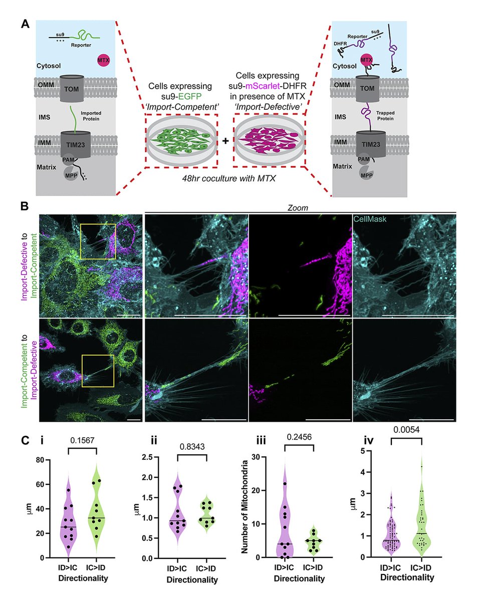
Emily Glover, Kevin Wilkinson, Ian Collinson et al. @BristolUni show that cells subject to mitochondrial protein import failure induce Intercellular Mitochondrial Transfer via tunnelling nanotubes. https://t.co/YZ3xCddMSE
#Organelles#Biochemistry#Mitochondria
Yasushi Izumi and Mikio Furuse @SEIRIKEN1977 show that the exclusion of apical aPKC by septate junction proteins in #Drosophila enteroblasts is required for stem cell homeostasis via the Kibra–Yorkie pathway. https://t.co/1QPtpTg9gG
#Adhesion#StemCells#Development#Polarity
Mia S. Frier, Elizabeth Conibear et al. show that the VPS9 GEF complex VINE unexpectedly recruits a phosphatase that stimulates GAP-mediated Rab5 inactivation, revealing dual control of Rab signaling by a single complex. https://t.co/sVehc7wm4s
#GTPases#Organelles#Trafficking
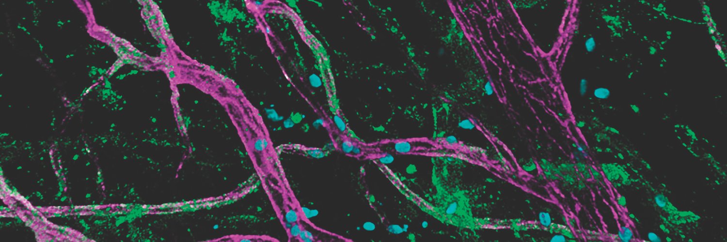
Our July issue is here! https://t.co/lVVkPOw7r6 The cover shows an immunofluorescence image of a TNFα-inflamed mouse cremaster muscle acquired w/ a Nikon A1 confocal microscope. Green, cortactin; magenta, CD31 (vessels); cyan, MRP-14 (neutrophils).
See https://t.co/jaPAD7gCuO
The discovery of a novel trafficking domain in unorthodox AQP12 by Chauvigné, Cerdà et al. yields insight into the channel’s native membrane function and its molecular regulation in intracellular yolk platelets and pancreatic zymogen granules. https://t.co/qYiSKT8R6F
Rajan, Yoon, Terman, Reisler et al. identify mechanisms for assembling & disassembling complex networks of multi-protein–bundled F-actin, including a synergism between different bundlers and disassemblers in these processes. https://t.co/249eW2MCjY
#Cytoskeleton#Actin
Alba Muniesa-Vargas, Hannes Lans et al. (Erasmus MC) use live-cell imaging to uncover how XPG activity is dynamically controlled during nucleotide excision repair. https://t.co/2YYWZStFu2
#DNAbiology#Genetics#DNArepair
Keegan, Dunleavy et al. show that, in #Drosophila, a new pool of constitutive #centromere protein CENP-C is loaded at the centromere in prophase of #meiosis I. This pool is critical for #kinetochore assembly and meiotic chromosome segregation. https://t.co/5a28x9VAW6
Thrilled to share this #lipidtime story in @JCellBiol where, led by Shiying Huang and in collaboration with Tamas Balla’s lab, we uncover plasticity in phospholipid metabolism to preserve plasma membrane identity and integrity in the face of disease-relevant perturbations!
How does the plasma #membrane preserve its #lipid identity under stress? The Baskin and Balla labs report a homeostatic circuit linking PI4P, phosphatidylserine, RhoB, and phospholipase D-driven phosphatidic acid. https://t.co/mTsss8ZZcX
@basktastic@weillinstitute@CornellChem
Jenkins, Botelho et al. show that the PIKfyve #lipid kinase extends its sphere of influence to modulate the architecture & dynamics of the ER by balancing phosphoinositide species at their inter-organellar sites. https://t.co/WeRlcklpMf
#Organelles#Membrane#ER_literature