@volcan85 Context of The findings is very important on this case. Looks like tubular cells, but,to have a more proper identification complementary information would be helpfull.
@Jefiner94 Ok, Thank you for The information. I requested it because maybe you are observing macrophages. Without The polarized light filters is difficult to proper identify oval fat bodies.
@VelezNephHepato@TELMALEMOS10 @CarlosMartnezF7 @TStehl1 I honestly have no idea. I would Go for artifact. But no clue on possible identification. First time observing something like this. Urine microscopy mistery!
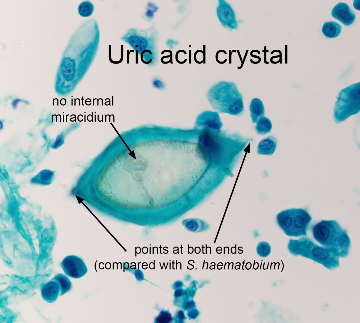
The answer to the #ParasiteCase 728 is now up: Uric acid crystals in urine– a tricky mimic of Schistosoma haematobium eggs! Read how to differentiate them here (and below): https://t.co/rZqKyV11qu #mayopath#pathology#CrittersOnTwitter#PathBugs
In brief:
1. Uric acid crystals vary in size and shape and are often much smaller than S. haematobium eggs.
2. Uric acid crystals have points on both ends instead of the single 'pinched-off' spine of S. haematobium eggs.
3. There are no internal parasite structures (i.e., miracidium) in uric acid crystals
4. Crystals often fracture and break, and may have irregular contours.
@zaheeramin1@jrseltzer So it seems to big to be bacteria. Maybe some free life protozoa. How was The urine sample collected? Maybe it is contamined with water from some source.