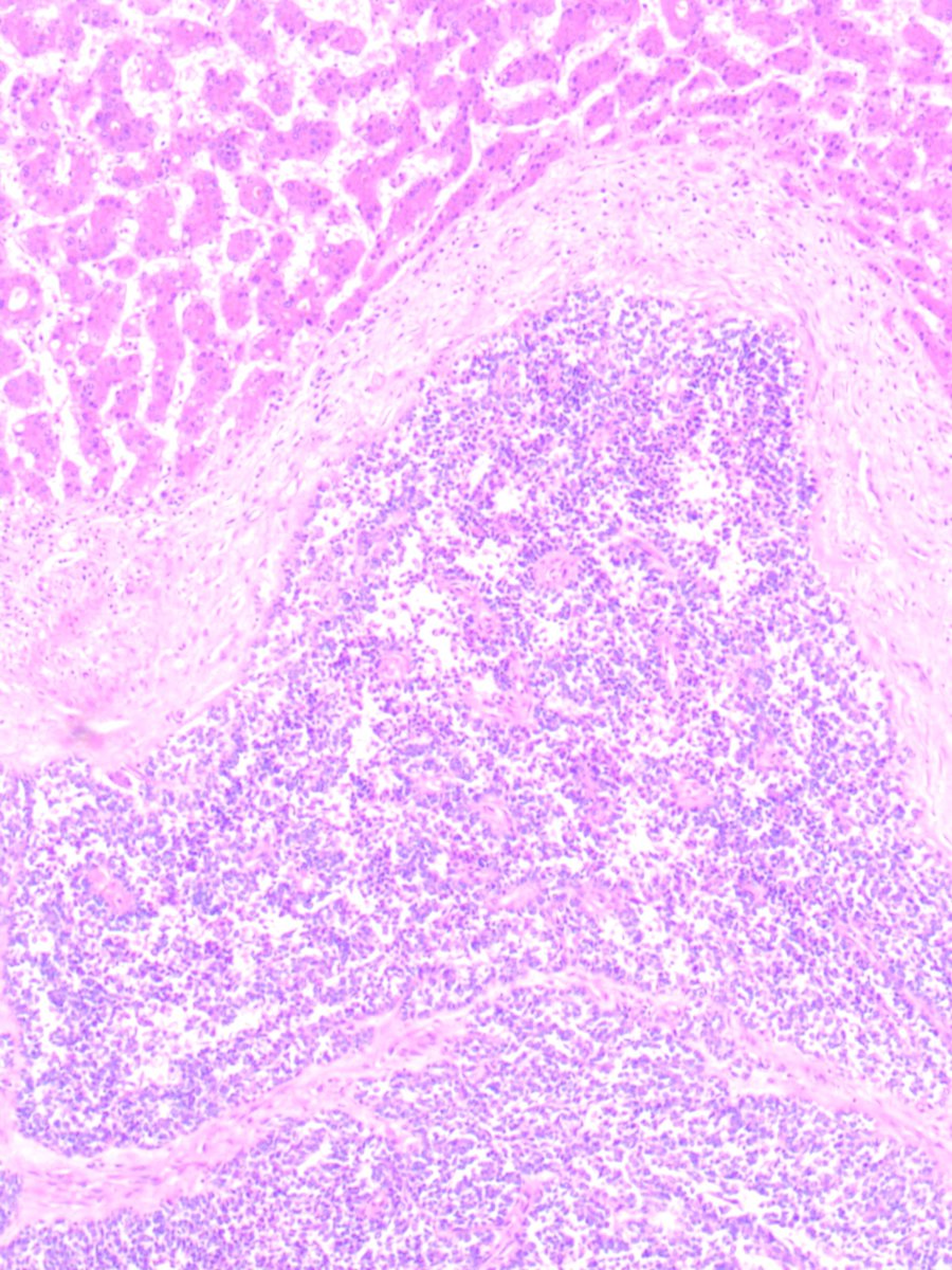
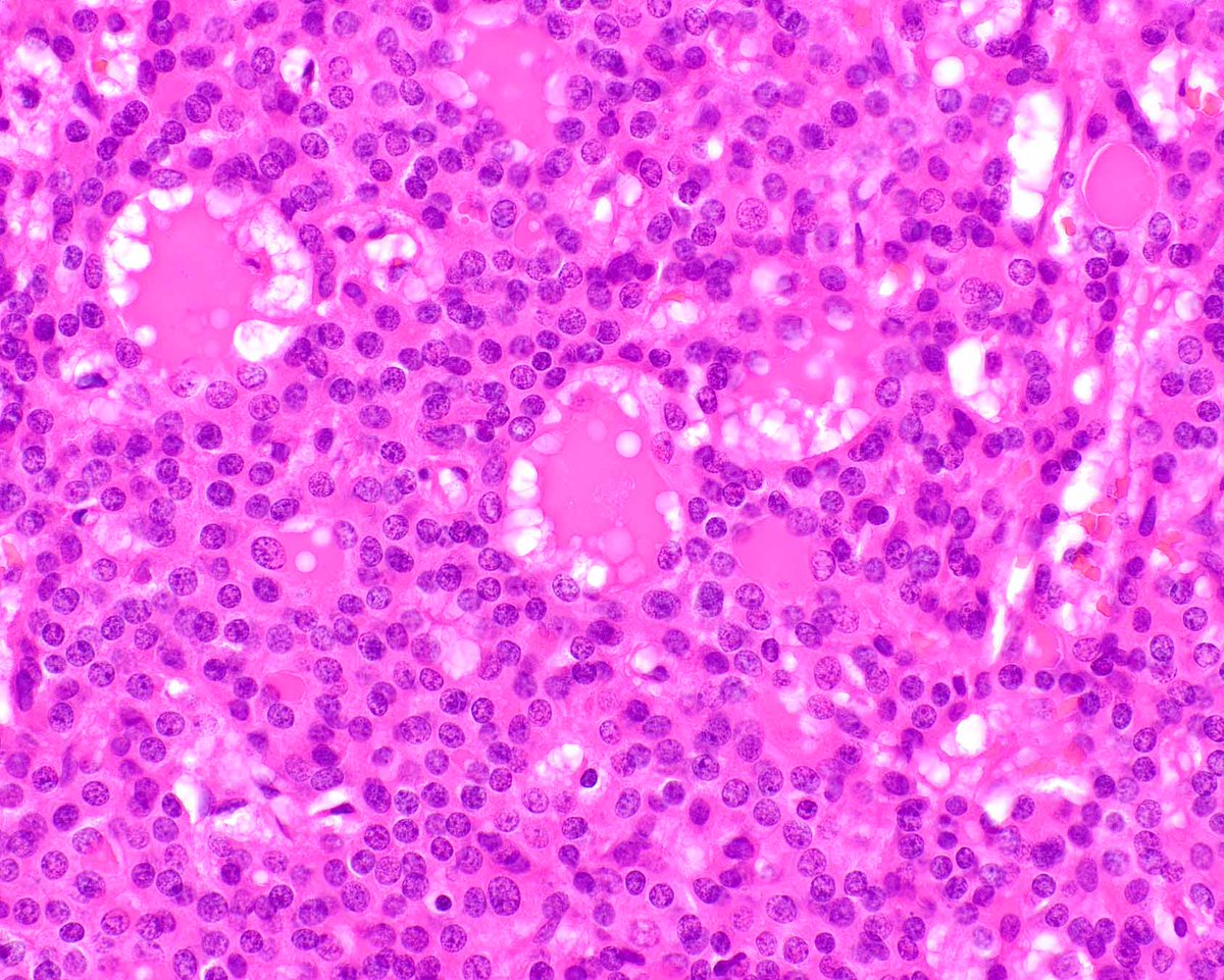
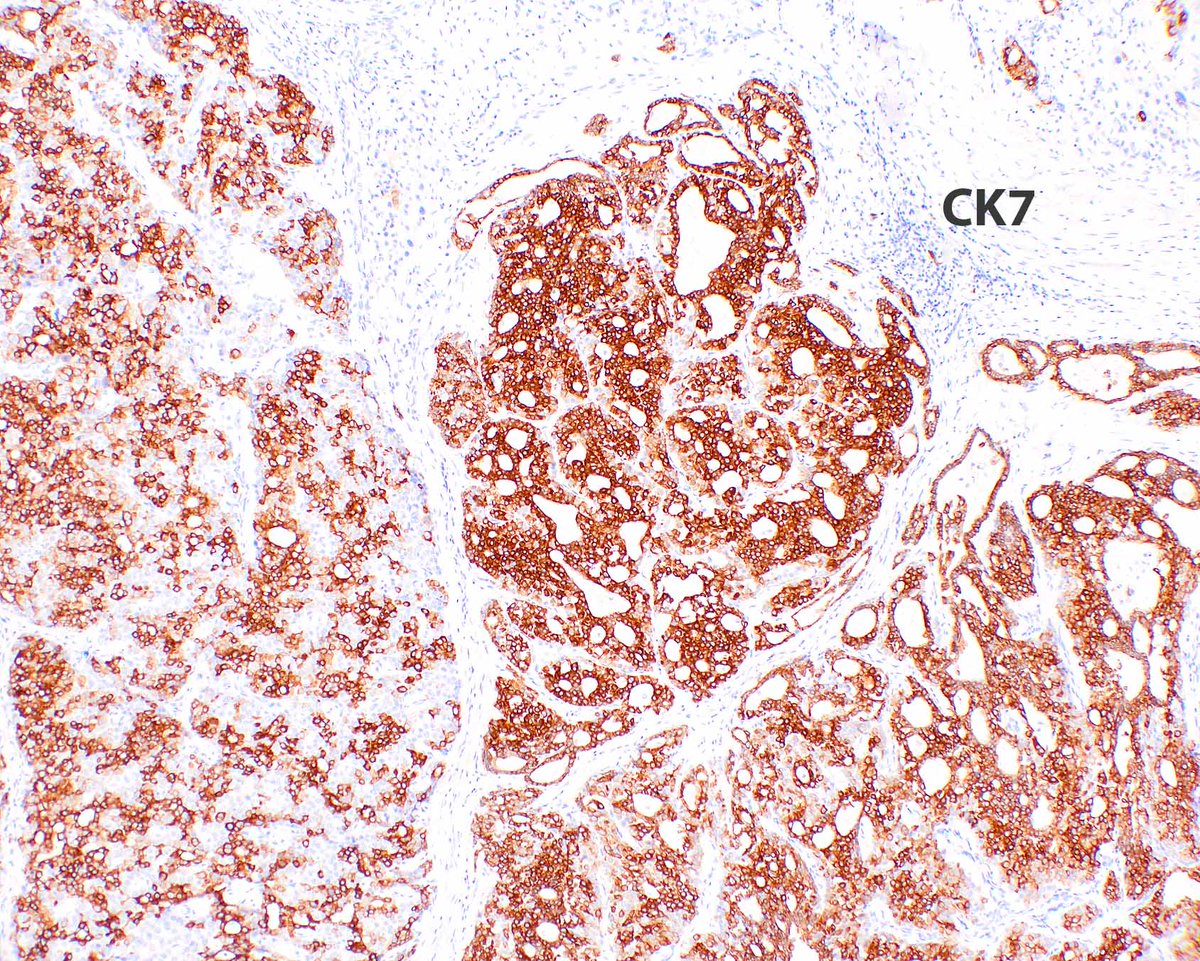
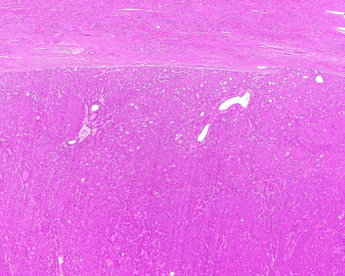
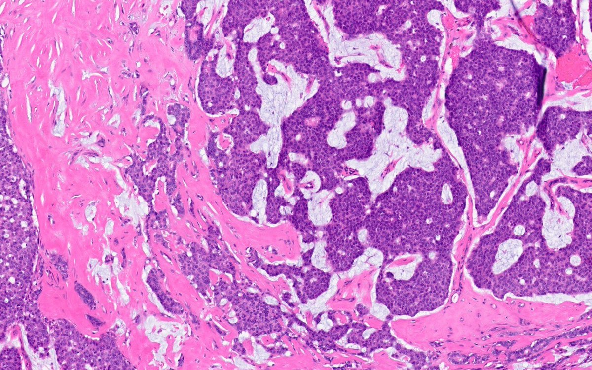
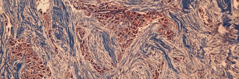
Cholangioblastic cholangiocarcinomas are really interesting - I am old so I like the old name!
Hissong E, et al. NIPBL::NACC1 Fusion Hepatic Carcinoma. Am J Surg Pathol. 2024 Feb 1;48(2):183-193. PMID: 38047392; PMCID: PMC11238093.
Happy Friday, everyone. Here's a cool case to think about over the weekend (yeah right😀)
Central duct excision for bloody nipple discharge. No previous biopsy.
What is your diagnosis?
#breastpath
Astounding intratumoral variability in grade and pattern within breast carcinoma is sometimes seen. This core shows low-grade tubule-forming IDC, apocrine IDC with markedly pleomorphic nuclei, and IDC with micropapillary pattern, all in the same tumor.
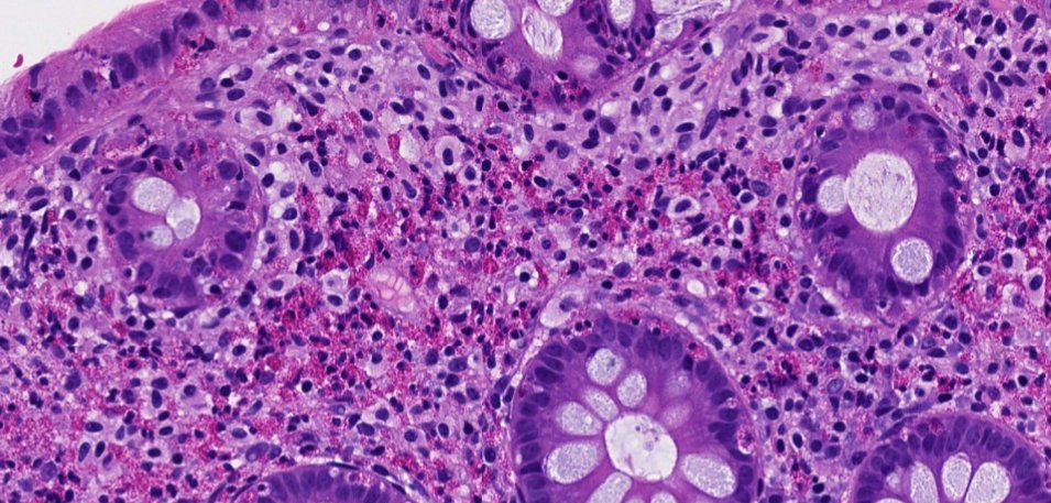
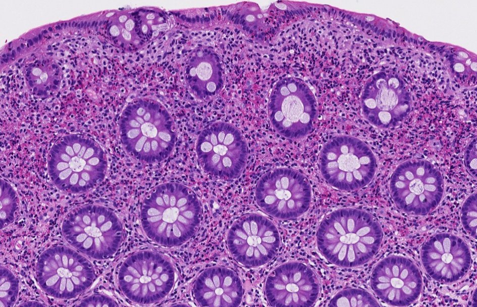
Colon - eosinophils
Increased eosinophils in colon should not be dismissed.
There are cells in the lamina propria that do not appear to be normal lymphocytes or plasma cells, which prompted a CD117 (KIT) stain.
Mast cells show positive staining for CD117
This is a case of systemic mastocytosis involving colonic mucosa
Increased eosinophils in colon, possible etiologies include:
• Drugs / medications
• Inflammatory bowel disease (increased lamina propria lymphocytes, plasma cells and eosinophils)
• Parasitic infections
• Systemic mastocytosis
Dr. Krasinskas - Tips and Hints in Surgical Pathology #USCAP #pathology #PathX
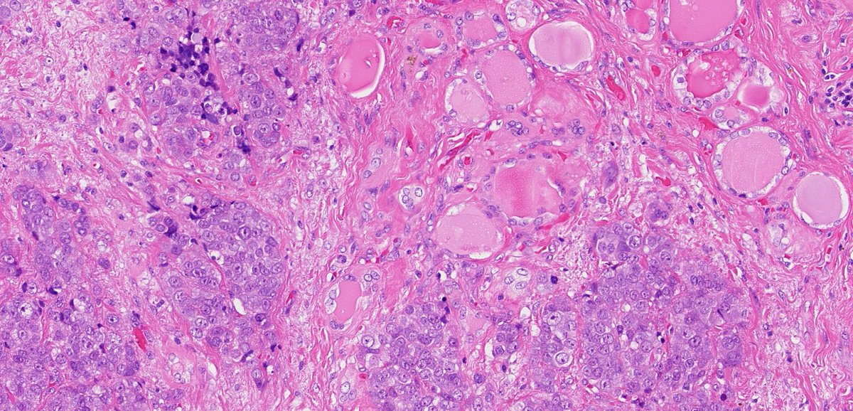
Head & Neck & Endocrine Pathology Residents - Quiz Time!
An elderly woman presents with an expanding left thyroid mass invading strap muscles, the recurrent laryngeal nerve, and the esophagus. Lobectomy is performed. What’s your dx? (Follow the thread!)
I think that's all I have to say for this fascinating case. DCIS with mucinous features, predominantly flat, with focal microinvasion, incidental in a prophylactic mastectomy.
Pretty insane stuff.
I'm sure this is not a case everyone would agree about, but this is my thought process, and my need to understand the life story of a lesion rather than going by rigid criteria that don't adequately explain the process unfolding in front of our eyes.
Thank you all, and I'll skip a few days and see you Monday for #BCCW10!
#breastpath #PathX @washu_pathology@washupathedu
"Bile duct brushing" is the last specimen in Cytology that I want to look at.
These ductal cells look horrendously worrisome compared to the benign control (arrow) but 3 features favor benign - Clinical hx of impacted stones - PMNs - Nuclei with multiple nucleoli/chromocenters.
Metastatic papillary thyroid carcinoma to the kidney #GUpath #EndocrinePath #ENTpath
➡️Metastatic PTC to the kidney is exceptionally rare, with < 50 cases reported
➡️May present as solitary or bilateral renal masses, often mimicking primary renal tumors
➡️Nuclear grooves/pseudoinclusions and colloid in this tumor
➡️The presence of renal metastasis generally indicates advanced disease and may be associated with a poorer prognosis
(PMIDs: 28588771; 28242987)
Happy Friday everyone!
Let's tackle our Breast Consult Case of the Week 5 #BCCW#breastpath
This one is a bit of a head scratcher because of incongruous cytology and architecture.
First of all, we have what appears to be a proliferative lesion with a population of uniform cells involving multiple spaces, which sounds like a very good definition for an atypical/neoplastic proliferation. The confusion here is that the cytology is predominantly columnar, if not tall columnar, and the pattern of involvement is pagetoid (you can see the flattened layer of residual native epithelium lining the luminal aspect of the proliferation).
Pagetoid involvement of spaces is typically seen with lobular neoplasia and higher grade examples of DCIS, while the worst columnar cell proliferation we know of is columnar cell hyperplasia with atypia, which this isn't.
In other words, this is an atypical columnar cell proliferation growing like a D/LCIS. Though I do not claim to have an answer to this diagnostic conundrum, I believe the pattern of growth and involvement of multiple contiguous spaces by a seemingly clonal population of cells in a pagetoid pattern should dictate what we call this, which is why I called it DCIS. Granted, not all DCIS cases are created equal, and one might argue that this columnar pattern of DCIS may carry a smaller risk than classic examples of low grade DCIS, just because the cells are "less abnormal". So I would not be opposed to calling this ADH, though a diagnosis of ADH is essentially inaccurate because this is not ADH by strict diagnostic criteria.
Finally, I would not go down the LCIS route because the cells are columnar, which means that their cellular scaffolding is robust, unlike the plasmacytoid cells of lobular neoplasia that suffer from a disrupted internal architecture.
That's all. Such cases with unclear diagnoses make us think more intently about our criteria and understanding of the neoplastic processes of mammary epithelium.
See you all Monday for BCCW 6!
@washupathedu@washu_pathology #PathTwitter #PathX
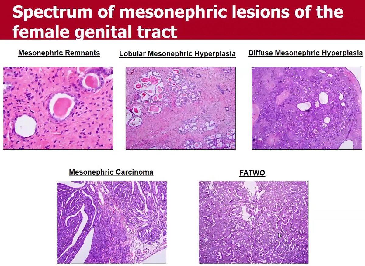
Spectrum of mesonephric lesions in the female genital tract
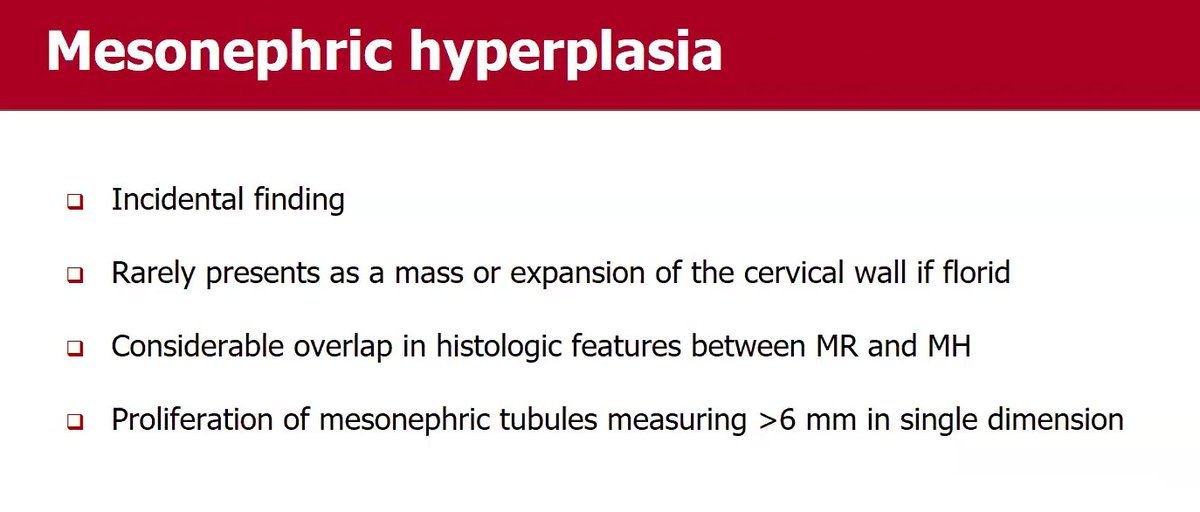
Mesonephric hyperplasia: Proliferation of mesonephric tubules measuring >6 mm in single dimension
Mesonephric hyperplasia vs carcinoma: There is no back to back tubular proliferation or architectural complexity in hyperplasia. If you see those features, then you favor carcinoma
Dr. Mirkovic-Mesonephric and Mesonephric-like Pathology #ISGyP #pathology #Gynpath #PathX
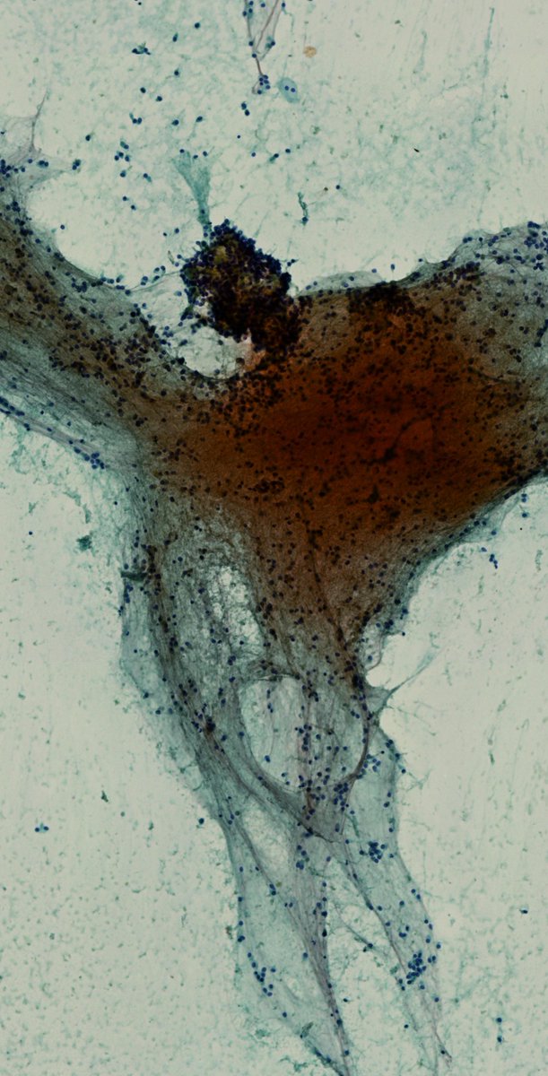
25-year-old, abdominal CT reveals a well-circumscribed, partially cystic and solid mass in the tail of the pancreas. An EUS-FNA is performed. What is your impression?
A) Benign
B) Malignant
C) Gimme a cell block
#CytoPath#PathTwitter#Pathology#PancreasPath