What I love about science is the feeling of discovery - that quiet moment when something you measured, observed, or calculated reveals a truth that was always there, waiting to be seen.
This work was possible due to an amazing collaboration with the Raphael Morscher Lab at the University of Zurich.
Co-led by Simona Ulrich and Victoria da Silva-Diz - teamwork truly makes the dream work! 🙌🏼
Huge thanks to my advisor Daniel Herranz for his mentorship and support. 🙏
Super excited to share the preprint of my PhD work! 🧵👇 🎉
We present the first in vivo dietary pan-amino acid dropout screen in cancer and discover a previously unknown metabolic vulnerability in T-ALL: Histidine.
https://t.co/eqoH7M1v7I
The most exciting part - Our findings may have therapeutic relevance beyond leukemia, including other cancers, immune disorders, and possibly hypercholesterolemia.
https://t.co/wSkeeHC0xS is a domain name that I own. AI companies that might want to capitalize on this extremely pertinent name should DM. It’s for sale!
context : https://t.co/ByqYkPbdH1
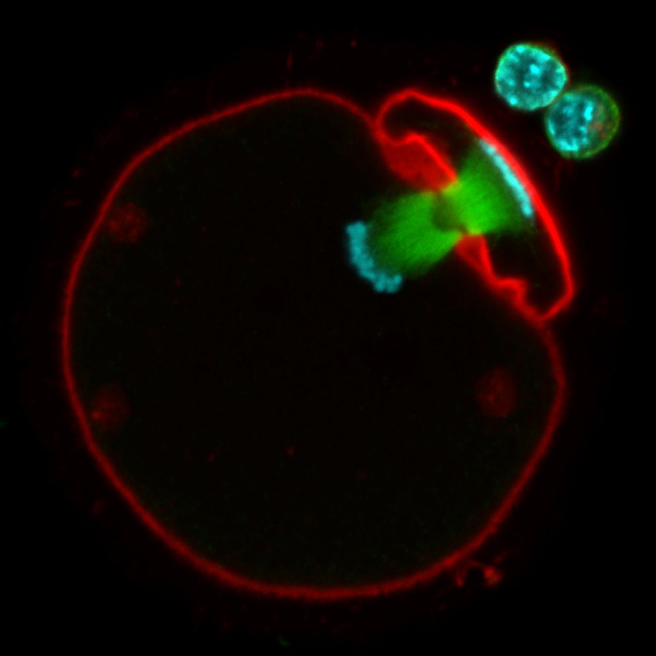
Women are born with all their oocytes, which need to stay viable for decades to ensure fertility. How are they maintained for that long?
Our latest research in @NatureCellBio reveals that oocyte maintenance involves exceptional protein longevity. https://t.co/ZiByc9F4hS (1/9)
A fascinating work is published today @Nature by Zhang & Zhang et al. on a serendipitous discovery of haemoglobin expression outside the RBC cells--in chondrocytes--and their critical role in keeping our cartilage alive under an oxygen-deprived environment.
https://t.co/DPhuZN1JGE
Our school teachers taught that haemoglobin (Hb) is an oxygen-transporting protein expressed exclusively in the red blood cells. Although there exist occasional sporadic reports of Hb expression outside the RBCs (e.g. neurons, retinal cells etc.), no one would have guessed Hb would play a critical role (required for survival) outside the RBCs.
The discovery
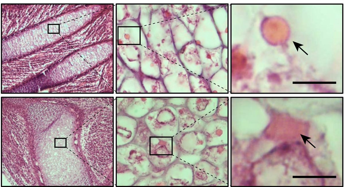
When studying the cartilage growth plate of neonatal mice, the authors noticed eosin-positive structures in the chondrocytes (cartilage cells) that resembled structures seen in RBCs. Out of curiosity, the researchers went on to stain and examine the chondrocytes of different cartilage tissue types both from mice and humans. They realized that no matter the source or species, the cells always displayed eosin-positive structures under the microscope.
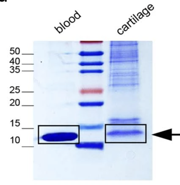
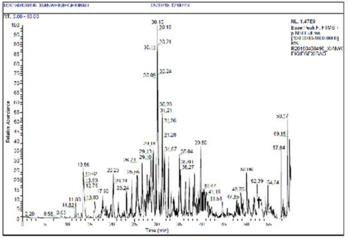
The curious researchers were determined to find out what these structures are made of. They carefully dissected these structures out and studied the protein components using mass spectrometry and were surprised to learn the results: the top hits were Hb proteins.
Unable to believe the results, they went on to study the proteins using different methods--western blotting, immunohistochemistry--and every time they ended up with the same results: the cytoplasm of cells was loaded with organelles-like bodies made of haemoglobin proteins. Finally, they came to the realization that Hb is abundantly produced in chondrocytes. They named these cytoplasmic Hb bodies as "Hedy".
Structure of Hedy
The authors studied the structure and formation of Hb bodies floating in the cytoplasm. Are they like an organelle? Do they have a membrane? Through various experiments, the researchers found that the Hedy structures do not have a membrane. The Hb proteins condense together by phase separation to form organelle-like structures in the cytoplasm. This condensation is itself an evolved process, requiring specific sequence structures of the Hb protein.
Globin switching
We know that there are different forms of Hb each expressed during different developmental stages: embryonic, fetal and adult Hb. There exists a sophisticated molecular machinery (which was believed to be RBC-specific) that switches one Hb type to the other at appropriate times. Using gene silencing experiments, the authors were further awestruck to find that the chondrocytes too switched their Hb types from embryonic to fetal to adult stages, just like RBCs!
Regulation of chondrocyte Hb production
It is well known that hypoxia induces Hb production via upregulating hypoxia-inducible factors (HIFs), an evolutionarily conserved molecular mechanism (Nobel Prize 2019; https://t.co/Sifi8LuYIX). But it turned out that chondrocytes have evolved to increase their Hb expression not via HIF proteins, but through a different protein, the same protein that is required for fetal to adult Hb switching: KLF1 (https://t.co/Zn0X8GpYdq).
How important is Hb for chondrocytes?
Such a high Hb expression in chondrocytes with similar globin switching behaviour as RBCs would mean that this Hb is critical for chondrocytes' survival. By deleting the Hbb gene specifically in the chondrocytes, the authors found that without Hb the chondrocytes die killing the animal a few days after birth.
Conclusion
Continuous oxygen supply is a prerequisite for the survival of cells in all tissues. The only way the cells can receive oxygen is through RBCs in the blood, which requires the tissue to be highly vascularized. When demand exceeds the supply, the cells evolve to survive an oxygen-depriving environment.
Muscles evolved to produce their own globin--myoglobin which has a higher affinity to oxygen than Hb thereby withholding O2 during oxygen excess states and releasing it back during oxygen-deprived state (during exercise). Likewise, the brain has its own globin: neuroglobin (https://t.co/6v78zvHajP).
Today, we are learning that cartilage (an avascular tissue), too, has its own globin. But unlike muscle and brain, have evolved to store oxygen not by making a new type of globin but by making just the same type as the ones in RBCs, but with a higher affinity than RBC Hb.
When it comes to fundamental biology, we often assume that we have found everything and then one day a discovery like this drops, hitting us on the head to make us realize that there is a whole universe of hidden biological secrets waiting to be discovered.
Some recent posts:
1. Gene x sex interaction of PNPLA3 I148M variant (https://t.co/RFegHHRS2z)
2. Effect of consanguineous marriage on the risk of common diseases in offsprings (https://t.co/dTkPsl935K)
3. Whole genome vs. Whole exome sequencing. Which is more cost-effective for genetic association studies? (https://t.co/BCwMzw4g16)
Just watched Oppenheimer. With zero sense of irony, in a movie about the making of the atomic bomb, there is a “smoking kills” disclaimer on screen for most of the running time (thanks to Indian government regulation of film screening).
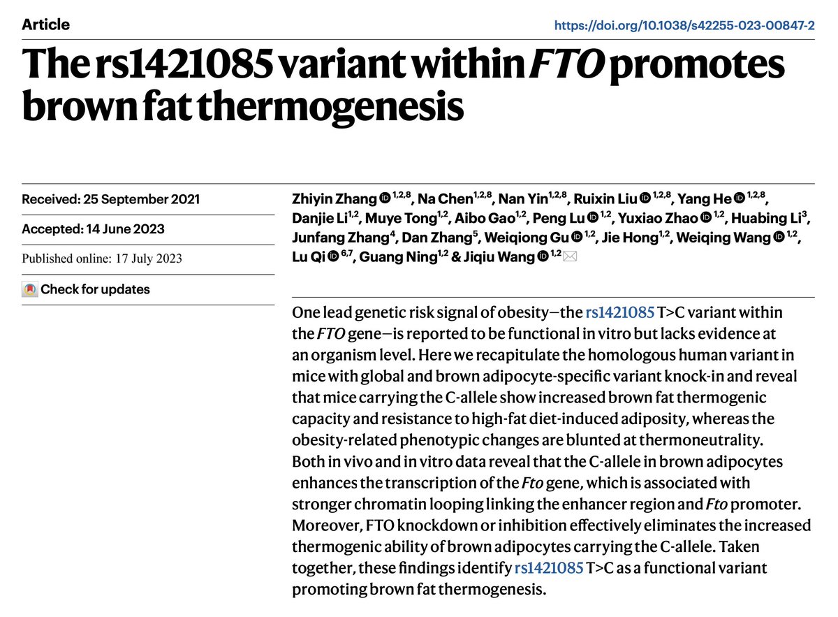
Buckle up! We're in for a wild ride today. A new @NatMetabolism paper by scientists from China adds a surprising twist to the long-known FTO GWAS story.
The FTO locus (16q12.2) is the first ever GWAS locus to be associated with obesity and even after 16 yrs now, scientists appear to be scratching their heads trying to make sense of this locus. Non-coding intronic variants within FTO strongly associate with BMI, where individuals homozygous for the top risk variant weigh ~3kg more than non-carriers (https://t.co/vh4ceXVQaQ).
Since its discovery in 2007, there have been tremendous efforts to identify the causal gene(s) at this locus. Given that the risk variants are sitting right within a gene, FTO was of course the primary suspect. How do you find out if FTO has an effect on BMI? Delete it in mice and see if the animal gains weight. And that's what scientists did and found out that Fto knockout mice were stunted and lean, and the leanness was mainly due to burning too much fat (https://t.co/DkKIGLvD0P). That's great. So FTO must be the causal gene.
But then contradicting findings appeared. If you knock out the Fto only in adipose tissue (https://t.co/W5A8JwT2Hx) or globally after the animal has grown (https://t.co/uJ69bO8A9x), the mice actually gain weight!
Amidst this confusion came an even bigger one: two landmark papers, one in Nature (https://t.co/Kq3uqKrAg1) and the other in NEJM (https://t.co/Ztqf4FTOYT), said, forget about FTO, the causal genes are located far away. The FTO locus is an enhancer that folds in the 3d space and touches the promoter of distant genes IRX3 and IRX5. And deleting Irx3 in mice resulted in weight loss.
Then scientists were like, you know, a proper experiment would be not to knock out Fto or Irx3, but to delete the homologous noncoding region in mice. So, they deleted an 82 basepair-homologous region in mice and showed that without this region, the mice don't gain weight when fed with a high-fat diet, and deleting this locus increases Irx3 and Irx4 expression (https://t.co/NFFL2OQNL8). So, the causal genes are IRX3 and IRX4 then.
And now, in the current paper, the scientists argue, you know, the most appropriate way to study the FTO locus is to recreate the exact genetic variant in mice and study the consequences.
What did they find? The exact opposite of what was found in humans. The risk allele that increased weight in humans, decreased weight in mice. Can it be because of some off-target effects of the CRISPR experiment? No. Even if you do the knock-in in an old-fashioned way, the results are the same. The weight loss is mainly via over energy expenditure via brown adipose tissue. Wait, there is another twist.
There is an interesting difference between humans and mice. Humans have brown adipose tissue only during infancy and then lose it as they grow into adults, which isn't the case in mice. The brown adipose tissue helps mainly during cold temperatures. The experiment mice are usually housed at around 20-22 degrees Celsius, which is an ambient temperature for humans but not for mice. It's substantially cooler than mice's "thermoneutral zone (29-31 degrees)".
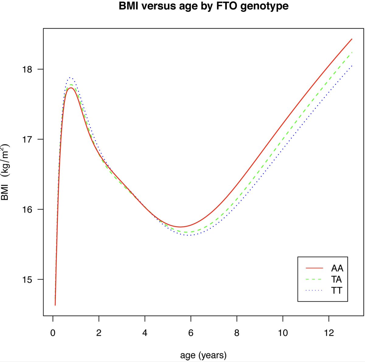
So what happens when you repeat the experiments in what might be the mice's ambient temperature--30 degrees? All the weight loss effects that were previously seen at 20-22 degrees are now blunted. So, the FTO locus effects are strongly dependent on two things: temperature and the presence of brown adipose tissue. And it turned out, in fact, it was previously shown in humans that the FTO variant has an age-dependent effect. It lowers the body weight in infants and then increases the body weight in adults, which aligns with the current finding. (https://t.co/S7iKVj0p5d)
Overall, the current paper is an impressive work and will stand as a landmark in the long twisty road of FTO story. But above all, this paper is a remarkable example to show case the challenges behind translating mice physiology to human physiology.
https://t.co/i8IIr2bSFc