@manruipa@ThorHessdalen4@mecfsskeptic I have also been on Valacyclovir for 5 years prophylactic- I used as needed, prior to Covid. I still get a few minor symptoms initially when I have been exposed to viruses and have a bit of a milder systemic reaction/reactivation most of the time. Covid reactivated EBV for me.
We have great news from @putrinolab🙏🏻 They just submitted a paper about the initial results of a novel device they tested for Long COVID related cognitive impairment!
Thank you for sending this video message, Dr. Putrino. 🙏🏻 #LC2026HopeVideos#LongCovidAwarenessDay2026
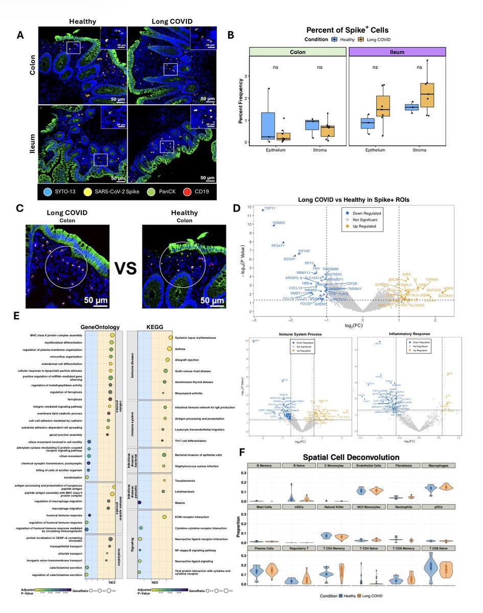
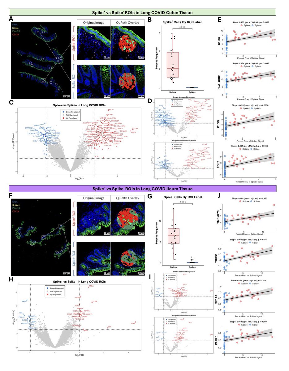
@vipintukur Plasma antigen decays, but gut biopsies show persistent Spike transcript + protein in *every* Long COVID case, triggering immune storms (57 DEGs!). Exactly why the plasma curve is lying to us: https://t.co/Tkge64Hy0h
Can we say dysautonomia? HIV patho 101 = virus in the small intestines causes dysautonomia presentation.
HIV can significantly impact the small intestine, contributing to dysautonomia through several mechanisms. The virus may directly infect the mucosal lining of the small intestine, causing inflammation and damaging the enteric nervous system responsible for gut function. This disruption affects the autonomic regulation of gastrointestinal processes. Additionally, HIV compromises the immune system, making individuals susceptible to opportunistic infections and conditions like chronic enteropathy, which further exacerbate inflammation and nerve signaling issues. Changes in the gut microbiome due to HIV can lead to systemic inflammation, impacting the autonomic nervous system and manifesting dysautonomia symptoms. Furthermore, infections and damage in the small intestine can result in malabsorption of essential nutrients necessary for proper nervous system function, worsening dysautonomia.
Seems like another mechanism that SC2 shares is this.
The persistence of SARS-CoV-2 spike protein in the gut indeed raises fascinating questions about its role in #LongCOVID and immune response. Could this be a factor contributing to prolonged symptoms in some patients? How might this discovery impact treatment and management strategies for #LongCOVID? For a deeper dive into such biomedical questions, Sci-Quest offers a comprehensive platform for generating reviews and insights: https://t.co/4Y9Imt8uIJ. #Medicine #Immunology
Persistent spike-positive areas in the colon showed increased immune cell activity, including:
• Macrophages
• Plasma cells
• Regulatory T cells
Suggesting an active local immune response in the gut.
➡️ Researchers also found disrupted expression of key immune-signaling genes, indicating impaired immune coordination and chronic inflammation in gut tissues. 2/
Take-home message:
👉 Persistent viral proteins in the gut may contribute to long-term immune dysregulation in Long COVID, helping explain ongoing symptoms. 5/5
https://t.co/KNLK8Zvlm5
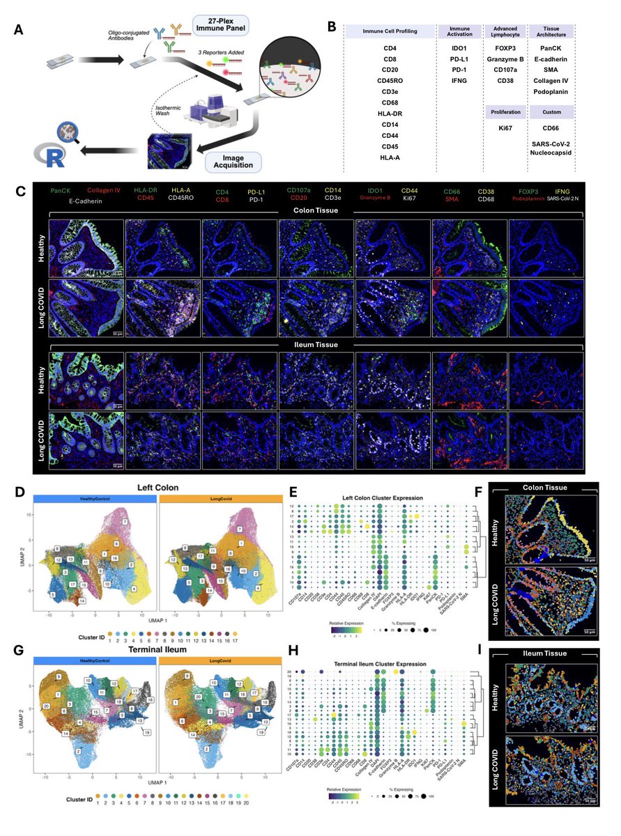
SARS-CoV-2 persistence is a proposed driver of Long COVID (LC), but the in-situ relationship between residual viral antigen and immune dysregulation remains poorly defined.
➡️ This NEW study provides robust evidence that persistent SARS-CoV-2 Spike protein detection in the gut is not immunologically inert.
➡️ Instead, it is actively associated with distinct, immune cell composition shifts and a dysfunctional pro-inflammatory transcriptional profile, supporting the hypothesis that retained viral antigen drives chronic immune dysregulation in tissue of LongCOVID subjects. 3/
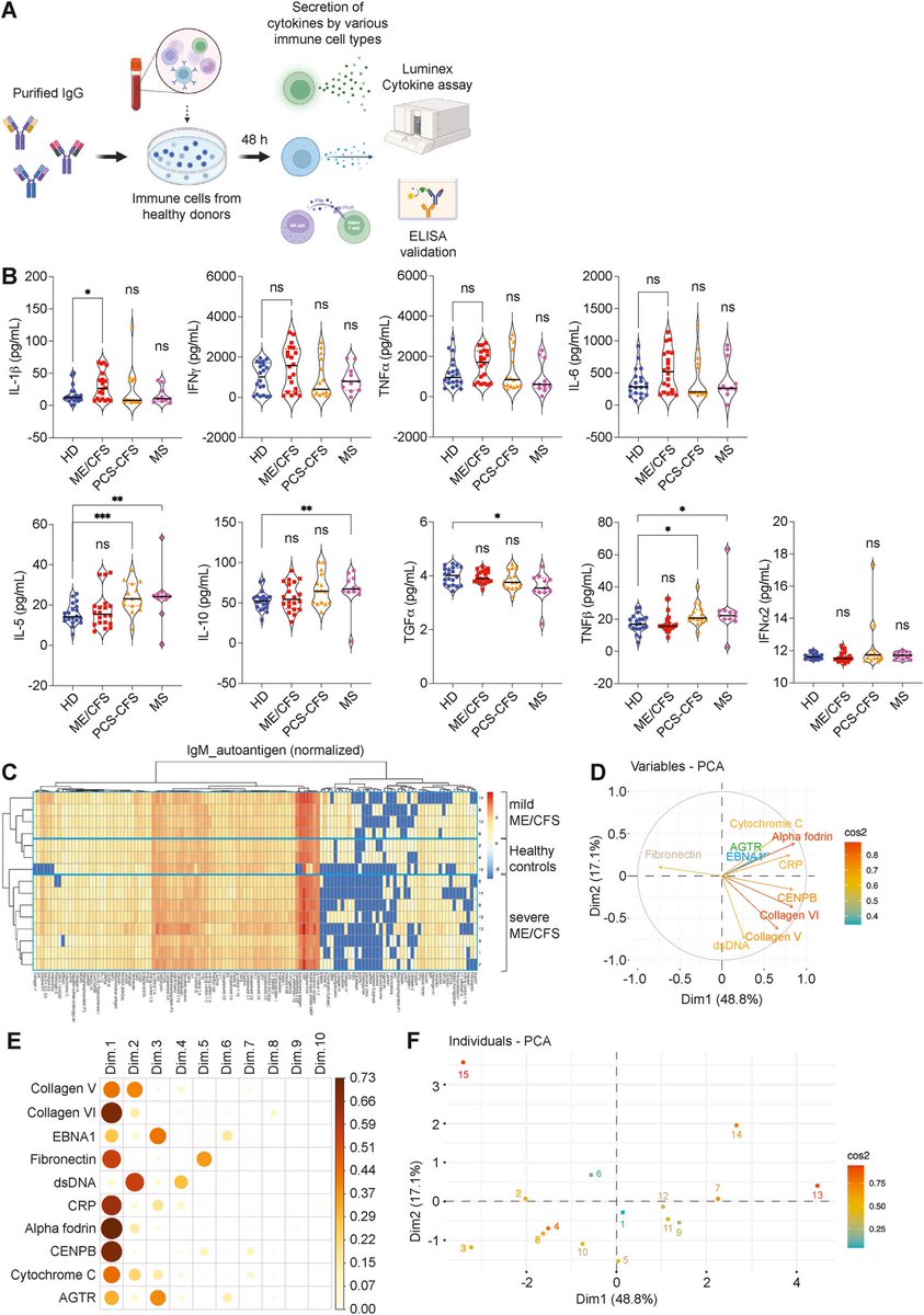
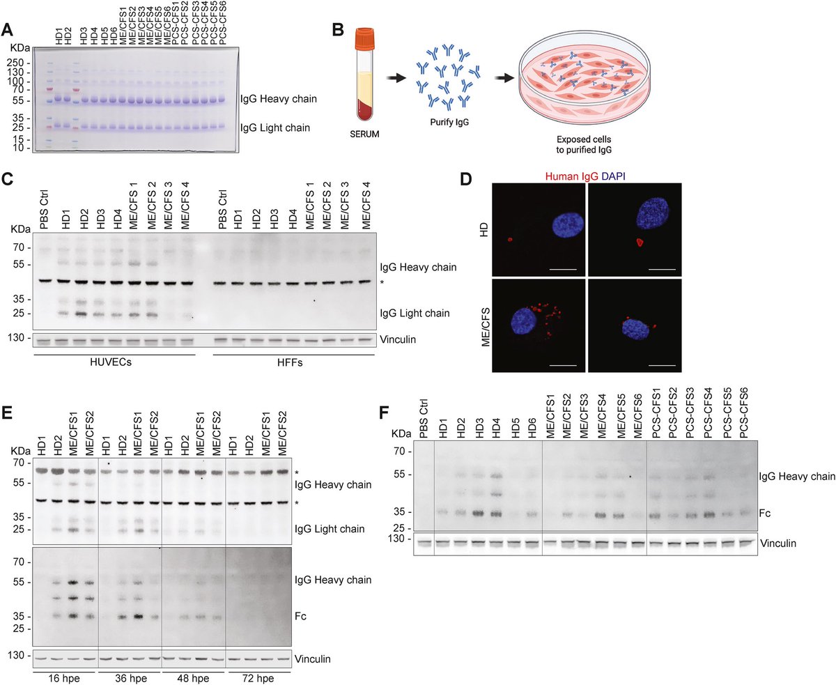
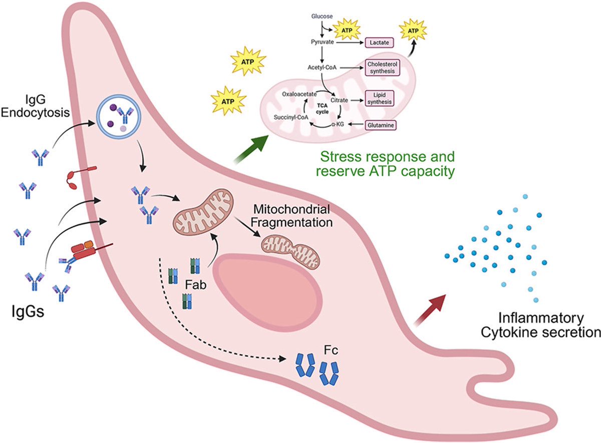
The study also suggests that IgGs from ME/CFS patients may induce a chronic, protective stress response, promoting mitochondrial adaptation through fragmentation, while preserving the cells’ ability to produce ATP. 3/3
H/T: @CatchTheBaby
https://t.co/9rw6dujiiV
Interestingly, the molecular signatures differed between ME/CFS and LongCOVID.
➡️ In ME/CFS, immune complexes primarily affected extracellular matrix organization—the structural environment surrounding cells.
➡️ In contrast, in LongCOVID, the changes were more closely linked to hemostasis and blood-clotting pathways. 2/
A potential new direction for treating #LongCOVID and ME/CFS.
➡️ A NEW study suggests that IgG antibodies—and even their Fab fragments—from patients with ME/CFS, including those who developed the condition after COVID-19, can directly affect human endothelial cells.
➡️ The researchers found that these antibodies can alter mitochondrial structure and function, leading to mitochondrial fragmentation and changes in cellular energy metabolism. 1/
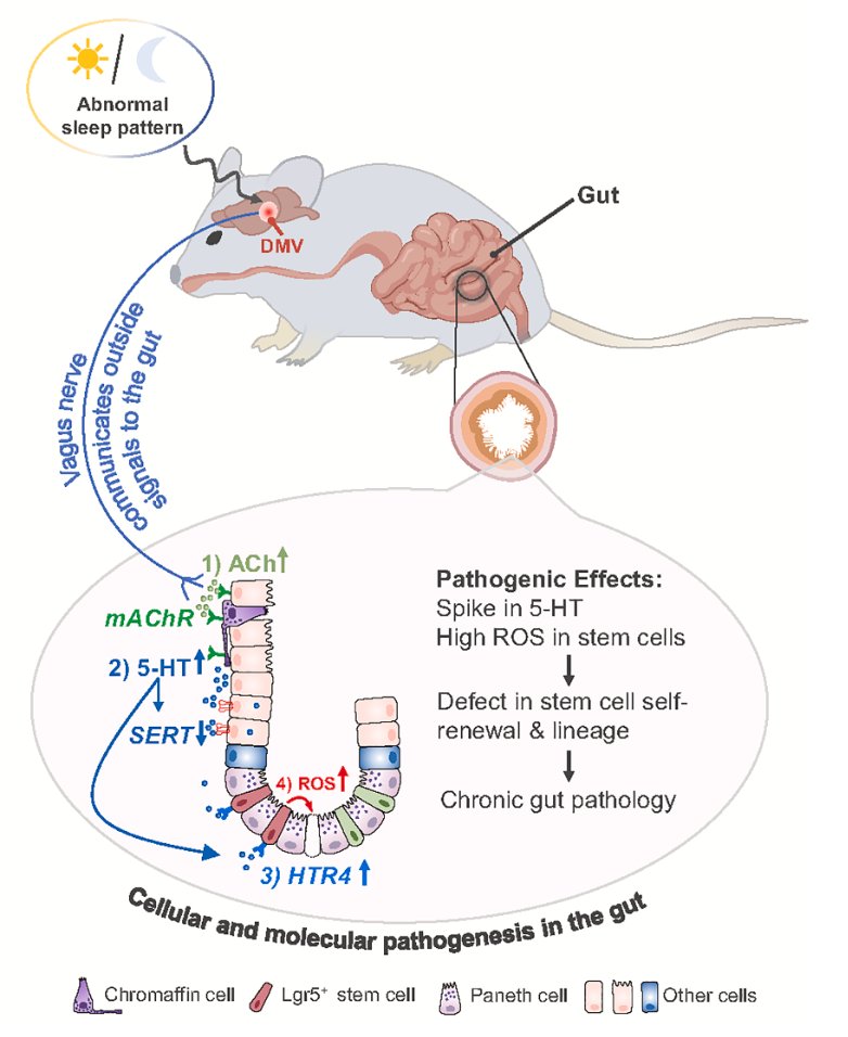
The vagus nerve (again), here connecting the dots between sleep disturbance and gastrointestinal dysfunction
https://t.co/8uopfjlX0S
More on this remarkable circuitry https://t.co/J2ZQBhE1eF
@DawgNation@TrinidadChambl1 I hope you take it all the way @TrinidadChambl1 ! This Dawg fan of 59 yrs is pulling for you! I can’t wait to continue to watch you evolve into an NFL legend! God bless!
The dysregulated immune system in #LongCovid with #MECFS. with sex-specific changes, increased inflammation (as seen by cells, chemokines and cytokines), and disrupted hormone levels https://t.co/1QRYwa6UJb
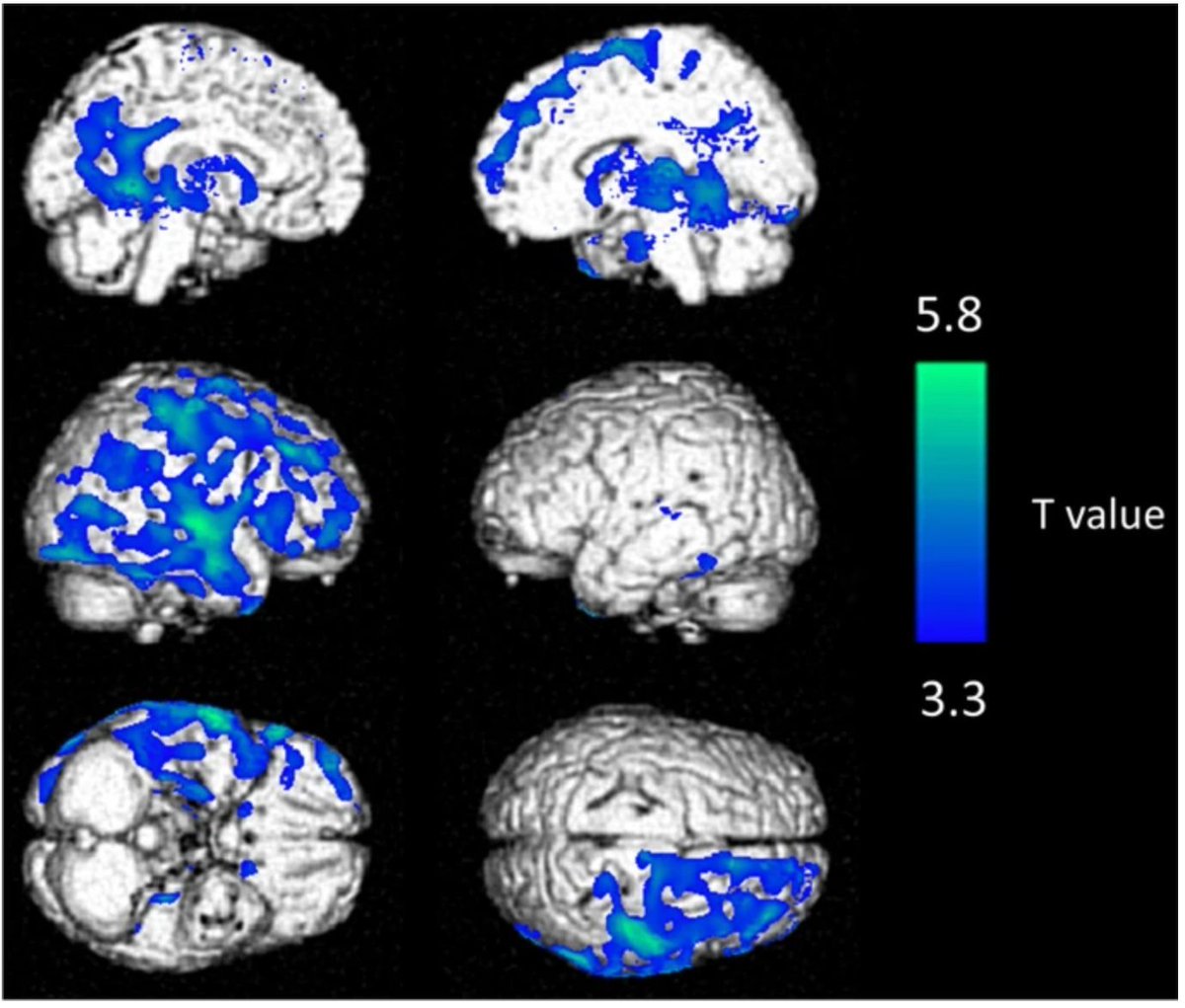
Long COVID patients have reduced metabolism in the right frontal and temporal lobes of the brain. Is this brain fog being visualized?
\ The Discovery:
Researchers from the Department of Nuclear Medicine at Université de Lorraine, in Nancy, France, intravenously injected a radioactive tracer in those with Long COVID and healthy controls. The tracer, 18F fluorodeoxyglucose (18F-FDG), is structurally similar to glucose, a sugar molecule metabolized by cells, including neurons.
\ How 18F-FDG Works:
Once 18F-FDG is transported into neurons, it becomes trapped. Here, the fluorine-18 isotopes decay, emitting light detected by the PET (positron emission tomography) scanner. More light = more glucose uptake.
A healthy brain would light up uniformly. But the brains of Long COVID patients light up in patches.
\ Significance:
This is important because sugar is like the gasoline that keeps the brain's engine running. Brain regions that metabolize less sugar suggest cellular (and cerebral) dysfunction.
\ What 🧠 Regions Were Affected?
Reduced glucose metabolism was found in the right frontal and temporal lobes, including the orbitofrontal cortex and hippocampus. This could contribute to brain fog by disrupting:
- memory
- attention
- decision-making
We at the Brain Inflammation Collaborative are a joint effort of patients, clinicians, and researchers working together to advance the understanding, diagnosis, and treatment of neuroinflammatory diseases, including Long COVID.
This older study is a great reminder that Long COVID is a widespread, debilitating medical condition that demands action!
Please give it a like and subscribe for more.
https://t.co/YiTjvqN6YH
Focal neuropathologies in the brain of COVID-19-infected humans: inflammation, primary gliovascular failure and microglial dysfunction | Signal Transduction and Targeted Therapy https://t.co/NvWLqJ0sUI
COVID-19 and the brain: a new study.
What the study looked at -
Researchers examined autopsies of 13 people who died with COVID-19 vs 23 controls. Goal: to uncover what exactly happens inside the brain during COVID-19.🧵
Identifying a potential causal role of mitochondrial dysfunction in neurodegenerative disease, in multiple mouse models
@NatureNeuro
https://t.co/MBdOwBuuOr
“The [vagus nerve] is immune privileged, but very microbially exposed… we haven’t begun to understand what those microbes are & what their byproducts might be,” said Dr. Melanie Walker. “We might be able to really narrow down our search for the origins & progression of [Alzheimer’s & Parkinson’s] if we understood the vagus nerve better.”