🚨 José Mourinho: “I was expecting to find a different La Liga compared to the past… they told me NO, I wanted to believe that was the case”.
“But it’s the SAME. It has not changed”.
The subclavian artery becomes the axillary. At the Teres major it becomes the brachial artery and divides into radial and ulnar arteries and deep palmar arches continuing as digital arteries
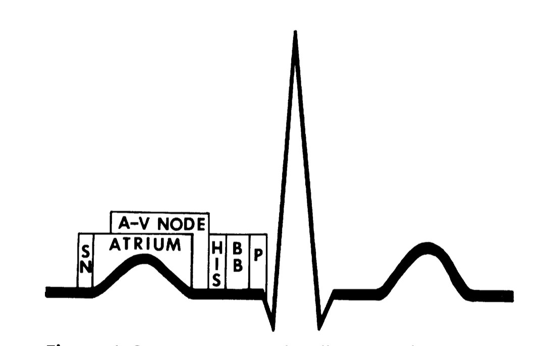
How the Electrical Impulse Travels
The ECG is easier to understand when you stop seeing it as a collection of waves and start seeing it as a map of electrical activation.
The sequence is:
SA node
⬇️
atria
⬇️
AV node
⬇️
His bundle
⬇️
bundle branches
⬇️
Purkinje fibers
⬇️
ventricles
The P wave represents atrial depolarization.
After the atria depolarize, the impulse reaches the AV node, where conduction slows. This delay gives the ventricles time to fill before ventricular activation.
The impulse then travels through the His-Purkinje system, producing rapid ventricular depolarization and the QRS complex.
Finally, ventricular repolarization produces the T wave.
So when you look at a normal ECG:
P = atrial depolarization
PR interval = atrial-to-ventricular conduction
QRS = ventricular depolarization
T = ventricular repolarization
The key concept is that the ECG is not simply recording the heartbeat.
It is recording the timing and sequence of electrical activation through the heart.
That becomes clinically important when conduction is delayed or blocked. For example, abnormalities anywhere from the sinus node through the AV node and His-Purkinje system can produce characteristic ECG changes and bradyarrhythmias.
And this is why understanding the normal pathway comes first.
Before you diagnose a conduction abnormality, know the normal conduction pathway.