La #PIM c'est un large univers !
Elle est en effet aussi membre de :
- @MICA_Microscopy, réseau local de plateformes de microscopie & cytométrie
- EMBRC-France @EmbrcFrance@EMBRC_EU qui regroupe au niveau national un éventail de services en lien avec les organismes marins
5/5
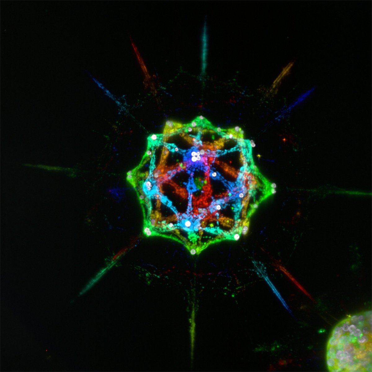
Quel type de microscopie est proposé par la #PIM ?
- microscopie à épifluorescence
- microscopie confocale
- microscopie à feuillet de lumière
- microscopie électronique à balayage
4/5
📢A voir !
Découvrez à l’occasion de notre série sur X :
🫧 « Les plateformes scientifiques @IMEV_mer à la loupe🔍» !
Volet Numéro 2 du Lundi 22 au Vendredi 26 Avril consacré à la #PIM
1 semaine, 5 chapitres !
@CNRS@Sorbonne_Univ_@EmbrcFrance@MICA_Microscopy
@openmicroscopy our monthly Tips & Tricks
@Univ_CotedAzur@EmbrcFrance
Omero Tips November edition : How to make the most of your global image measurements - Omero.parade - Part III Plot layout
MICA is proud to be part of this interdisciplinary project with GL Lippi
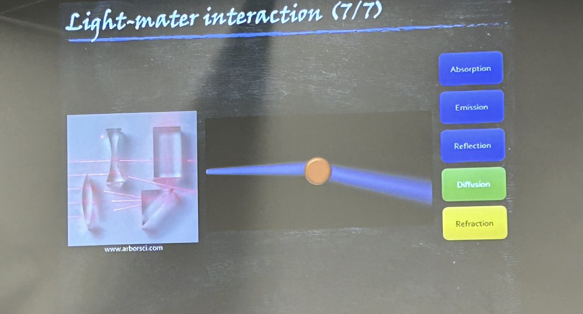
Nanoscatterer-Assisted Fluorescence Amplification Technique https://t.co/7l73WF3NYh #mdpinanomaterials via @nano_mdpi@ipmc#inphyni
@openmicroscopy our monthly Tips & Tricks
@Univ_CotedAzur@EmbrcFrance
Omero Tips October edition : How to make the most of your global image measurements - Omero.parade - Part II Filter measurements using the heatmap
@openmicroscopy our monthly Tips & Tricks
@Univ_CotedAzur@EmbrcFrance
Omero Tips September edition : How to make the most of your global image measurements - Omero.parade - Part I Filter image display using measurements
Back at it again!
@openmicroscopy our monthly Tips & Tricks @Univ_CotedAzur@EmbrcFrance
Omero Tips February edition : Populate Metadata script : Part I format the .csv file
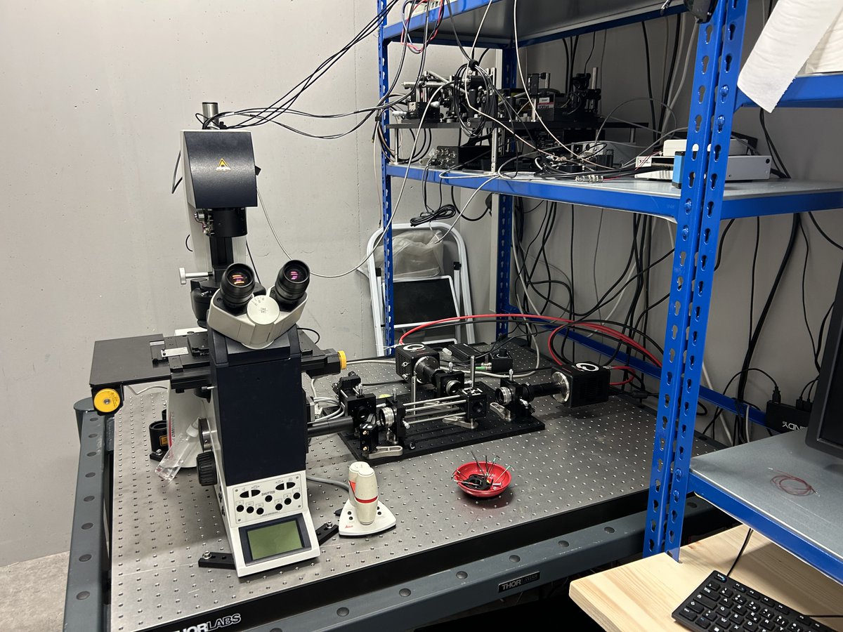
Easing batch image processing from #OMERO: a new toolbox for #ImageJ. We are proud to announce that the @MICA_Microscopy facility (partner @IPMC_sophia) and the @GReD_Clermont @UCAAuvergne have developed a new interface to automate the analysis of images managed through OMERO.