Feeling bipolar about bilateral thalamic lesions?
Unfortunately, the differential for bilateral thalamic lesions isn’t binary!
But here’s an easy mnemonic to help: THALAMIC!
T = Tumor (glioma)
H = Hypoxic/ischemic encephalopathy
A = Artery of Percheron Infarct
L = Loss of thiamine (Wernicke’s)
A = ADEM
M = Metabolic/Toxic
I = Internal cerebral vein thrombosis. Infection (west nile)
C = Creutzfeld-Jacob
Now you’ll never bypass a bilateral thalamic diagnosis with this easy mnemonic!
We see more and more lateral dural tear #CSFleaks with small epidural fluid missed because the correct MRI sequences were not obtained. 3DT2FS really shines in this context. Sometimes the 🔑 isn't new technology, but re-understanding how to look at existing data.
When you’re asked to localize the lesion in a patient with aphasia, do you suddenly feel speechless?
At a loss for words to categorize the type of aphasia?
Never fear—here is the decision tree for patients w/aphasia and the associated anatomic correlates
Three main questions:
1. Fluency? Nonfluency indicates damage to the FRONTAL language regions anterior to the fissure of Rolando
2. Comprehension? Impaired comprehension indicates damage to the TEMPOROPARIETAL language regions posterior to the fissure of Rolando
3. Repetition? Impaired repetition indicates damage within the core PERISYLVIAN language zone
The answers will lead you both to the type of aphasia and the location of the lesion.
Keep this figure with you for quick reference—and then when you’re asked about a patient with aphasia, you will have a lot to say!
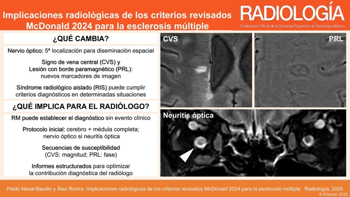
🧵 Los criterios McDonald 2024 son la mayor revisión del diagnóstico de #EsclerosisMultiple desde su creación — y cambian el papel del radiólogo. 👇
https://t.co/YMDlhlVqzG
#Neurorradiología#RM#OpenAccess
In Alzheimer’s imaging, there are some things you just can’t forget!
New Alzheimer’s treatments are changing the way we look at these scans!
Removal of amyloid beta proteins from vessel walls by anti-amyloid antibodies leads increased vascular permeability.
This can cause edema or hemorrhage, called ARIA (Amyloid Related Imaging Abnormalities)
Grading of the degree of ARIA is important bc it can change management. Asymptomatic ARIA is usually treated by pausing treatment if it’s moderate to severe
For ARIA-E (edema) or ARIA-H (microhemorrhage), if the edema measure > 5 cm or number of microhemorrhages are >5, it is moderate
For superficial siderosis, 2 regions is considered moderate.
Remember it by this little rhyme:
--If ARIA findings are great than 5, then the medication deprive
--Or if siderosis is 2, then medication break for you!
Hopefully, now the grading of ARIA will stay in your memory!!
RCT: Among patients with 5 to 20 brain metastases, stereotactic radiation targeting individual tumors improved symptom severity and interference with daily functioning compared to hippocampal-avoidance whole brain radiation.
https://t.co/drqdp2N9rX
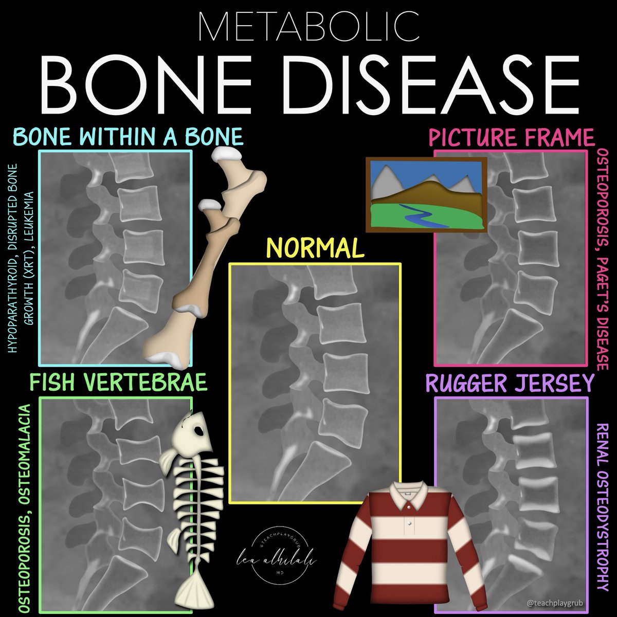
Can’t metabolize all the metabolic bone diseases?
When you see weird bones, do you just say, “suggestive of metabolic bone disease” and hope no one asks questions??
Is trying to memorize all their patterns disrupting your homeostasis?
Metabolic disease are complex but there are a few that are a slam dunk
Bookmark this figure for the for the 4 classic xray patterns you NEED to know!
The three major global threats to population health are non-communicable diseases (#NCDs), infectious disease outbreaks, and environmental degradation.
A Lancet Commission provides a set of priority recommendations to address these threats.
Read now 👉 https://t.co/fJMXEylZF2
Debates sobre vocación, heroísmo…¿en qué momento nos olvidamos de la vida? La fisiología no.
Sostener un sistema 24/7 con guardias que fuerzan este estado -noche tras noche-durante años, no es resiliencia, es una deuda biológica. Y el cuerpo lleva la cuenta
#HuelgaMedica
Un meme que invita a pensar.
👩✈️En aviación, fatiga🟰 riesgo
👩⚕️En sanidad, fatiga 🟰dedicación
La seguridad del paciente también depende del médico descansado. El nuevo Estatuto Marco parece una oportunidad perdida para mejorar.
#HuelgaMedica#EstatutoMédico
The data are clear.
Night shifts stress the same systems — memory, metabolism, cellular energy.
Mitigation isn’t a complaint; it’s part of doing the job well.
More knowledge should guide better habits, not just accumulate.
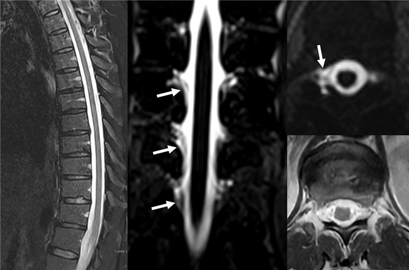
💡 New study: In MRI of multiple sclerosis, a simple switch in sequence nearly DOUBLED the number of active lesions we can detect.
👉 Open access:
https://t.co/LjbdUKsH6n
#MultipleSclerosis#MRI#Neuroradiology
This week’s SPIN-POV: Intracranial hydatid cysts classically present with floating membranes, daughter cysts, and no enhancement. On MRS, look out for the pyruvate/succinate peak at 2.4 ppm—a specific clue to intracranial cestodal infection.
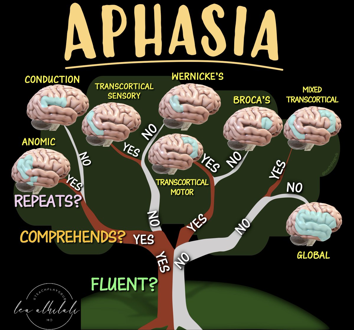
When you’re asked to localize the lesion in a patient with aphasia, do you suddenly feel speechless?
At a loss for words to categorize the type of aphasia?
Never fear—here is the decision tree for patients w/aphasia and the associated anatomic correlates
Three main questions:
1. Fluency? Nonfluency indicates damage to the FRONTAL language regions anterior to the fissure of Rolando
2. Comprehension? Impaired comprehension indicates damage to the TEMPOROPARIETAL language regions posterior to the fissure of Rolando
3. Repetition? Impaired repetition indicates damage within the core PERISYLVIAN language zone
The answers will lead you both to the type of aphasia and the location of the lesion.
Keep this figure with you for quick reference—and then when you’re asked about a patient with aphasia, you will have a lot to say!