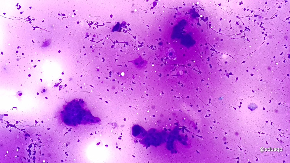
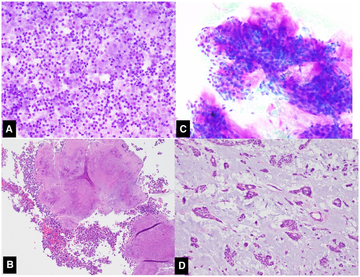
Almost always a reassuring finding - The eye-catching rhomboid “Amylase Crystals” are typically a feature of benign salivary gland disease, in this case, aspirated from a case of chronic sialadenitis. (Parotid FNA)
New #MilanSystem research! ✨ 🔬 Investigators from Japan conclude that the modified @MilanSystem as well as the @WHO system may be a useful cytopathologic classification tool for both bone and soft tissue lesions.
https://t.co/mJ2FAIWkLd
CC: @MichiyaNishino#CytoPath
The latest Milan system for #salivarygland#cytopath has a refined ROM, added risk of malignancy, a new chapter summarising the use of imaging studies, latest ancillary technics and an updated nomenclature in accord with the recent blue book.⬇️
https://t.co/pNP8njQJkr
How the Milan System for Reporting Salivary Gland Cytopathology works in cytopathology practice: Meta‐analysis of prospective studies and comparison with retrospective studies - @CancerCytopath https://t.co/YUQwPDbIMI… with @IvanaKholova ; @ZMaleki_cyto ; Henri lagerstam
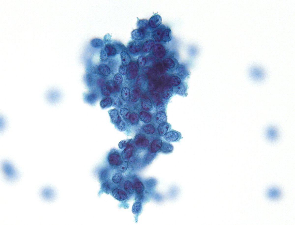
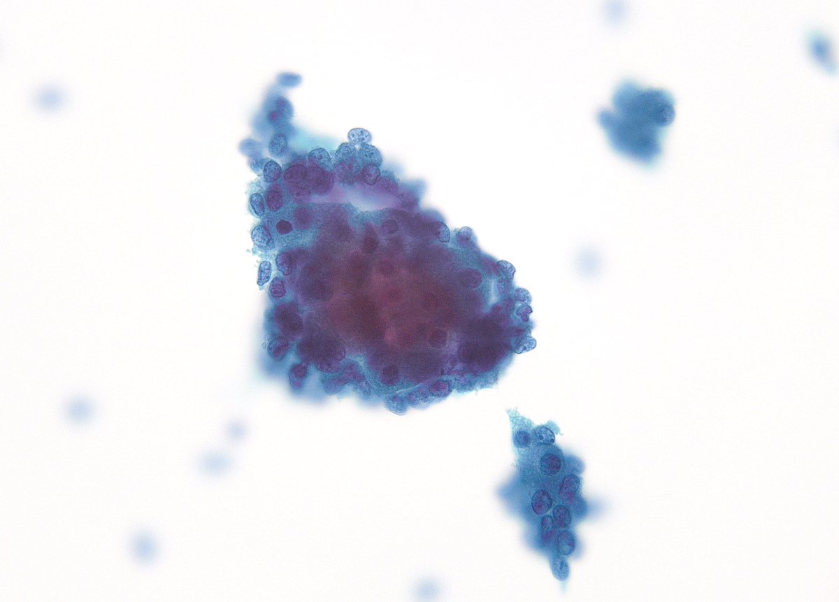
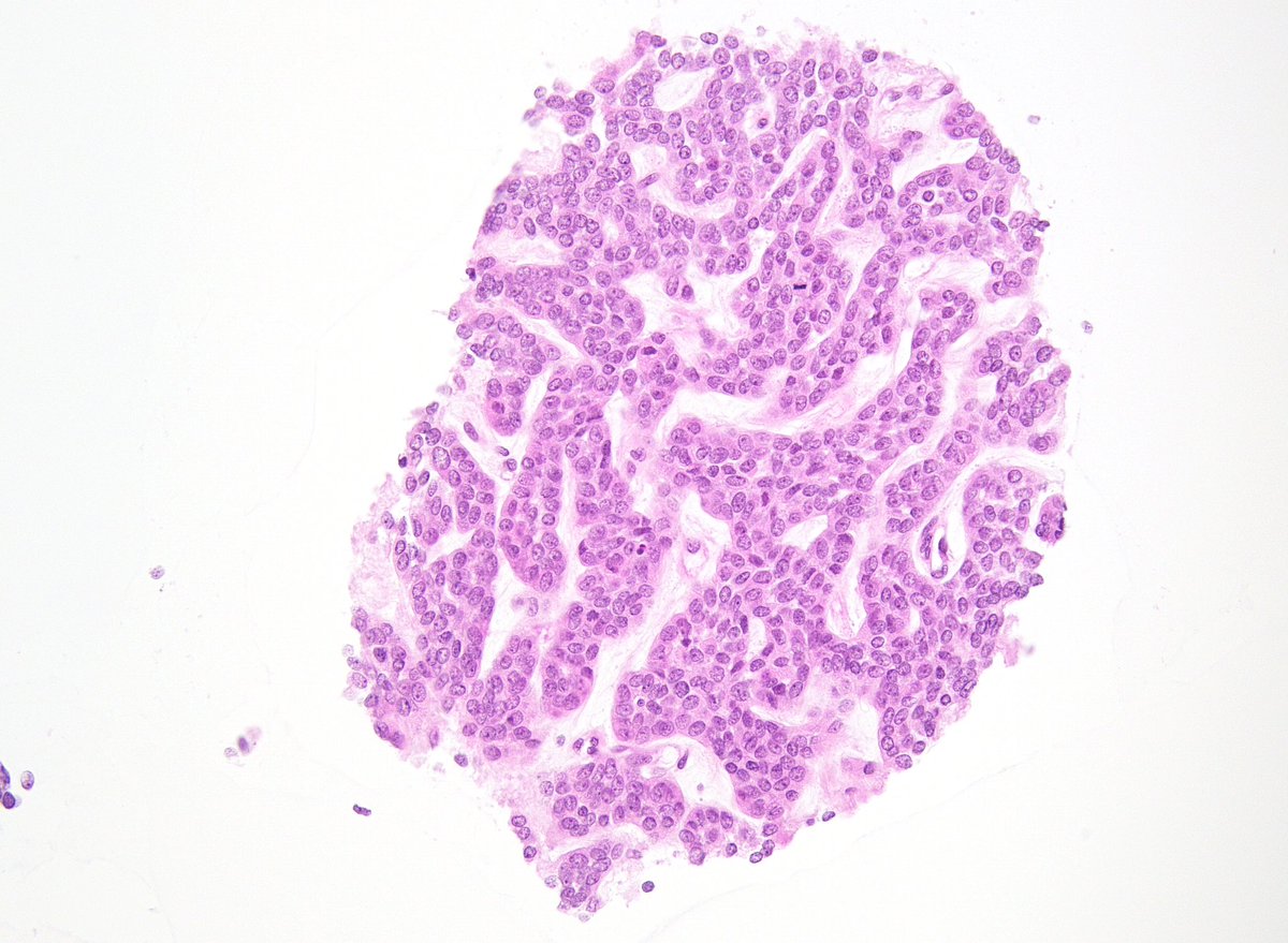
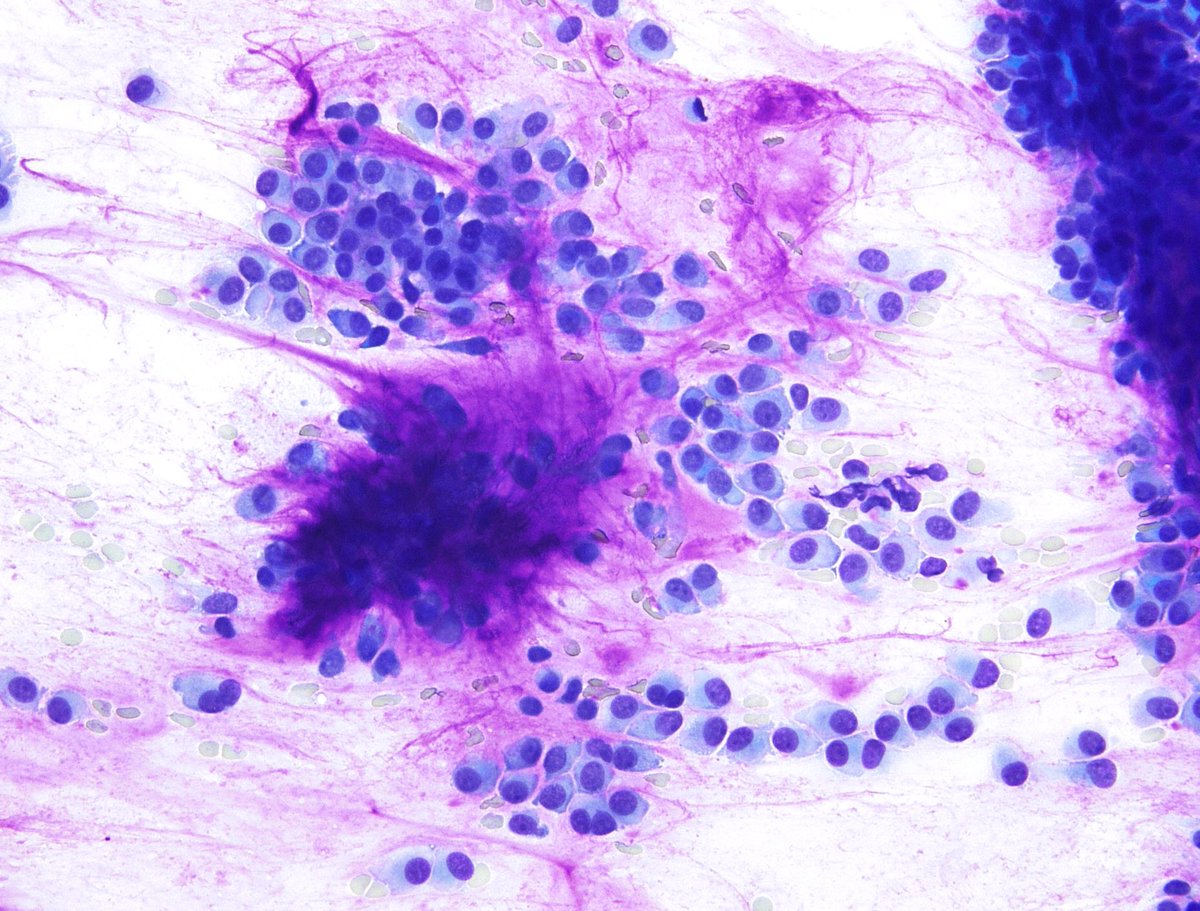
I personally find it harder to interpret salivary gland FNAs on liquid-based preps without a Giemsa-stained direct smear. Here is a nice example of a recurrent Basal Cell Adenoma with cell block. Without the history and cell block -> SUMP/Basaloid @DianaEstherossi (Parotid)
#USCAP2024 | At 1:30pm today, our Editor-in-Chief @bfaquin hosts an Interactive Microscopy session on the #MilanSystem. Dr. Faquin and a team of experts published a review of the 2nd edition of the Milan System. Hear from Dr. Faquin below 👇 https://t.co/i05yxqzjgI
👀 Revisit this systematic review and meta-analysis of the literature published after the introduction of the MSRSGC which validates the applicability of this reporting system #MilanSystem#CytoPath#EntPath
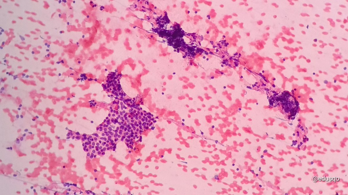
Chronic sialadenitis. Category II - Non-Neoplastic of the Milan System for salivary gland #CytoPath:
▫️Ductal & acinar epitelial elements (atypia can occur in the ductal component)
▫️Chronic inflammatory background
▫️#CellBlock + #IHCPath for IgG4-RS, lymphoma
#FNAFriday#ENTPath
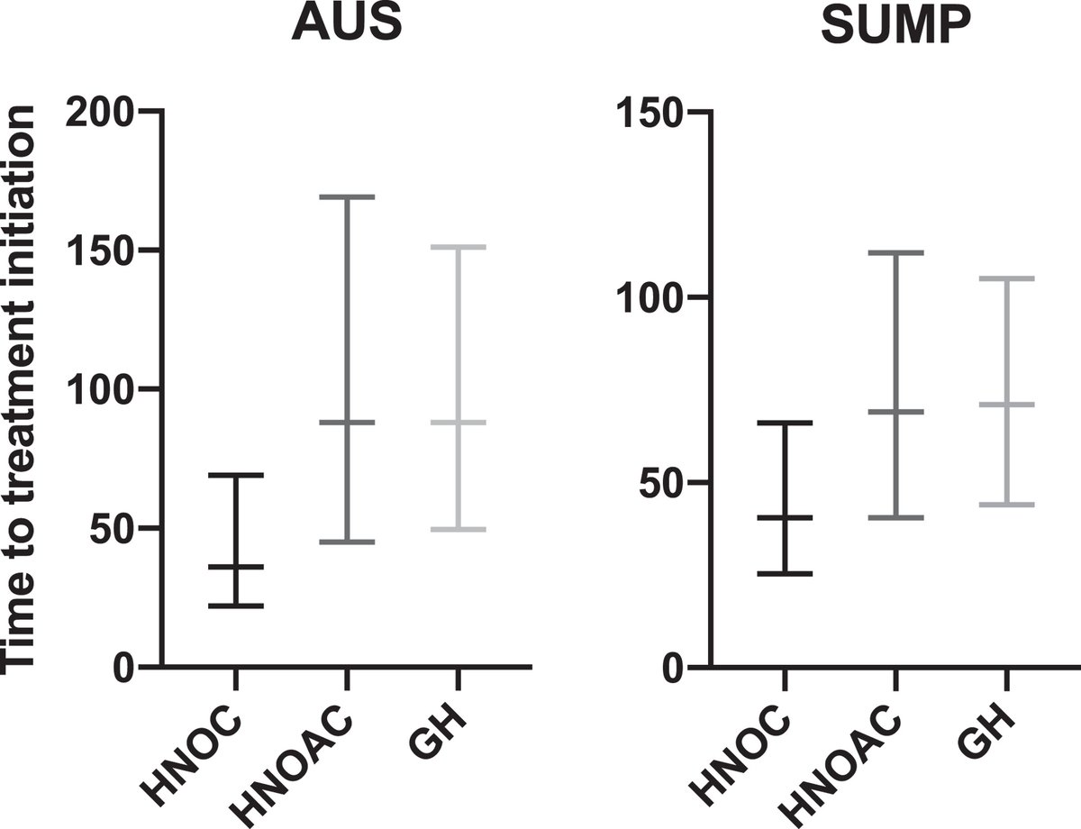
Standardized systems like Milan 2.0 are beneficial in many ways. What benefits have you seen in your practice? See the study from @radboudumc on how Milan categories provide a framework for examining the lag time between FNA Dx and surgical Rx: https://t.co/Swftuvrbp1 #CytoChat
Which Milan images do you find most helpful? @dkurtycz and his team believe the quality of both the submitted images and the resulting published figures in Milan 2.0 have improved. Do you agree, and what figures would you like to see in the future? #CytoChat
🔬 Wondering when to use the AUS category? Christopher Cormier DO and @ShwetaA61079880 from @UNMPathology evaluated the utility and performance of the #MilanSystem with a focus on the cytomorphology of lesions diagnosed as AUS. Revisit it here: https://t.co/qrAYVQMK22 #CytoChat
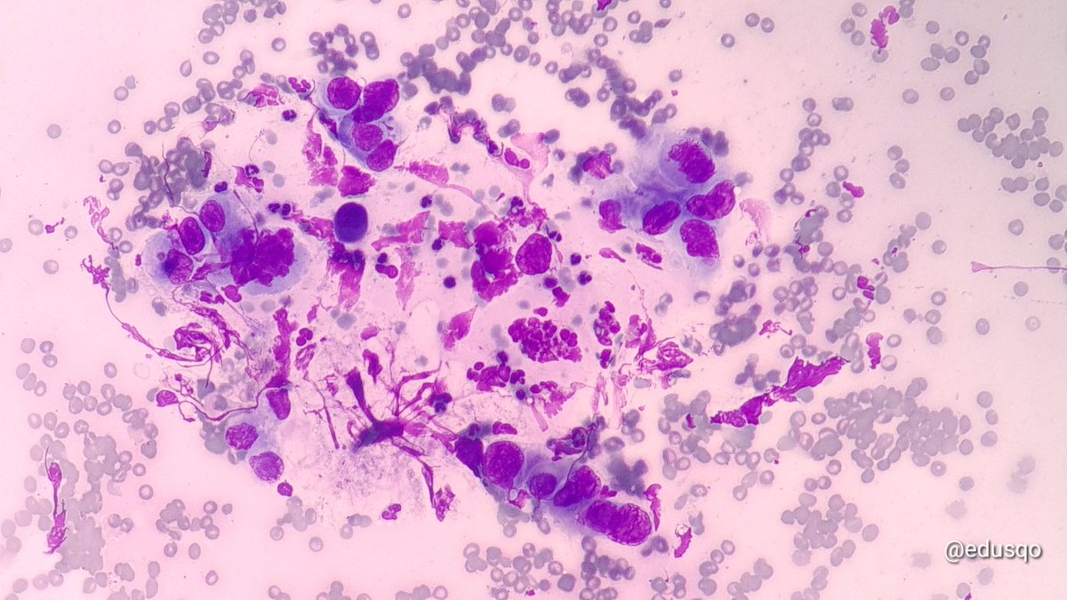
I always find Salivary Gland Pleomorphic Adenoma stained with Diff Quik to be most photogenic. Here, a fragment of myxoid matrix is surrounded by myoepithelial cells. (Parotid FNA)