Science
Bad memories, bad sleep
This Science article, “Bad memories, bad sleep,” discusses the close two-way relationship between memory and sleep. Sleep is not just a resting state; it is a period when the brain actively processes memories. The article focuses on how negative or traumatic memories can disrupt sleep quality, and how poor sleep may in turn affect the way those memories are stored or reactivated.
In simple terms, the article argues that bad memories and bad sleep can reinforce each other. Understanding the brain mechanisms behind this relationship may help explain sleep problems linked to stress, fear, or trauma, and could eventually guide new approaches for treating trauma-related sleep disturbances.
The full Science page was access-restricted, so this summary is based on the accessible title, snippet, and indexed description.
https://t.co/m5jqWoXFNa
The effect of high-protein vs. low-protein diets on CRP, IL-6, TNF-α, and liver enzymes in adult participants: A systematic review and meta-analysis https://t.co/vdgCRB8nuu
Evidence of UPF harms in children keeps mounting: UPF intake from 6 to 72 mo inversely associated with brain subcortical volume. Affected regions play key roles in reward processing, emotional regulation, motivation, and sensorimotor integration. https://t.co/4E9fcjwFs1
Cumulative ultra-processed food intake from 6mo to 6y – NO association with cognitive performance at 2y or 6y. BUT: higher UPF → smaller subcortical volumes at 6y (accumbens, amygdala, pallidum, putamen, thalamus; FDR p<0.05). Each 10% higher UPF → 1.9% smaller volume.
It’s not the eating disorder alone-it’s the weight of depression that rewires the brain. In binge-type EDs, severe depression (not mild) drives disrupted connectivity in body-image, reward, and impulse control circuits, and slows neural timescales.
https://t.co/W5fgyhJ8ZU
Short sleep ages you. Long sleep usually means something is already aging you.
A new paper in Nature this week mapped sleep duration against biological aging across 9 organ systems in half a million UK Biobank adults aged 37 to 84. Junhao Wen's lab at Columbia used 23 different aging clocks built from MRI scans, plasma proteins, and metabolites. The relationship is U-shaped. The slowest measured aging sat between 6.4 and 7.8 hours per night. Outside that window in either direction, organs looked older than chronological age would predict.
The two arms of the U are not the same biology.
On the short-sleep side, the causal story is well established. Sleeping under 6 hours raises systemic inflammation, impairs next-morning glucose tolerance, suppresses NK cell activity, and tracks with markers of poorer overnight brain waste clearance. Mendelian randomization in this paper supports a direct causal effect of short sleep on aging biology. Short sleep drives the wear and tear.
On the long-sleep side, the picture flips. Consistently sleeping over 8 or 9 hours is a well-documented marker of underlying disease, not a damaging behavior in itself. It tracks with major depression, undiagnosed sleep apnea, hypothyroidism, chronic inflammation, and neurodegenerative disease. The authors note that Mendelian randomization could not strongly support reverse causality, but they explicitly could not exclude it either. Decades of sleep medicine argue that for most long sleepers, the long sleep is the body compensating for something already wrong.
This matters because the practical advice for the two groups is opposite.
If you sleep under 6 hours, the levers are direct. Total sleep opportunity. Consistent timing. Morning light. Caffeine cutoff after lunch. Alcohol stopped at least three hours before bed. Sleep extension trials adding 45 to 90 minutes a night have shown measurable improvements in metabolic and cardiovascular markers.
If you consistently sleep 9 or more hours and still wake unrefreshed, the right move is to investigate what your body is recovering from. Home sleep study to rule out apnea. TSH, free T4, ferritin, CRP, vitamin D, B12. Depression and anxiety screening. A review of medications that increase sleep need.
One scenario gets lumped in with long sleepers that shouldn't be. Athletes in heavy training blocks, adolescents, people recovering from infection, and people in their first trimester genuinely need 9 to 10 hours. The paper looked at habitual sleep in adults aged 37 to 84, not acute recovery states. The curve does not apply to them the same way.
The cleaner way to state the finding: there is a window in the middle where the body looks youngest on every clock the authors built. Both sides of that window correlate with faster organ aging. The reasons differ. Short sleep does the damage. Long sleep usually shows the damage is already underway.
Here, higher adherence to the Dietary Approaches to Stop Hypertension (DASH) diet, the Mediterranean diet, and the Mediterranean-DASH Intervention for Neurodegenerative Delay (MIND) diet was associated with improved cardiometabolic risk factors in individuals with type 2 diabetes.
Visceral obesity may be missed by BMI alone.
A new study validates EASO criteria in Chinese adults and proposes a simpler tool for identifying visceral fat–related metabolic risk.
👉 https://t.co/vCiEnunYL1
#Obesity#VisceralFat#MetabolicHealth
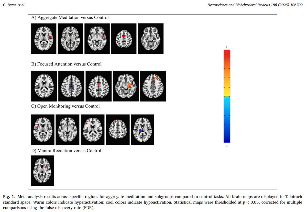
We have now mapped the most robust whole-brain characterization of meditation to date.
Our new paper in Neuroscience and Biobehavioral Reviews, “The Functional Neuroimaging of Meditation: A Quantitative Whole-Brain Meta-Analysis and Systematic Review,” provides a systematic and rigorous whole-brain characterization of meditation and establishes a foundational reference for future research in this domain.
Using quantitative whole-brain meta-analytic methods, we synthesized findings across the growing neuroimaging literature to move beyond individual experiments and identify the most reliable and reproducible brain activity common across meditation techniques as well as activity specific to different meditation techniques.
From 34 studies and over 700 individuals, we assessed neuroimaging data during focused attention, open monitoring, mantra recitation, and loving-kindness meditation. We found that:
– Across practices, meditation consistently recruits brain regions including the rolandic operculum, insula, superior temporal gyrus, supplementary motor area (SMA), and hippocampus.
– Functional decoding further linked whole-brain activation patterns to self-monitoring, reappraisal, motivation, experience, and awareness states., highlighting meditation’s role in engaging domain-general cognitive processes that may develop through intentional training.
Also, different meditation practices demonstrated dissociable neural signatures:
– Focused attention meditation recruited brain regions implicated in attentional control and monitoring, including the insula, SMA, superior frontal gyrus, and hippocampus.
– Open monitoring meditation engaged brain regions associated with salience, attentional awareness, and present-moment monitoring, including the insula, inferior frontal gyrus, middle temporal gyrus, and superior frontal gyrus.
– Mantra recitation was linked to distinct activation in the putamen/insula, regions associated with sustained attentional and psychomotor integration.
– Loving-kindness meditation activated aspects of prefrontal, cingulate, and affect-related brain systems associated with compassion and socioemotional processing.
Together, these findings provide robust evidence that meditation is not a single, unitary brain state, but rather a family of practices that engage overlapping yet distinct neural systems, which highlights the need for greater specificity in how meditation is studied and interpreted.
Our new research informs the personalization of clinical applications that seek to use meditation as an intervention to promote psychological well-being. In addition, it establishes a baseline for future research studying meditative development and advanced meditation. It also identifies promising neural targets for targeted clinical neuromodulation protocols.
Congratulations and gratitude to first author Caitlin Baten and our co-authors Arielle Keller @ArielleKeller and Chris Miller!
The full preprint is included below ⤵️
May our work benefit many 🙏
Sleep is vital for brain development—especially in children with neurodevelopmental disorders. Disruptions can affect brain function, hormones, metabolism & inflammation, potentially worsening conditions like autism & ADHD. More research = better care. #SleepScience #Neurodevelopment
https://t.co/NhPu38RrEi