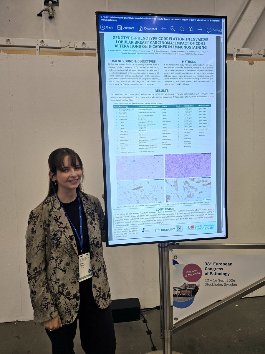
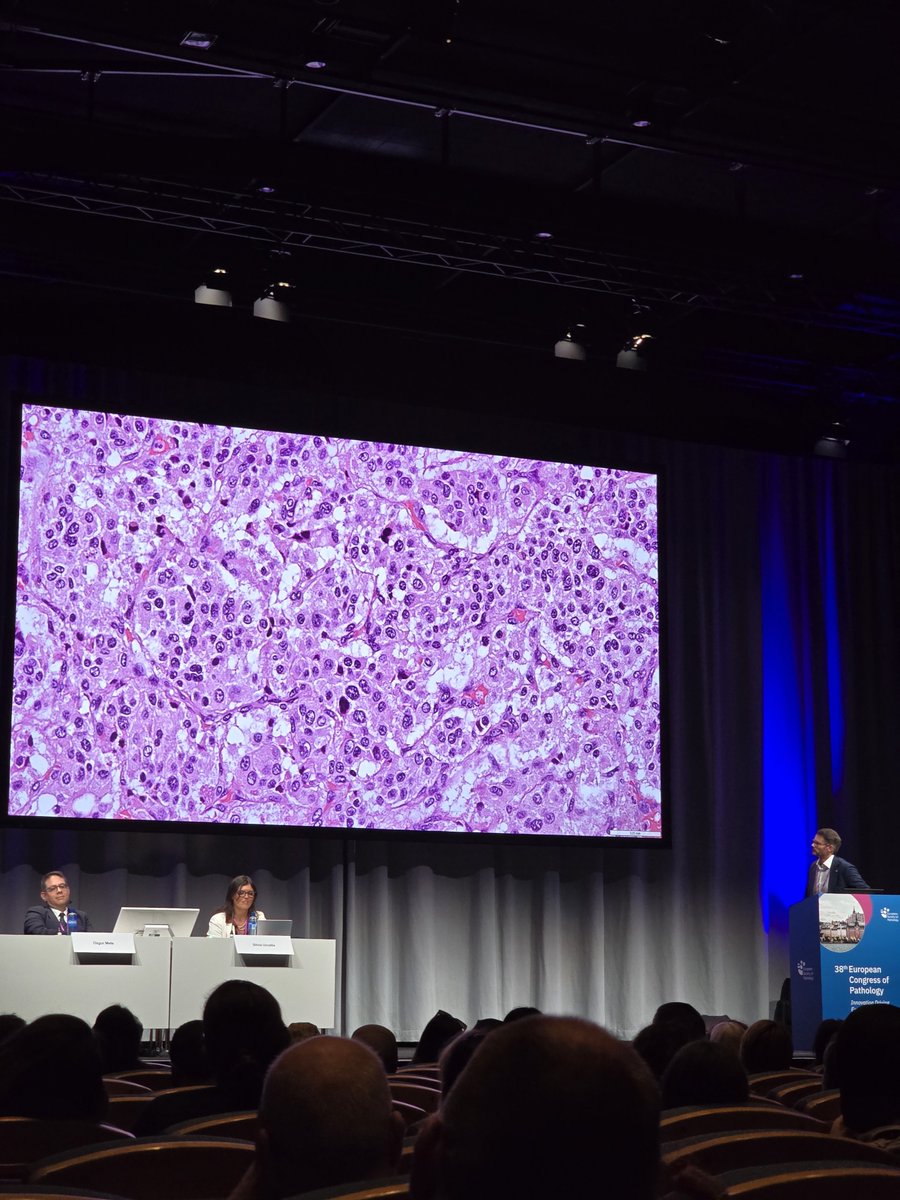
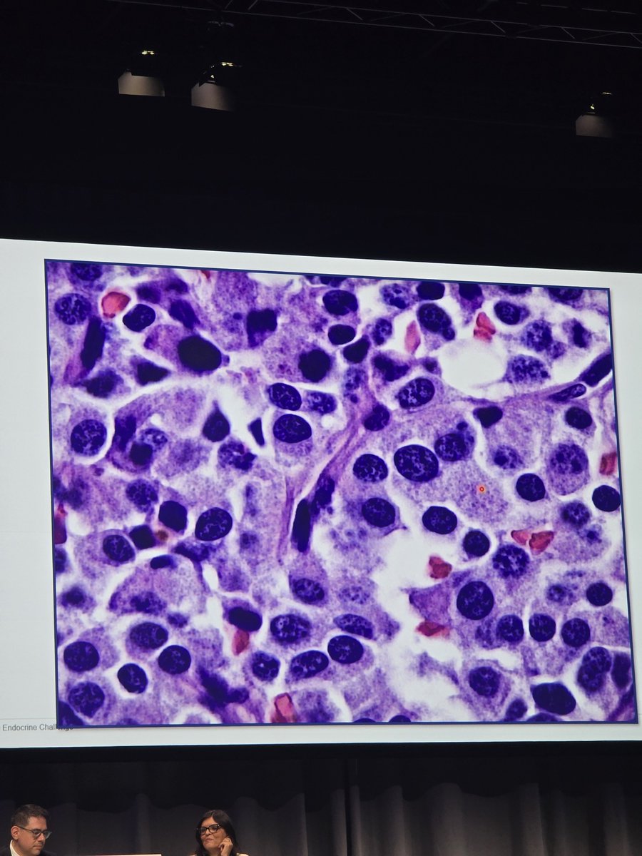
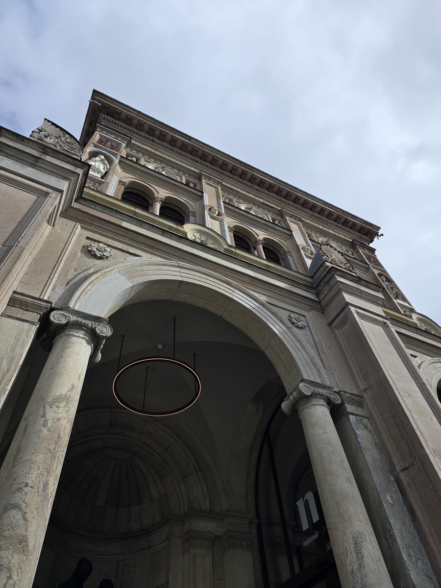
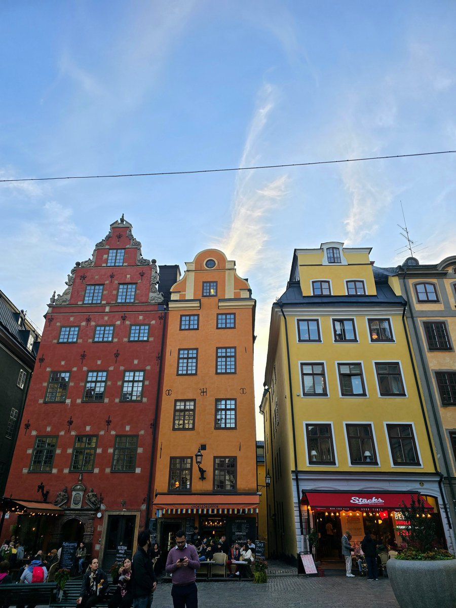
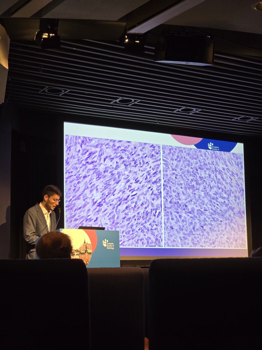
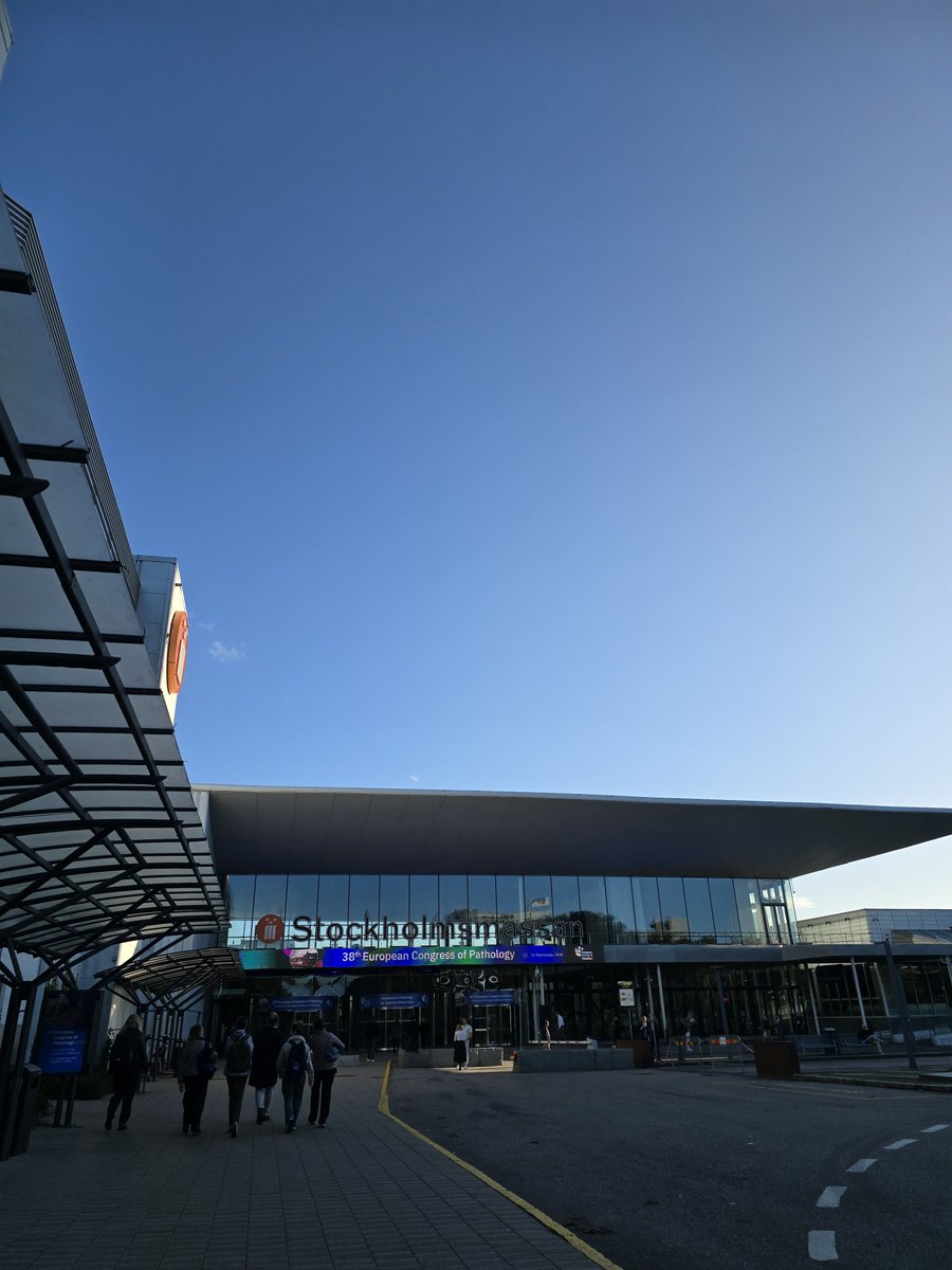
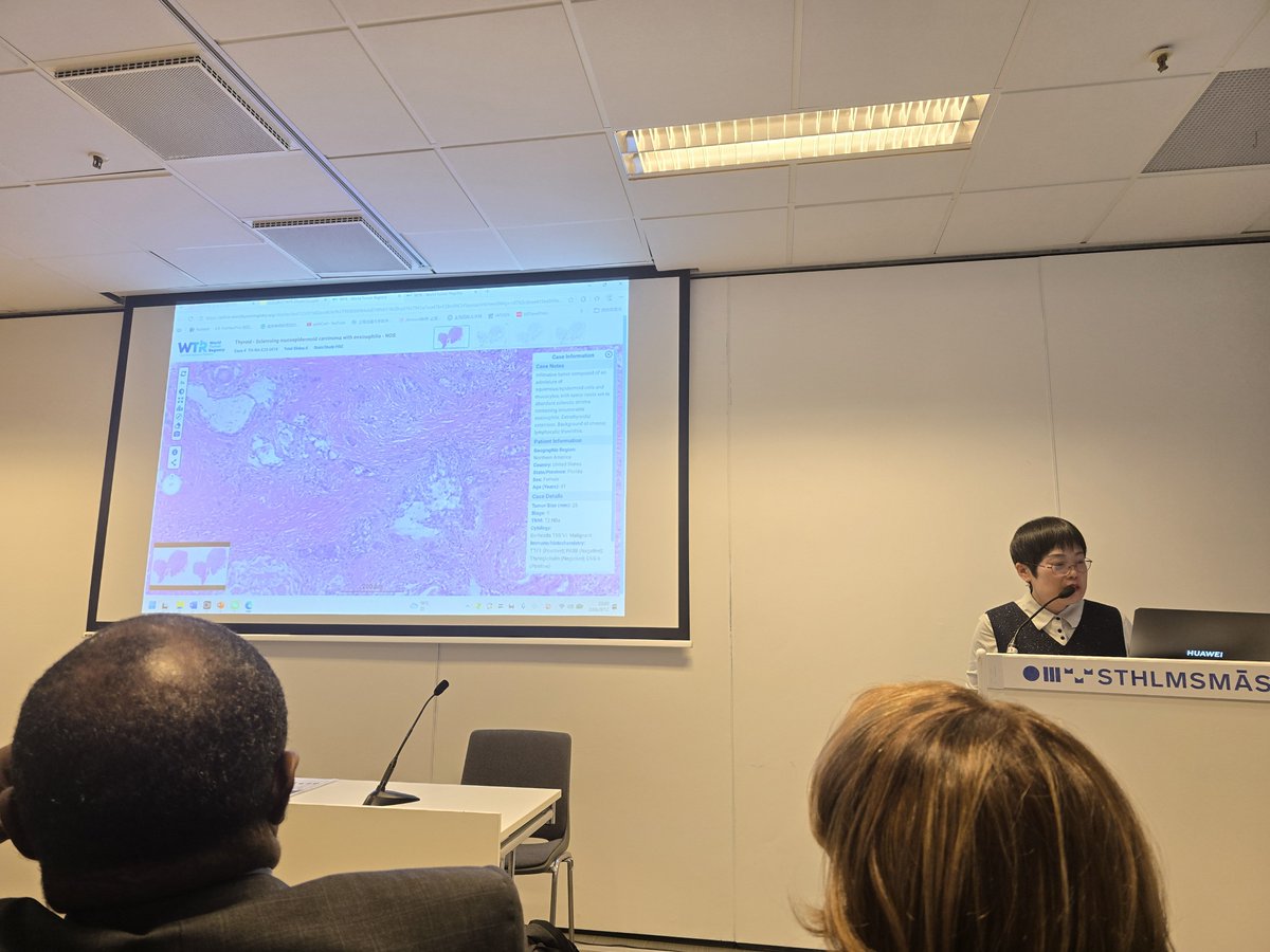
It is always a pleasure to participate in the #ESPCongress , such a great way of keeping up with #pathology updates and meet amazing people! I am also very thankful for the #ECP bursary program that enables residents to attend it. See you in #Maastricht (hopefully)!
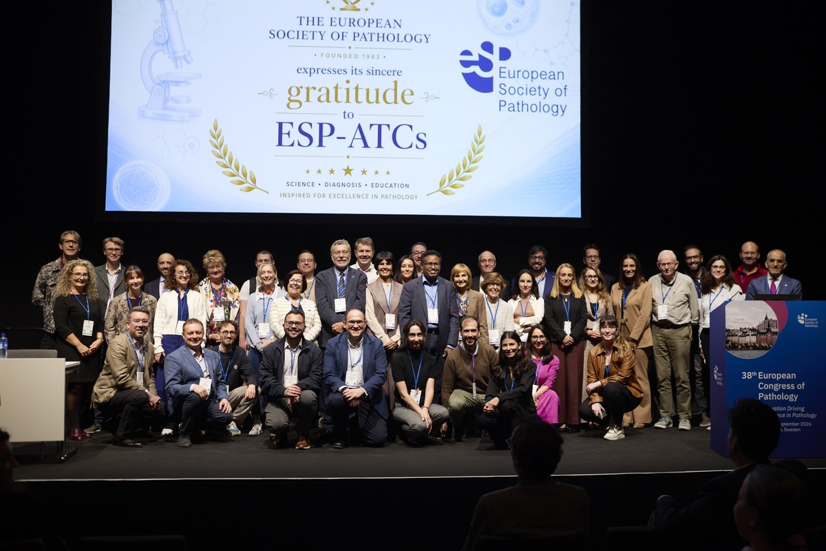
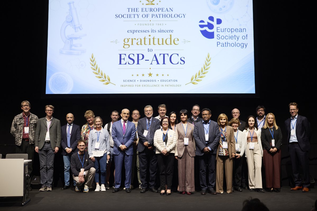
At #ESPCongress and the session of the #education Committee we organised a brief ceremony to acknowledge the work of the ESP Advanced Training Centres!
This was a small recognition of their work and for training more than 150 Giordano Fellows thus far!
#pathology#PathTwitter
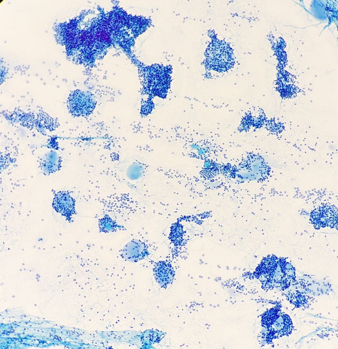
Diagnostic Approach to Mediastinal Tumors
For mediastinal lesions and tumors, most cystic lesions and neoplasms show a predilection for a particular mediastinal compartment and are relatively uncommon in others. In routine pathology practice, once the precise location of a mediastinal lesion or tumor has been established by imaging studies, the differential diagnosis can be systematically narrowed according to its anatomic compartment. This diagnostic pathway is intended to provide a practical framework for daily surgical pathology practice and to help develop a comprehensive, compartment-based approach to the diagnosis of mediastinal lesions and tumors.
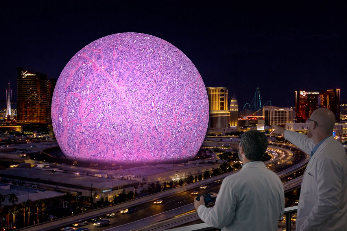
Pleased to announce that Dave McClintock and I have displayed and navigated a whole slide image on the Las Vegas Sphere — 580,000 sq ft of H&E at 256 megapixels. Press release follows. 1/
#digitalpathology#PathTwitter@dpatweet@SphereVegas
5/
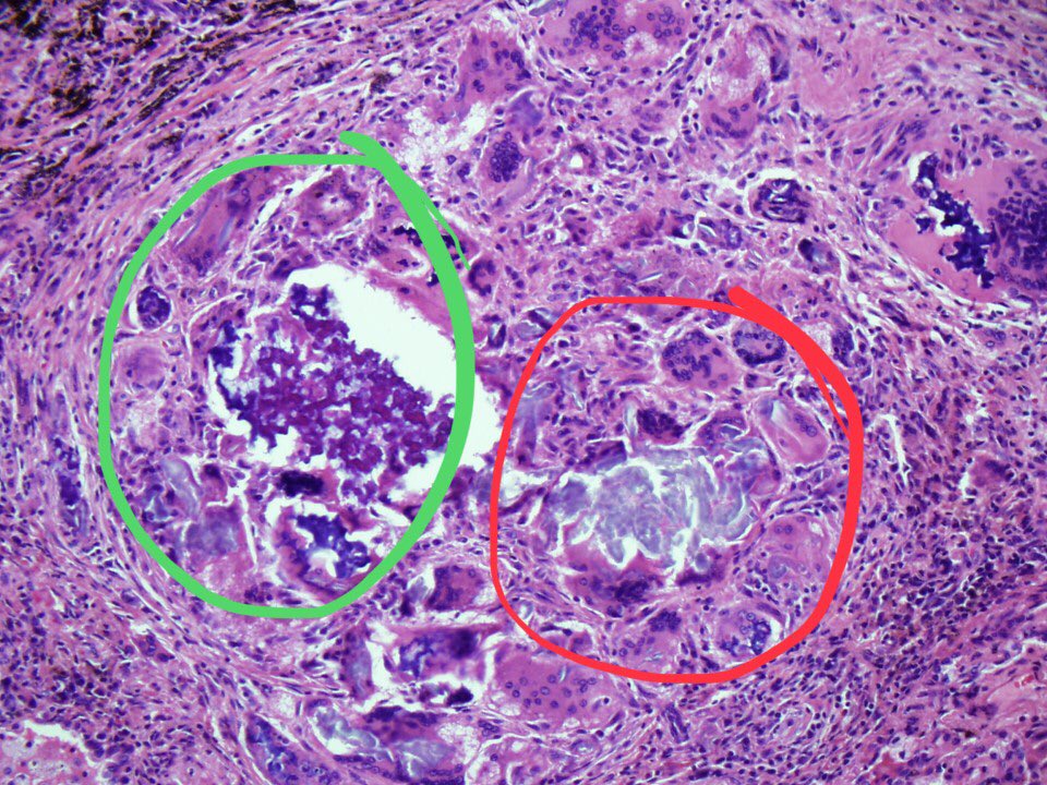
It’s because there are 2 types of pill fillers (excipients) in this image. Microcrystalline cellulose (red circle) and crospovidone (green circle). The former is colorless and brightly birefringent. The latter is purple but not birefringent. Both provide bulk to oral pills.
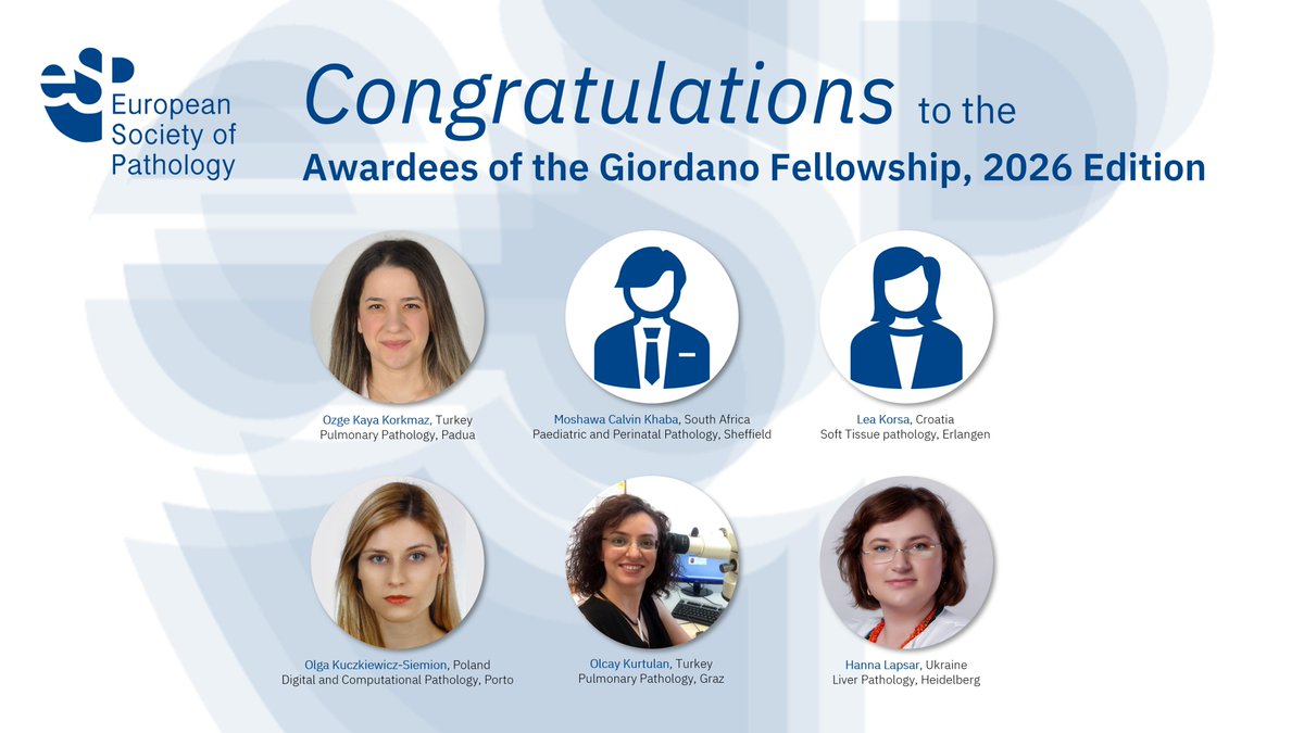
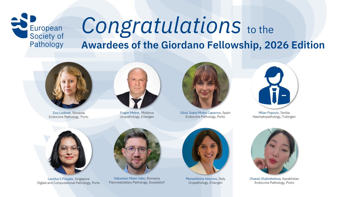
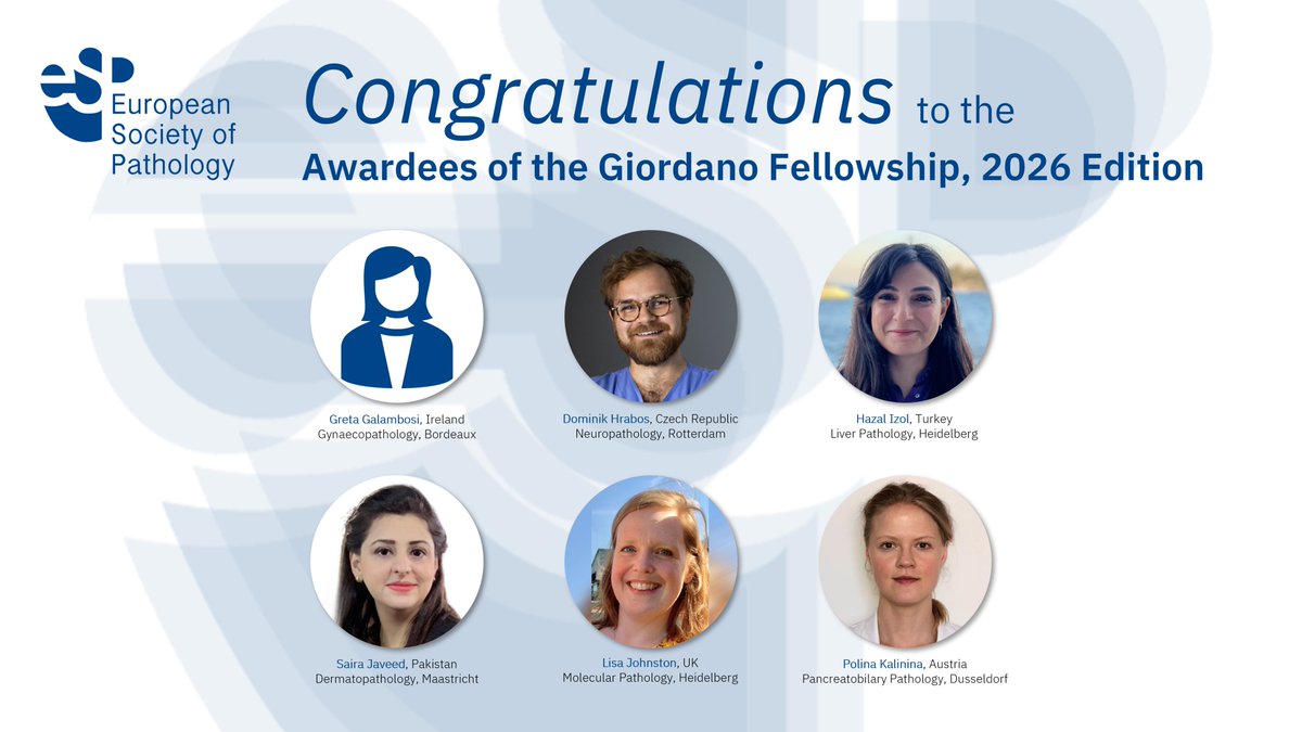
🔬🏅Having completed 10 wonderful years of Giordano Fellowship, we are pleased to announce the 2026 edition of Giordano Fellows!
Congratulations to all awardees and best of success at their Fellowships!
#education#pathology#PathTwitter#Pathx
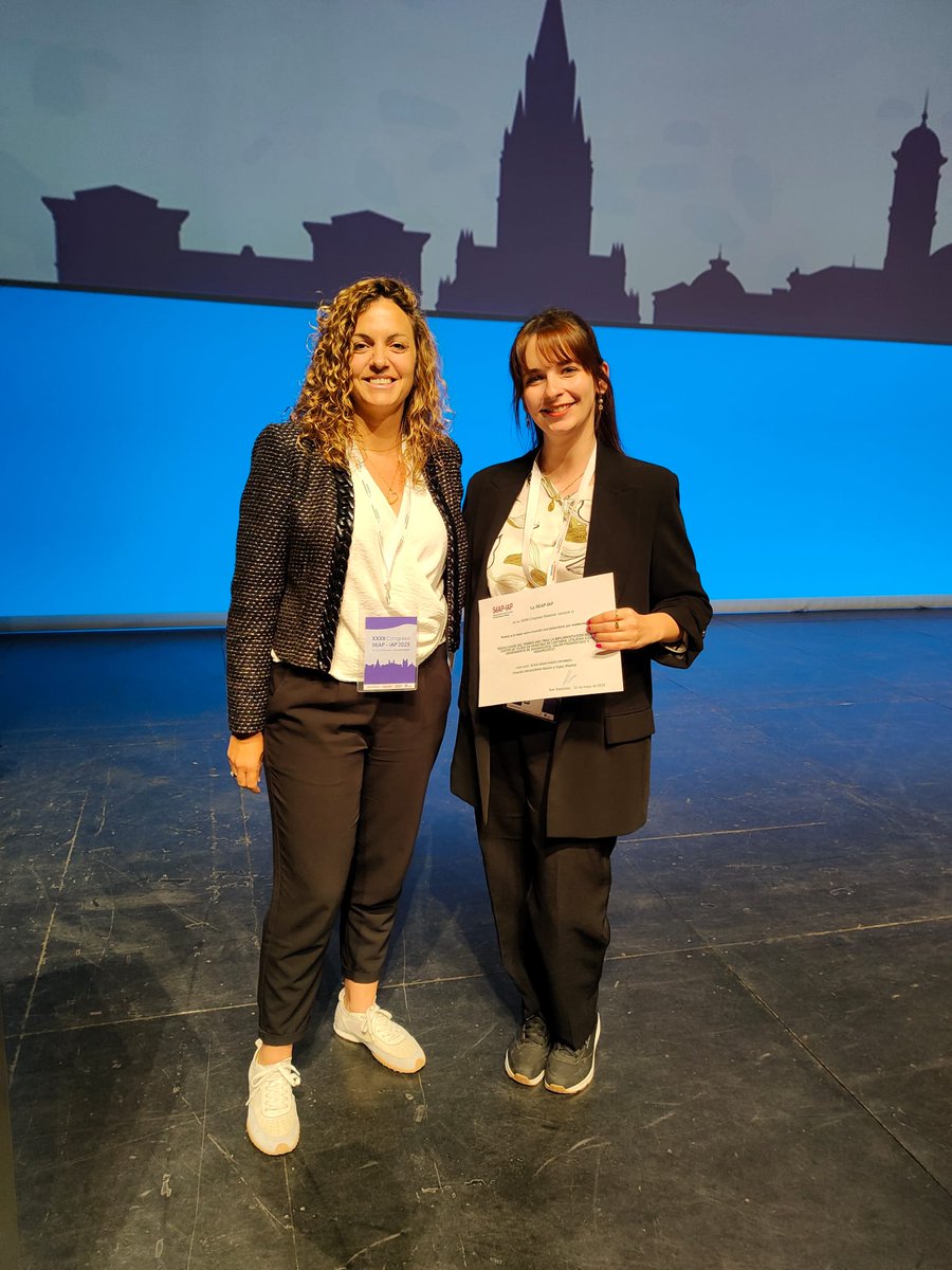
Premio a la mejor comunicación oral presentada por residentes: “RESULTADOS DEL PRIMER AÑO TRAS LA IMPLEMENTACIÓN NGS A PARTIR DE TEJIDO EN PARAFINA DE LINFOMAS. UTILIDAD COMO HERRAMIENTA DE DIAGNÓSTICO, VALOR PRONÓSTICO Y TERAPÉUTICO”. Autor: SILVIA JOANA MOLÉS CAPARRÓS