Iconic Traitors series 3 contestant Linda Rands has a special good luck message exclusively for GCU students sitting their exams during the current assessment diet 💙
Linda encourages you to be faithful to your fellow students during the exam period, supporting each other throughout with words of comfort and advice! 🤝
Good luck to all our students currently sitting their Tri B exams! 🍀
Band 5 Summer 2025 radiographer vacancies at @UHSFT
We offer a 14 week supervised induction to build your confidence as part of our year long preceptorship.
Come join the SOR SE region 2022 team of the year.
As part of her MSc Dissertation Julia is looking to explore forensic radiography experiences with diagnostic radiographers across the UK. You do not need to be working as a forensic radiographer to take part. Please contact Julia ([email protected]) to find out more.
Glasgow Caledonian has been named the UK’s Modern University of the Year in the Daily Mail University Guide 2025 👏
Read more on our website: https://t.co/BRpXz8ybjs
📢Hi fellow Diagnostic Radiographers, we're looking for international Radiographers working in Scotland to participate in our research project. Please consider getting in touch with Sam and sharing this post! @GCUMScDI@SCoRScotland
The SoR has launched the Student Placement Survey, to understand the challenges faced on placement. Read more to find out how the Society can help develop the support you need 👉 https://t.co/H25MfvwO2o
Heading back north of the border after a really inspiring two days at #ARE2023. Coming home with lots of ideas, particularly regarding Inclusion, placement capacity and assessment
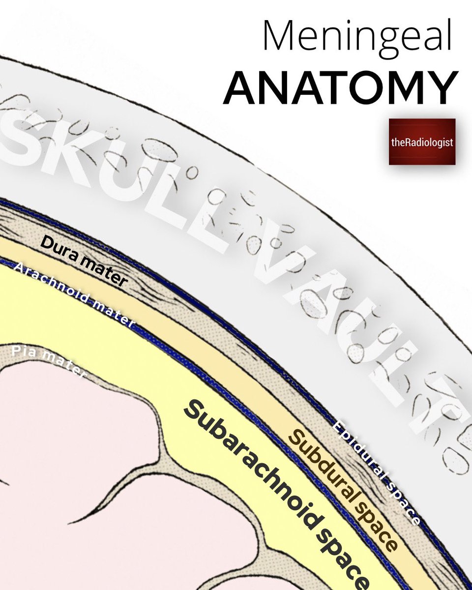
Basic meningeal anatomy
There are three layers of meninges:
1️⃣Dura mater: thicker outer layer consisting of two layers of connective tissue (periosteal and meningeal layer). The dural venous sinuses are found between the two layers. The meningeal layer folds upon itself to make up the dural reflections which divide the brain into compartments including the falx cerebri (separates the cerebral hemispheres) and the tentorium cerebelli (separates cerebellum from occipital lobes)
2️⃣Arachnoid mater: made up of connective tissue and is a vascular unlike the dura and pia. The ‘subarachnoid space’ lies below the arachnoid and contains CSF.
3️⃣Pia mater: thin layer overlying the brain surface
.............
Different location of brain bleeds:
1.SUBARACHNOID: look for blood within the basal cisterns, especially the penatagon shaped suprasellar cistern. Linear streaks of blood within the sulci represent subarachnoid blood. Perimesencephalic SAH is a subset of SAH where no bleeding point can be found on angiography and the epicentre of the bleed can be seen anterior to the midbrain so checking between the 'Mickey Mouse ears' of the midbrain is always an important review area - a small focus of high density here can be the only abnormality on an acute CT head and I have seen these missed before.
2.SUBDURAL: blood between the dura and arachnoid mater. Usually due to a venous bleed with tearing of subdural cortical bridging veins. Blood will be peripheral and crescentic. Unlike epidural/extradural bleeds, these can cross sutures and they don't cross the midline
3.EXTRADURAL: blood between the skull and outer periosteal layer of the dura. Caused usually by injury to the middle meningeal artery and you may see a temporal bone fracture. Unlike subdural bleeds these are biconvex and are restricted by the skull sutures
Are you a student radiographer and interested in forensic radiography ? Did you know we offer a discounted student membership with access to a wide range of CPD ? Interested ? Look at https://t.co/S3zP0mEWTd or email us at [email protected] for more details
We've recruited Diagnostic Imaging's Elsie Llewellyn to pass on more #Tips4You to new students 💙
"Participating in societies or clubs at the Student's Association is an excellent way to relieve stress, especially during exam season."
Follow @GCUstudents to find out more ⬅️