Worried that hydrocephalus is hiding in plain sight?
Normal pressure hydrocephalus (NPH) can mimic volume loss
How can you tell the difference?
Many findings can be seen with NPH—you may use 1 or more. But do you know how well your sign performs?
Here are how common NPH signs perform:
Evans index
SENS: 100
SPEC: 74
Cingulate sulcus sign:
SENS: 63
SPEC: 61
Callosal angle:
SENS: 49
SPEC: 78
Disproportionately enlarged subarachnoid spaces (DESH):
SENS: 94
SPEC: 41
Anteroposterior diameter of the lateral ventricle index:
SENS: 98
SPEC: 100
For the SINPHONI trial, a combination of Evans index > 0.3 & DESH had a 70-80% PPV for shunt responsiveness.
Now you know the signs of NPH & how good they may or may not be. Hopefully, you can now set the bar high when it comes to hydrocephalus!
Just because you don’t see it, doesn’t mean it’s not there!!!
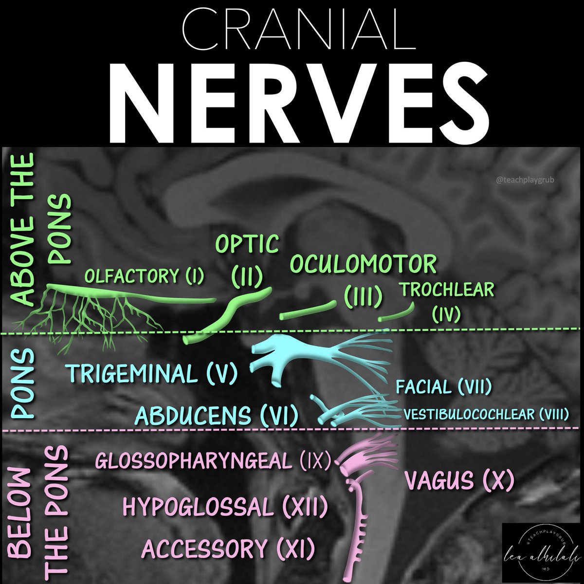
Just because you don’t see all cranial nerves on MRI doesn’t mean they aren’t there!
You must know their anatomy so you know when pathology might affect them.
Here is a figure w/the anatomy you NEED to know!
➡️Just remember this easy rule of four from Peter Gates:
There are:
🔸 4 cranial nerves from above the pons (including 2 from the midbrain)
🔸4 from the pons
🔸And 4 from the medulla oblongata!
Now you won't have be nervous when it comes to this nerve anatomy!
Need a better strategy to find strategic infarcts?
Strategic infarcts are infarcts located in structures directly related to cognition.
Damage to these structures results in dementia without any other significant volume loss.
These infarcts are like a ninja. Just one shot right to the heart to take it down.
Just one infarct in one of these important structures can cause dementia like a shot to the heart.
So which structures are these?
Well, there are many structures that have been implicated in strategic infarct dementia.
But the main ones are hippocampus, internal capsule (ant & genu), thalamus (paramedian) & caudate.
I remember this w/the mnemonic:
One HIT CAUses dementia
H = Hippocampus
I = Internal Capsule
T = Thalamus
CAUD = Caudate
Now you can be a ninja looking for these ninja infarcts!
To call it or not to call it? That is the question!
Do you feel a bit wacky & wobbly when it comes to calling normal pressure hydrocephalus on imaging?
You don’t want to overcall it, but you don’t want to miss it either!
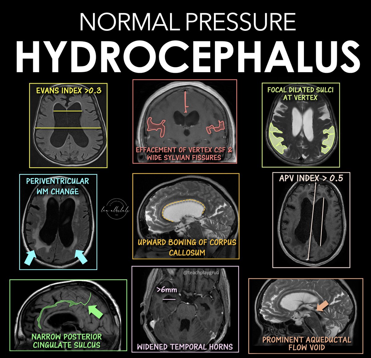
Here are the signs of NPH
🔹 Evans Index > 0.3
--Enlarged frontal horns relative to skull width → ventriculomegaly.
Remember it: “One out of three is too big for me!”
🔹 Periventricular White Matter Change
--T2/FLAIR hyperintensity from transependymal CSF flow.
Remember it: “Leaking pressure leaves a glow.”
🔹 Narrowing of the Posterior Cingulate Sulcus
--Tight high-convexity sulci from crowding at the vertex.
Remember it: “The top gets squeezed shut.”
🔹 Effaced Vertex CSF + Wide Sylvian Fissures
--Tight sulci up top but enlarged fissures laterally (DESH pattern).
Remember it: “Dry on top, wet on the sides.”
🔹 Upward Bowing of the Corpus Callosum
--Ventricular expansion pushes the corpus callosum upward.
Remember it: “If the corpus callosum is arched, the patient can’t march (wobbly)”
🔹 Temporal Horns > 6 mm
--Early temporal horn dilation from hydrocephalus.
Remember it: “Big horns before big vents.”
🔹 Focally Dilated Sulci at the Vertex
--Patchy enlarged sulci despite surrounding crowding.
Remember it: “Holes in a leaky roof = too much water on the brain!”
🔹 APV Index > 0.5
--Increased ventricular-to-parenchymal proportion suggesting hydrocephalus.
Remember it: “Glass half full is too much!”
🔹 Prominent Aqueductal Flow Void
--Increased CSF flow through the aqueduct on MRI.
Remember it: “If the aqueductal flow is super black, the ventricles are out of whack.”
Classic NPH imaging theme: enlarged ventricles + tight high convexities + disproportionate CSF redistribution.
Hopefully now you won't wobble on the diagnosis of NPH!!
1/Does your ability to remember temporal lobe anatomy seem, well, temporary?
Or are you feeling temporally challenged when it comes to this complex region?
Here’s a thread to help you remember the structures of the temporal lobe!
Before you diagnose CIDP:
✅ Make sure to check if the motor conduction velocity is truly in the demyelinating range. Upper limb CV should be slower than 35 m/s and lower limb less than 25 m/s. Be careful with amplitude-dependent slowing.
✅ Consider whether it's a variant — if so, be aware that these cases are more prone to misdiagnosis.
✅ Exercise caution with elevated CSF protein levels, as this finding is non-specific and should not be solely relied upon for diagnosis.
✅ Look for the presence of monoclonal gammopathy, which could indicate POEMS syndrome, AL amyloid, or IgM/Anti-MAG neuropathy.
✅ If any of these are present consider an auto-immune nodopathy: markedly elevated CSF protein (>200 Mg/dl), distal predominant weakness, CNS demyelination, nephrotic syndrome, marked hypertrophy of nerve roots and plexi, respiratory failure, or cranial neuropathy
✅ Finally, verify your diagnosis against the EAN/PNS guidelines to ensure accuracy here: https://t.co/xQI59awxQU
When you’re asked to localize the lesion in a patient with aphasia, do you suddenly feel speechless?
At a loss for words to categorize the type of aphasia?
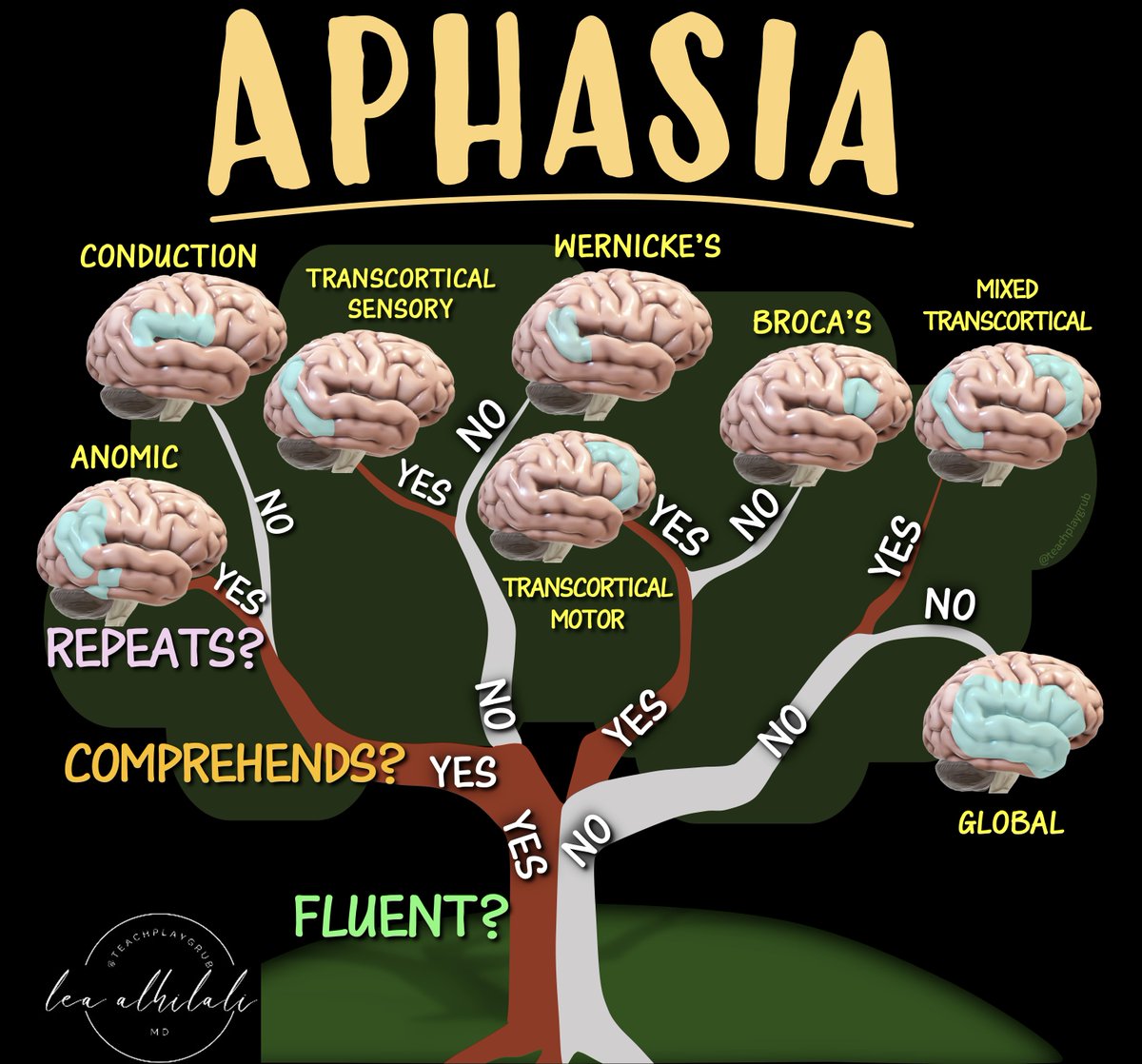
Never fear—here is the decision tree for patients w/aphasia and the associated anatomic correlates
Three main questions:
1. Fluency? Nonfluency indicates damage to the FRONTAL language regions anterior to the fissure of Rolando
2. Comprehension? Impaired comprehension indicates damage to the TEMPOROPARIETAL language regions posterior to the fissure of Rolando
3. Repetition? Impaired repetition indicates damage within the core PERISYLVIAN language zone
The answers will lead you both to the type of aphasia and the location of the lesion.
Keep this figure with you for quick reference—and then when you’re asked about a patient with aphasia, you will have a lot to say!
In Alzheimer’s imaging, there are some things you just can’t forget!
New Alzheimer’s treatments are changing the way we look at these scans!
Removal of amyloid beta proteins from vessel walls by anti-amyloid antibodies leads increased vascular permeability.
This can cause edema or hemorrhage, called ARIA (Amyloid Related Imaging Abnormalities)
Grading of the degree of ARIA is important bc it can change management. Asymptomatic ARIA is usually treated by pausing treatment if it’s moderate to severe
For ARIA-E (edema) or ARIA-H (microhemorrhage), if the edema measure > 5 cm or number of microhemorrhages are >5, it is moderate
For superficial siderosis, 2 regions is considered moderate.
Remember it by this little rhyme:
--If ARIA findings are great than 5, then the medication deprive
--Or if siderosis is 2, then medication break for you!
Hopefully, now the grading of ARIA will stay in your memory!!
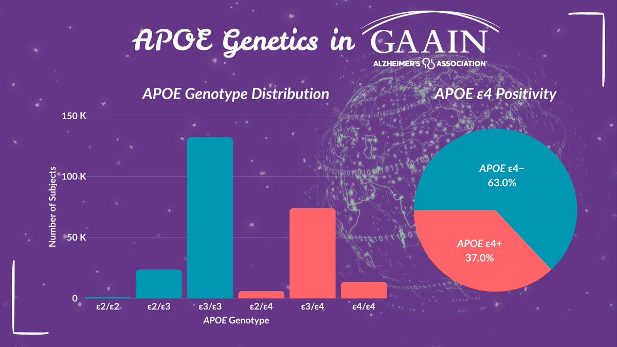
Of the subjects in GAAIN with #genetic data, here is the breakdown by #APOE genotype and APOE e4 positivity, which is a major risk factor for #Alzheimers disease.
Explore more data in the #GAAINInterrogator:
🧬https://t.co/GwtPKsiCOd
Is trying to remember toxic encephalopathy patterns making you toxic?
Does memorizing metabolic encephalopathy patterns put you into a metabolic coma?
Don't become encephalopathic!
Here is the cheat sheet of the encephalopathy patterns you NEED to know & how to remember them!
🔶Corpus Callosum:
--Lesions here are called cytotoxic lesions of the corpus callosum or CLOCC.
--Usually transient from cytotoxicity, especially viral infections.
--Remember they are commonly viral & transient bc CLOCC lesions are on the CLOCK
🔶Central Pons:
--Usually osmotic demyelination
--Remember this bc the T2 hyperintensity is in the shape of O for Osmotic
🔶Dentate:
--Usually flagyl toxicity
--Remember this bc the wavy appearance of the dentate looks like a FLAG for FLAGyl
🔶Corticospinal tract:
--Usually from cobalamin deficienty
--Remember cortiCOspinal for CObalamin
🔶Symmetric Gray Matter:
--Differential is EXTENSIVE
--Remember the differential list for deep gray is deep!
🔶Cortical Gray Matter:
--Typically hypoglycemia & hyperammonemia
--Remember it looks like the brain has been coated in shimmering sugar = related to glucose
--Hyperammonemia has about 1000 Ms in it & the undulated cortex looks like an M
🔶Symmetric White Matter:
--Commonly from chasing the dragon (heroin) & methotrexate
--Remember that the puffy white matter signal looks like the smoke from a dragon
--Remember methotrexate as methoTRACTSate = affects many white matter tracts
🔶Posterior subcortical edema:
--This is PRES or posterior reversible encephalopathy
--So if it’s all POSTERIOR, think POSTERIOR reversible encephalopathy
🔶Asymmetric White Matter:
--This is usually demyelinating from chemotherapy
--Demyelinating lesions have a C shaped pattern of enhancement
--So if you see a C enhancing think of C-hemotherapy
Now you know all the toxic/metabolic encephalopathy patterns. So hopefully now remembering the patterns of toxic/metabolic won’t seem so diabolic!
A new FDA-cleared blood test makes #Alzheimer biomarker screening possible in primary care, but experts say it’s important to understand what such tests can and cannot do. https://t.co/gHiI6zRofi
This #NeurologyEd Teaching Neurovisual by Sutherland and Gummerson details the Head-Impulse-Nystagmus-Test-of-Skew exam, which uses special maneuvers to identify central etiologies of acute vestibular syndrome with greater sensitivity than hyperacute MRI: https://t.co/3RMvuWVoZw
8/So now you know where to look in the lumbar spine when a patient tells you the pain radiates down their leg—and hopefully remembering the lumbar radicular distributions won’t cause you pain any longer!
The hardest lessons after training aren’t always in the OR or clinic.
🙏 I wrote this essay for anyone navigating their first year as an attending.
“Unseen Scars” in @JAMA_current
🔗https://t.co/0e09Zx1nb6
When you’re asked to localize the lesion in a patient with aphasia, do you suddenly feel speechless?
At a loss for words to categorize the type of aphasia?
Never fear—here is the decision tree for patients w/aphasia and the associated anatomic correlates
Three main questions:
1. Fluency? Nonfluency indicates damage to the FRONTAL language regions anterior to the fissure of Rolando
2. Comprehension? Impaired comprehension indicates damage to the TEMPOROPARIETAL language regions posterior to the fissure of Rolando
3. Repetition? Impaired repetition indicates damage within the core PERISYLVIAN language zone
The answers will lead you both to the type of aphasia and the location of the lesion.
Keep this figure with you for quick reference—and then when you’re asked about a patient with aphasia, you will have a lot to say!
Form follows function!!
Do you know functional neuroanatomy?
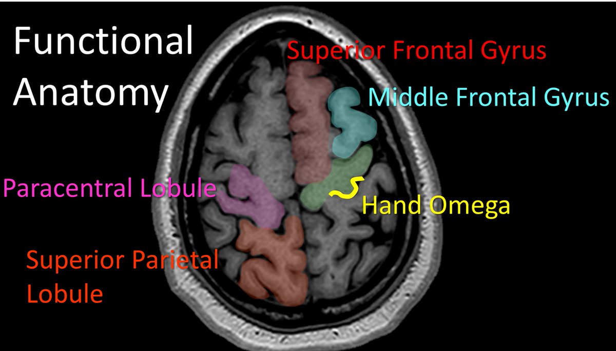
This post will help you to remember the functional neuroanatomy you need to function if you are reading brain MRIs!
Here’s how:
1. First start at the top
—At the top you will see a gyrus that looks like a thumbs sticking up. This is the superior frontal gyrus (SFG).
—Remember this bc you get a thumbs up when you do a superior job!
2. Next to the SFG is the middle frontal gyrus
—This looks like knuckles next your superior frontal gyrus thumb
—Remember this bc your MIDDLE finger is in your knuckles
—ALWAYS LOOK FOR THE KNUCKLES W/THE THUMBS UP!
3.Use the SFG to find the motor strip
—SFG has a motor & language component
—Motor component is first at the back (remember, you walk before you talk!)
—Motor component of SFG crashes into the motor strip
—Remember, when two cars crash, their MOTORS hit
4.Confirm its the motor strip by finding the hand omega
—Hand motor region looks like an upside down omega
—Remember Omega is a fancy watch brand you wear near your HAND!
Hopefully now you will be eloquent when it comes to this eloquent cortex!!!
1/They say form follows function!
Brain MRI anatomy is best understood in terms of both form & function.
Here’s a short thread to help you to remember important functional brain anatomy--so you truly can clinically correlate!
Excited to be presenting at #AAIC2025 this year! I’ll be sharing our latest work on a novel PRS method predicting tau pathogenesis in AD —hope to see you there! 🧠🧬 #Research#AD
1/Raise your hand if you’re confused by the BRACHIAL PLEXUS!
I could never seem to remember or understand it—but now I do & I’ll show you how!
A thread so you will never fear brachial plexus anatomy again!