Associate Professor Medicine Tanta University, Armed Forces College of medicine , Al Rayan National College ,consultant surgical Pathology and nephropathology
Our Gynecologic Pathology cases @UMiamiPathology are fascinating! Here’s a striking example of pilomatrix-like high-grade endometrial carcinoma (PiMHEC), a recently described entity mostly driven by CTNNB1 mutations. Look for solid basaloid growth with conspicuous central tumor cell necrosis, and pilomatrix-like keratinization with the hallmark ghost cells. This case also had a focal conventional FIGO grade 1 endometrioid component. IHC shows diffuse nuclear β-catenin with loss of PAX8 and ER in the pilomatrix-like component. These tumors are believed to behave aggressively.
Clear cell adenocarcinoma it is. 😊
The main differential diagnosis is florid nephrogenic adenoma.
But once you start seeing solid growth, striking atypia, and mitotic activity, it may be time to look beyond nephrogenic adenoma.
See attached two nice cases.
https://t.co/3UK8gLZKyc
https://t.co/bqqDzux6WE
#DxMindMap #BladderPath #GUPath #PathTwitter @JHUpath
Acute phlebitis in an interlobular vein. Although commonly seen in acute cellular rejection, the lesion itself is not diagnostic for rejection per Banff classification.
Not every “cancer” is cancer.
IgG4-related disease can present as a hard mass in the salivary gland, pancreas, orbit, kidney, lung, and many other organs—often mimicking malignancy on imaging and clinically.
Before calling it a tumor: sometimes the immune system is the culprit.
Half of cellular blue nevus are S100 negative but SOX10 positive
Potential pitfall and also a clue when evaluating these lesions
Dr. Folpe #USCAP2026#pathology#PathX#PathTwitter
Before making a diagnosis of invasive breast cancer in a core needle biopsy, be sure it is truly a primary breast tumor, particularly in the era of neoadjuvant chemotherapy
Metastatic serous carcinoma and metastatic melanoma to breast (pic 1)
Metastatic lung adenocarcinoma (pic 2)
Metastatic medullary thyroid carcinoma (pic3)
Of 2423 TNBC submitted to Caris for molecular profiling over 19 months there was a misdiagnosis of TNBC in 73 cases (3%). Half of those cases were non-small lung cancer (data submitted for publication)
Dr. Schnitt #USCAP2026 #pathology #PathX #PathTwitter
🔓 Open Access
"XVIIth Banff Conference on Allograft Pathology: The Banff Workshop Heart Report on Microvascular Inflammatory Burden in Heart Transplantation..."by Fedrigo et al
https://t.co/z5HkaRdAL2
#HeartTransplantation#BanffClassification#TransplantPathology
A sample from the pathology teaching slide set on Lupus Nephritis, jointly prepared by Arkana Laboratories and @goKDIGO. https://t.co/KwXMfV3nTh
#KDIGO#pathology#renal#kidneypath
Interesting finding of large multi-nucleated endothelial cells in a txp biopsy with chronic active AMR. IHC stain is ERG (endothelial cell marker). Probably just a manifestation of chronic endothelial cell injury in this setting #renalpath#pathtwitter#nephrology
ANSWER: D.
Tubulopathy with isometric vacuolization, acute tubular injury, acute arteriolopathy and TMA can be seen in acute CNIT.
#pathology#nephx#renal#renalpath
#KidneyQuiz Some of the acute changes associated with calcineurin inhibitor toxicity (CNIT) are:
A) Tubules with isometric vacuolization
B) TMA
C) Only A
D) A and B
#nephX#kidneypath#pathX#nephtwitter
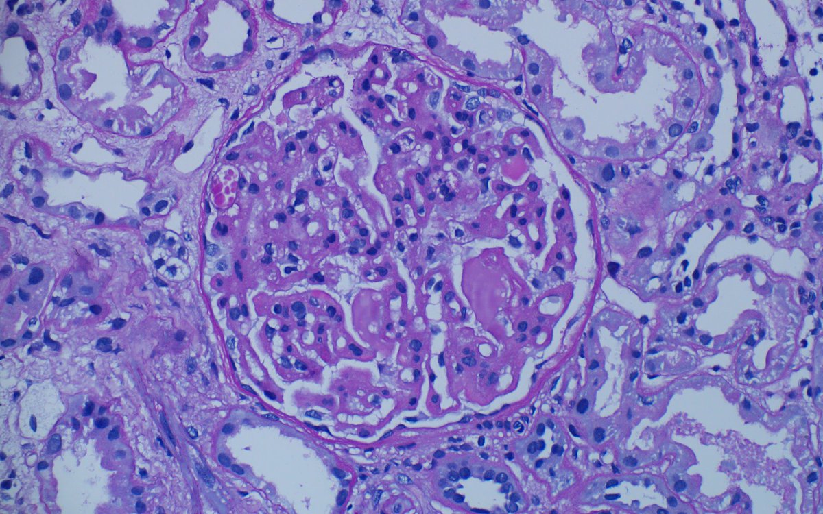
#renalpath young F, NS, normal SCr. The material filling the microaneurysms with double contours was seen in glomeruli. Did not receive any anti-VEGF therapy. Gene test showed MMACHC mutation.
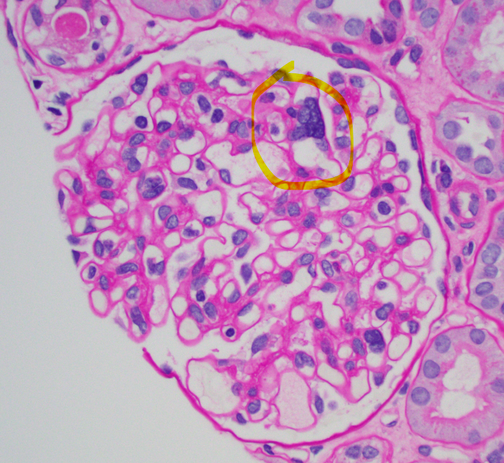
Multinucleated giant cells in glomeruli of 56 yo man with MPO positive crescentic GN. Usually more frequent in anti-GBM, these cells can also be seen in ANCA GN. Necrotizing arteritis noted in interlobular artery