In 109 adults with type 2 diabetes, higher liver disease activity on MRI went hand in hand with greater insulin resistance.
That is the finding of a study published this year in The Journal of Clinical Endocrinology and Metabolism by Ortiz Rocha and colleagues.
The question the authors asked: in people with type 2 diabetes seen in primary care, does liver disease activity, measured by iron-corrected T1 (cT1) on a LiverMultiScan exam, track with wider cardiometabolic risk?
What they found: higher cT1 was associated with more severe features of metabolic syndrome, higher insulin resistance (HOMA-IR) and greater adipose tissue dysfunction (all P < .01).
Their conclusion: cT1 may help early identification of individuals at greater risk of liver disease progression and cardiometabolic disease.
Tomorrow is World Diabetes Day, and the International Diabetes Federation’s theme for 2026 to 2027 is early action on diabetes. This study is a reminder that the liver is part of that picture, and that liver disease activity can be measured without a needle.
For researchers in diabetes, hepatology and imaging: in your cohorts, where does liver disease activity sit alongside the metabolic measures you already collect?
Ortiz Rocha, A. et al. (2026) ‘Severity of MASH activity by iron-corrected MRI predicts insulin resistance and cardiometabolic risk in type 2 diabetes’, The Journal of Clinical Endocrinology and Metabolism, 111(7), pp. e1813 to e1822. https://t.co/P4ifag85Hr
#Perspectum #WorldDiabetesDay #Type2Diabetes
We’re heading to @RSNA 2026 in Chicago, November 29 to December 3.
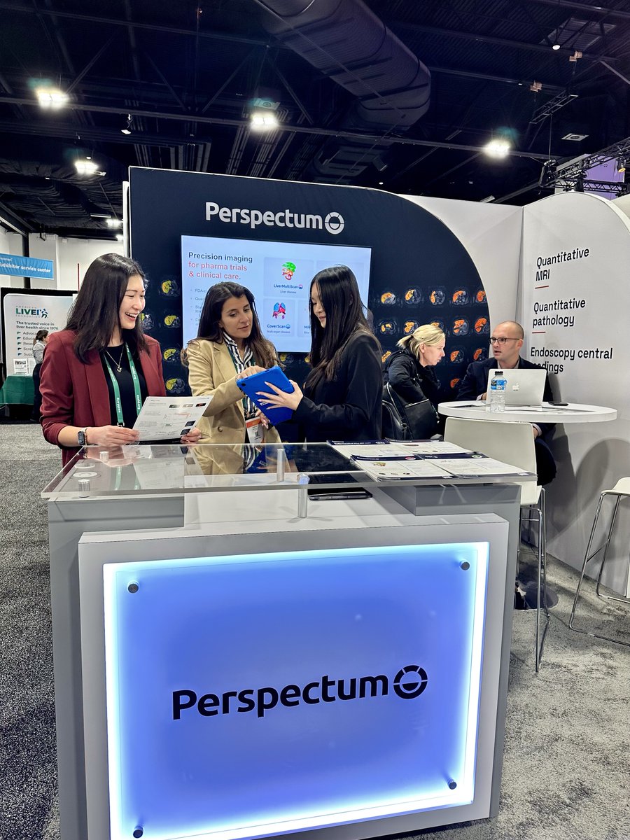
Eleven members of the Perspectum team will be at #RSNA26, and you’ll find us at booth #5129, South Hall Level 3.
For radiologists and imaging teams: see how LiverMultiScan supports non-invasive liver health assessment, with quantified metrics for liver disease activity (cT1), liver fat fraction (PDFF) and liver iron concentration to inform clinical decisions across diagnosis, treatment and monitoring.
We’ll share more on what we’re presenting in the coming weeks.
#Perspectum #RSNA26 #Radiology
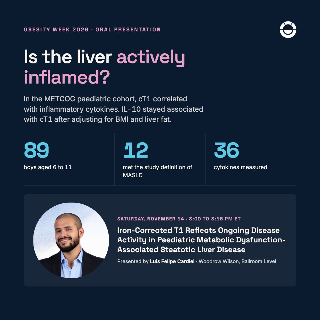
In 89 boys aged 6 to 11, cT1 correlated with inflammatory cytokines; its link with IL-10 held after adjusting for BMI and liver fat. Luis Felipe Cardiel presents at ObesityWeek 2026, Sat Nov 14, 3:00 pm ET. #Perspectum#MASLD
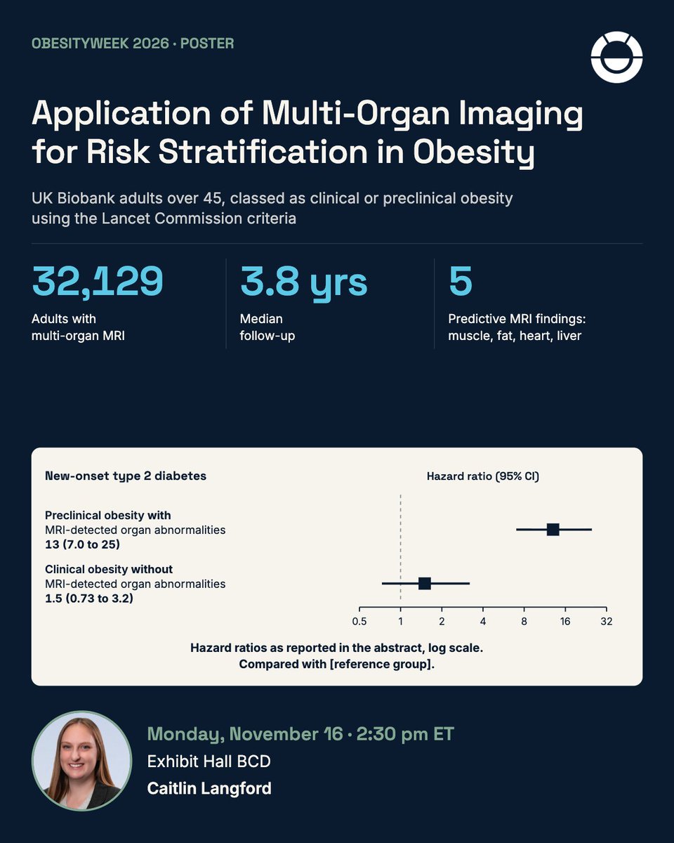
Preclinical obesity plus MRI-detected organ abnormalities: hazard ratio 13 (95% CI 7.0 to 25) for new-onset type 2 diabetes in 32,129 UK Biobank adults. ObesityWeek 2026 poster, Mon Nov 16, 2:30 pm ET, Exhibit Hall BCD. #Perspectum
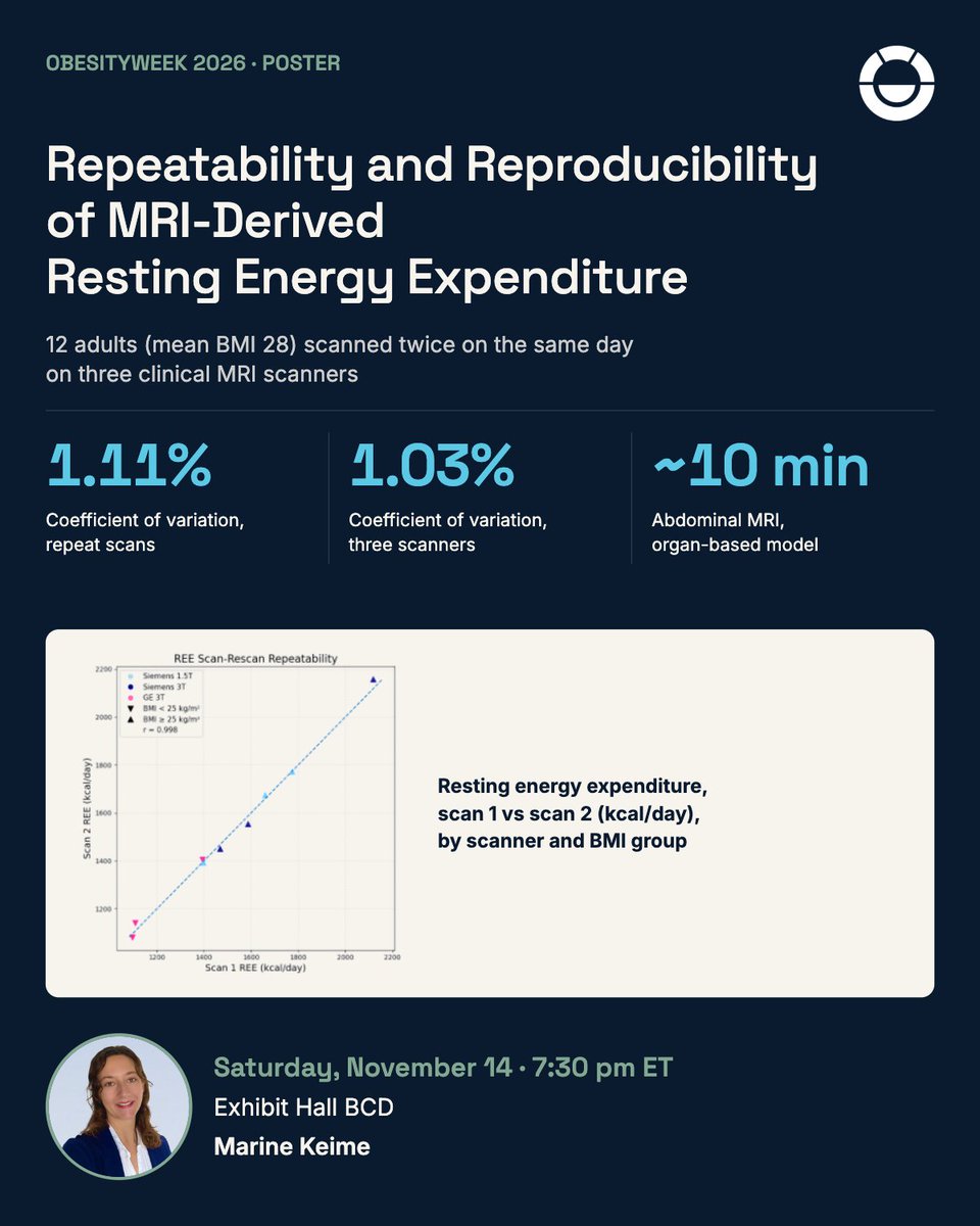
A roughly 10-minute abdominal MRI estimated resting energy expenditure with a coefficient of variation of 1.11% between repeat scans and 1.03% across three scanners. ObesityWeek 2026 poster, Sat Nov 14, 7:30 pm ET, Exhibit Hall BCD. #Perspectum
Two people can lose the same amount of weight and end up in very different physiological states. One loses visceral fat. The other loses muscle. Body weight alone cannot tell them apart.
That gap is where our conversations at ObesityWeek 2026 will start.
We will be at The Obesity Society’s 44th Annual Meeting, 14 to 17 November at the Gaylord National Resort and Convention Center, National Harbor, MD.
#Perspectum #ObesityWeek2026 #BodyComposition
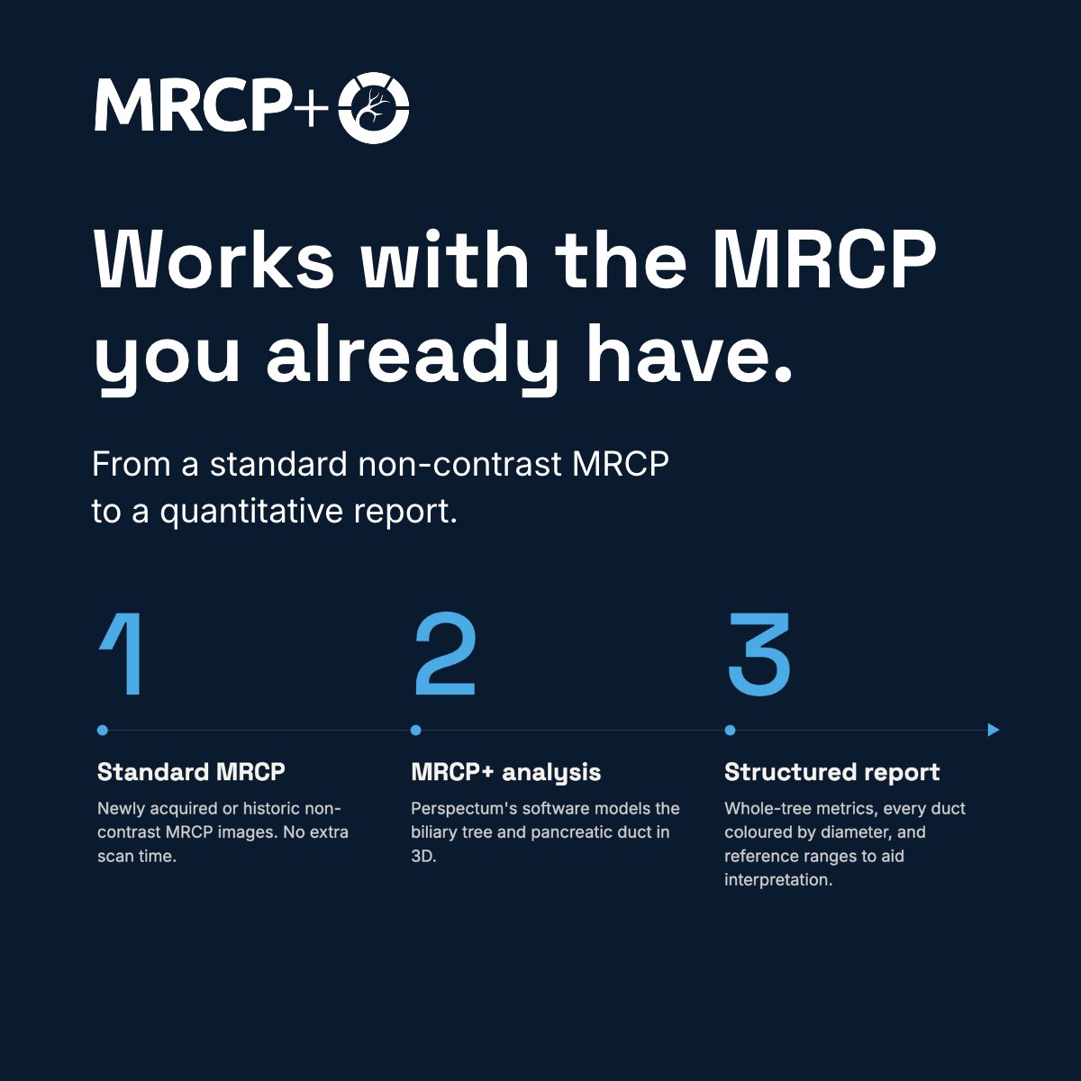
Whole-tree volume. Duct counts. Strictures. Dilatations. Quantified from one non-contrast MRCP scan.
MRCP+ reports metrics for the whole biliary tree and for individual ducts. Here is what each describes:
Whole biliary tree:
Biliary tree volume (mL): the volume of the modeled biliary tree.
Total ducts, strictures and dilatations (count): the number of narrowed and widened duct segments, and the number of ducts with either.
Percent of ducts with median width greater than 3 mm up to 5 mm (%): how much of the tree sits in that width range.
Individual ducts:
Duct-level widths, strictures and dilatations (mm): the median, minimum, maximum and interquartile widths, as well as locations of narrowed and widened segments, of select ducts
Each report pairs these metrics with 3D visualizations and reference ranges to aid interpretation, giving specialists and referring clinicians objective data to complement visual interpretation. MRCP is used in biliary conditions such as primary sclerosing cholangitis (PSC).
Which of these measurements would be most useful in your practice?
#Perspectum #TLM26 #Hepatology
Day 2 of @AASLDtweets The Liver Meeting in Denver. Find Perspectum at booth #1055 until 5:00 PM MT today.
This week our work was shared in five posters and one oral presentation, covering digital pathology and biopsy reading, liver disease screening, cirrhosis risk, liver and heart health in people with high blood pressure, and autoimmune liver disease in children.
If you’re at the meeting, come and say hello before we pack up. To book time with us about clinical trials, email https://t.co/[email protected].
#Perspectum #TLM26 #Hepatology #LiverDisease
Day two at @AASLDtweets The Liver Meeting, and we have three posters for you to stop by in one hour. Find us at booth #1055 in Denver.
Today’s Poster Session II runs from 1:00 to 2:00 PM MT.
Poster #2329: cT1 as the second step in the MASLD screening pathway, after a FIB-4 blood test. Led by Niharika Samala, Associate Professor of Medicine, Indiana University
Poster #2422: an MRI risk model for identifying cirrhosis in people with MASLD. Led by Charlie Diamond, Biomarker Scientist, Perspectum, with the ELEGANCE Consortium.
Poster #2443: MRI of the liver and heart together in adults with high blood pressure, with and without type 2 diabetes. Led by Yameng Deng, Scientist II, Perspectum with the REMODEL study team.
All three use cT1, an MRI measure of liver disease activity. We’ll share what each one found in its own post today.
The team is at booth #1055 through Sunday, November 8. To book time with us, email https://t.co/[email protected].
#Perspectum #TLM26 #Hepatology #LiverDisease
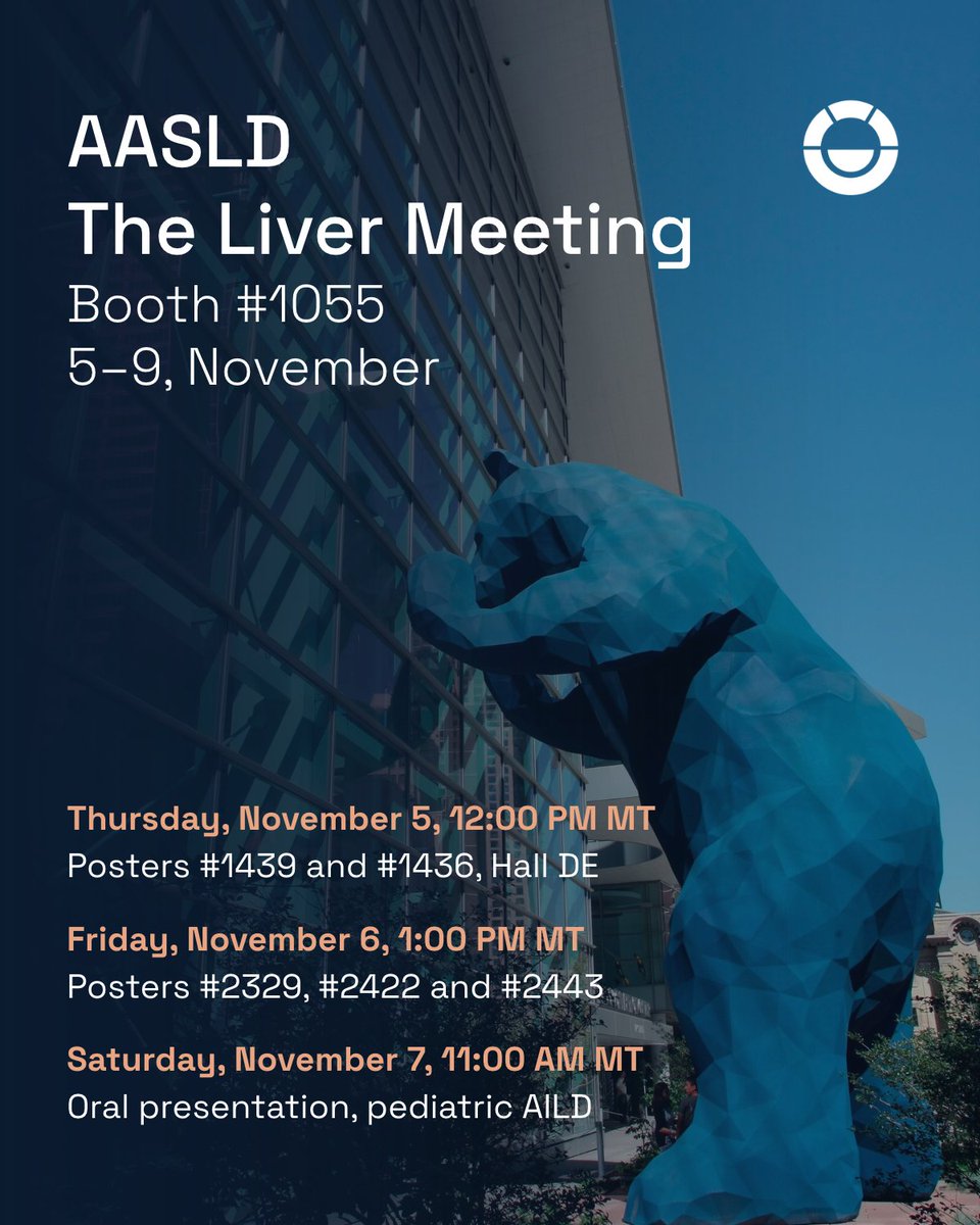
Day one at
@AASLDtweets
The Liver Meeting. Find Perspectum at booth #1055 in Denver.
Our team is here through Sunday, November 8, and we’d like to meet you. Stop by to talk liver imaging in clinical practice, or quantitative liver imaging biomarkers in liver disease trials!
Where to find our science this week:
Today, Thursday, November 5, 12:00 to 1:00 PM MT, Poster Hall, Hall DE
Poster #1439: digitized whole slide imaging for METAVIR fibrosis staging
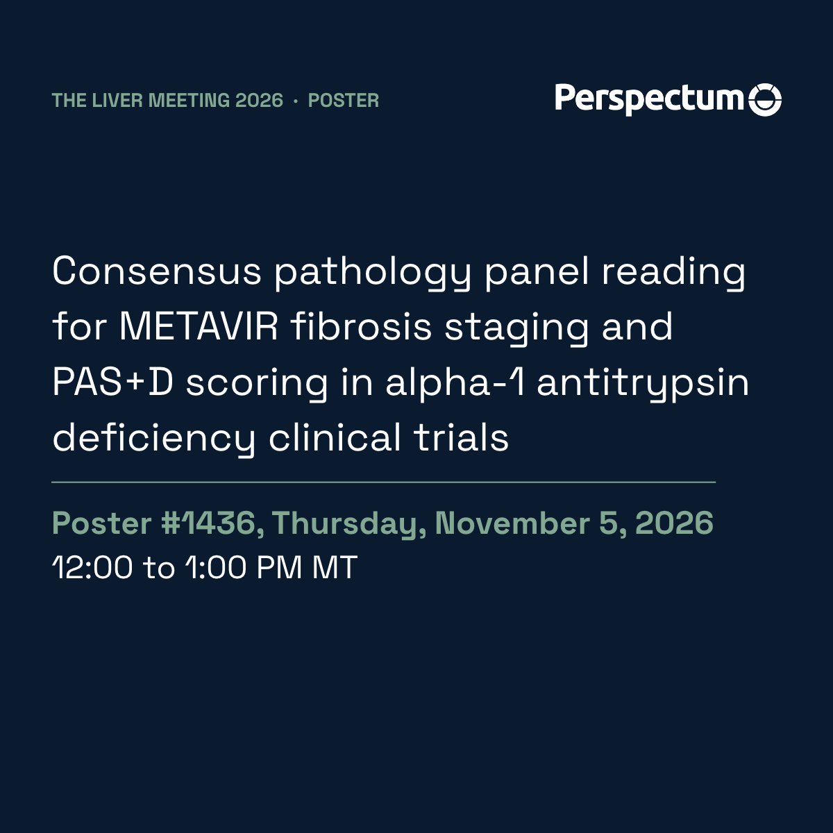
Poster #1436: consensus biopsy reading in a Phase III AATD program
Friday, November 6, 1:00 to 2:00 PM MT, Poster Session II
Poster #2329: cT1 as the second step in the MASLD screening pathway
Poster #2422: a multiparametric MRI risk model for identifying cirrhosis
Poster #2443: multi-organ MRI of the liver and heart in adults with hypertension
Saturday, November 7, 11:00 AM to 12:30 PM MT
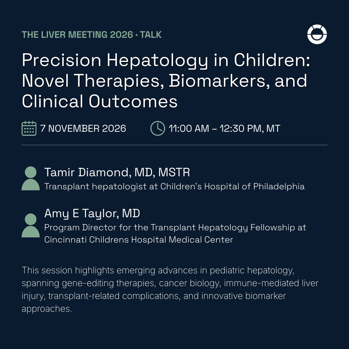
Oral presentation in Precision Hepatology in Children: Novel Therapies, Biomarkers, and Clinical Outcomes, on imaging and plasma biomarkers in pediatric autoimmune liver disease
Want time with the team? Come to the booth, or email https://t.co/[email protected] if you would like a conversation on clinical trials.
#Perspectum #TLM26 #Hepatology
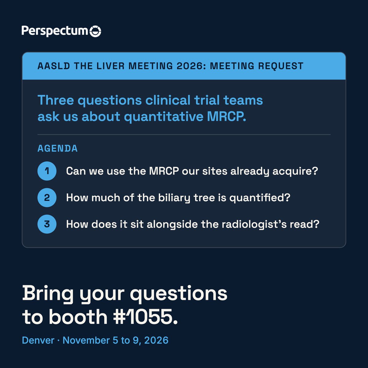
Three questions clinical trial teams ask us about quantitative MRCP, answered before The Liver Meeting.
Can we use the MRCP our sites already acquire?
Yes. MRCP+ can analyze newly acquired or historic non-contrast MRCP images, on a cloud-based platform with no additional hardware or software required.
How much of the biliary tree is quantified?
Quantitative assessment of biliary system structures across the intra and extra-hepatic biliary tree: bile duct widths, strictures and dilatations, plus the pancreatic ducts, gallbladder and regional volumetric analysis.
How does it sit alongside the radiologist’s read?
Quantified metrics, enhanced 3D visualizations and reference ranges to aid interpretation, designed to sit alongside visual review.
Our team is in Denver at AASLD The Liver Meeting from November 5 to 9. Book time with us at booth #1055.
Find out more: https://t.co/xHeMv8zlQy
#Perspectum #TLM26 #ClinicalTrials
Two posters at #ScientificSessions26: epicardial fat in type 2 diabetes (MDP0137, Sat, Nov 7, 9:15 AM CT) and coexisting heart and liver abnormalities in hypertension (REMODEL, MDP1904, Sun, Nov 8, 3:15 PM CT). Abstract data, not yet peer reviewed. #Perspectum
How can clinical trials ensure reproducible histology scoring between readers? That’s the focus of a poster we contributed to at #TLM26
Poster #1436
Thursday 5 November 2026, @AASLDtweets The Liver Meeting, 12:00 to 1:00 PM MT (Poster Presenter Hour)
Convention Center, Poster Hall, Hall DE
If you’re in Denver, stop by during the session.
Read the abstract in Hepatology: https://t.co/SFgBeM22cn
#Perspectum #TLM26 #AATD
Can digitized images be used for assessing METAVIR fibrosis? That’s the focus of a poster we contributed to at #TLM26
Poster #1439
Thursday 5, November 2026, @AASLDtweets The Liver Meeting, 12:00 to 1:00 PM MT (Poster Presenter Hour)
Convention Center, Poster Hall, Hall DE
If you’re in Denver, stop by during the session.
Read the abstract in Hepatology: https://t.co/SFgBeM22cn
#Perspectum #TLM26 #DigitalPathology
27 of 57 patients with pediatric-onset autoimmune liver disease showed features of PSC on MRI. Yet how that disease is read on MRCP can vary from one radiologist to the next.
That matters because bile duct injury often develops alongside autoimmune hepatitis in children, and it worsens prognosis.
Presented at @AASLDtweets The Liver Meeting, the team looked at which imaging and blood-based biomarkers detect large bile duct disease, and which predict future liver events.
What the abstract reports:
Quantitative MRCP metrics, such as maximal duct dilation, total duct length, and biliary volume, reached an AUC above 0.8 for identifying primary sclerosing cholangitis (PSC) features.
Liver stiffness by MR elastography, APRI, and taurine-conjugated bile acids predicted future liver events, with AUCs above 0.91. Ten of the 57 patients went on to have a clinical endpoint.
Most cytokines and chemokines tracked fibrosis rather than ductal disease.
For pediatric hepatologists, the open question is how imaging and blood markers fit together when following children with AILD over time.
Join the session:
Precision Hepatology in Children: Novel Therapies, Biomarkers, and Clinical Outcomes
Saturday, November 7, 11:00 AM–12:30 PM MT.
How does your center currently assess bile duct involvement in children with AILD?
#Perspectum #TLM26 #PediatricHepatology
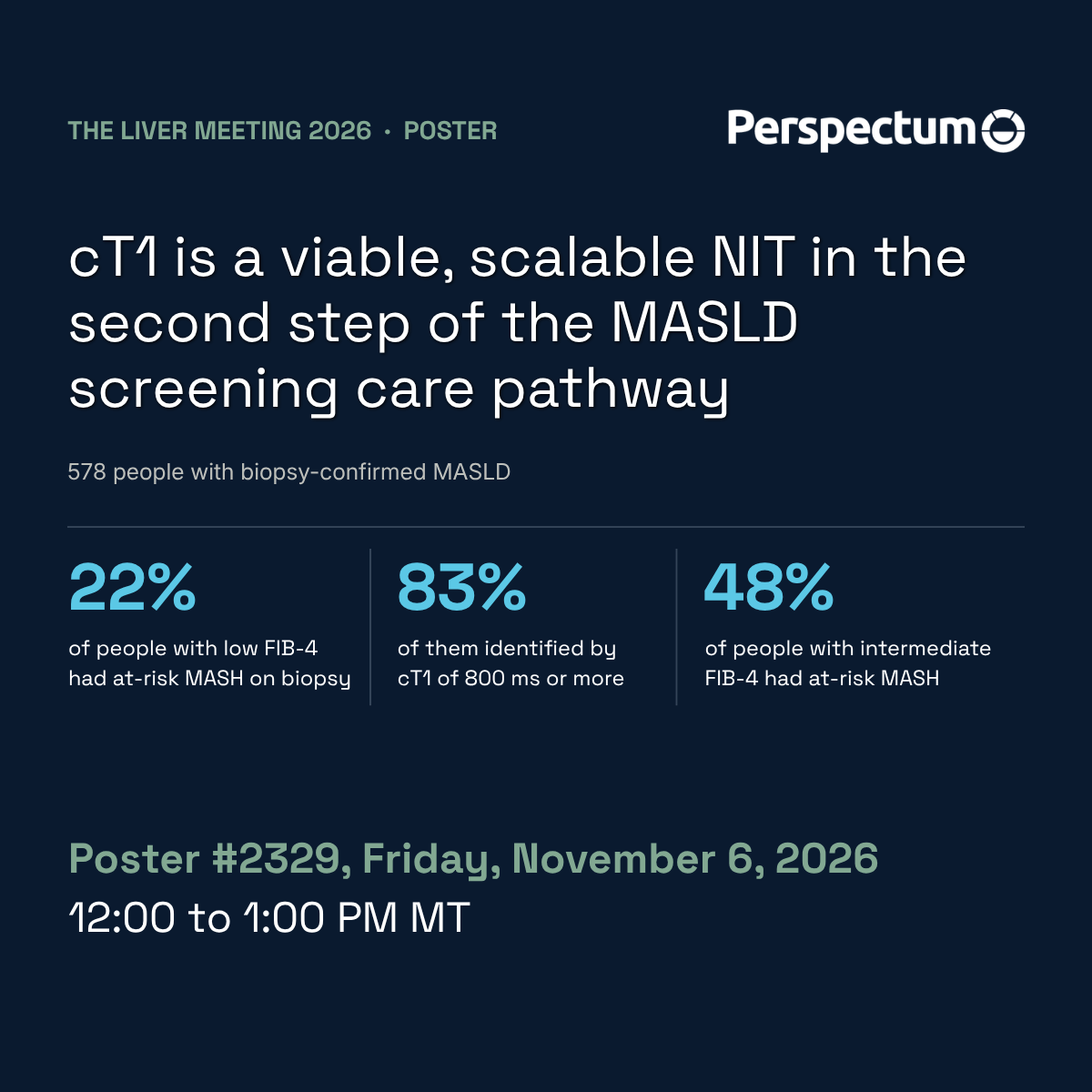
In people whose blood test score suggested low risk, 22% had at-risk MASH on biopsy. An MRI measure picked up 83% of them.
Screening for liver disease usually happens in two steps: a blood-based score called FIB-4, then a second test for people who need a closer look. Many patients never get that second test.
The authors pooled data from 578 people in the US, the Netherlands, Japan and Singapore who had all had a liver biopsy. They looked at cT1, an MRI measure of liver disease activity, as the second step. Among people with a low FIB-4 score, 22% had at-risk MASH, and cT1 of 800 ms or more identified 83% of them.
Visit poster #2329 at @AASLDtweets The Liver Meeing on Friday, November 6, 1:00 to 2:00 PM MT. The work was led by Niharika Samala.
#Perspectum #TLM26 #MASLD
Three MRI measures, one question: together, can they help identify cirrhosis in people with MASLD?
At @AASLDtweets The Liver Meeting in Denver this week, Perspectum and our collaborators are sharing five posters and a talk.
The team looked at cT1 (a measure of liver disease activity), liver fat content and spleen volume. On their own, each identified cirrhosis with an AUC of 0.64 to 0.72. AUC measures diagnostic accuracy, where 1.0 is perfect. Combined into one risk score, the AUC reached 0.86 in 368 people with biopsy-confirmed MASLD from Japan and Singapore. In a separate group of 261 people in Singapore, it held at 0.83.
Find us at poster #2422 on Friday, November 6, 1:00 to 2:00 PM MT
#Perspectum #TLM26 #MASLD #Hepatology
Almost 1 in 2 people living with diabetes have not been diagnosed.
That figure, from the International Diabetes Federation, is why this year’s World Diabetes Day theme is early action on diabetes: finding it sooner, and reducing the risk of complications.
This Diabetes Awareness Month, we want to highlight one organ in that conversation: the liver.
For people living with type 2 diabetes, liver health and diabetes are closely connected. Liver fat can accumulate silently for years, which makes it easy to overlook.
Early action can be a simple question at your next diabetes review: how does my liver health fit into my care?
If a liver scan is recommended, it helps to know what to expect. LiverMultiScan is a non-invasive MRI-based assessment of liver health. It is safe, pain-free and needs no contrast. Results come in patient friendly reports and must be interpreted by trained healthcare professionals.
For those working in diabetes care: how often does liver health come up in a routine review?
#Perspectum #DiabetesAwarenessMonth #LiverHealth
Heading to #ScientificSessions26 in Chicago, November 6 to 9? So are we. Masliza Mahmod, Luis Felipe Cardiel and Nicholas Hyde will be there to talk about a whole-system view of cardiometabolic health. #Perspectum