BOOK COVER REVEAL!!!!
About 4 years ago, @tony_breu and I set out to write a book that would inspire wonder in the remarkable resilience of the human body. That project became “Why Doesn’t Your Stomach Digest Itself?”, a book about the magic of homeostasis, how the body maintains and heals itself (and knows that it needs to heal itself), and can endure extreme challenges if necessary (think altitude acclimatization, freediving, ultramarathoning). The imperative to explore and understand this intrinsic physical resilience feels more urgent than ever in the age of AI and biohacking.
Coming March 16, 2027 from @wwnorton. We hope you enjoy reading it as much as we enjoyed writing it. The link to pre-order your copy is in my profile and it’s available from all booksellers!!
https://t.co/U2U4NJhHY3
The IMF mourns the passing of Dr. Robert A. Kyle, the “Father of Myeloma.” His life’s work helped transform myeloma research, treatment, and care, and his influence will continue for generations.
Read more: https://t.co/yTNOTalRZm
TACI × CD3 bsAb potently suppresses MM cell growth. TACI × CD3 bsAb represents a promising strategy to overcome BCMA escape in relapsed/refractory MM. Read the full article in Blood Advances: https://t.co/0EUpy9y78V #MM#MultipleMyeloma#BloodAdvances
1/14
I saw a patient last week with a hemoglobin of 4.3 g/dL.
Sitting comfortably.
Normal oxygen saturation.
Normal blood pressure.
Heart rate in the 70s.
How can the body tolerate such a profound reduction in oxygen-carrying capacity?
A physiology thread. 🧵
High-dose IV NAC is safe when administered as a continuous infusion in adults with #SCD at disease baseline. NAC decreases VWF multimer size, drastically decreases dense cell numbers, and improves systemic redox balance. Read in Blood RCI: https://t.co/TWnfTbL8YA
What has destroyed medicine is leaderships fixation on rankings. That’s all hospitals and universities care about. A meaningless list saying they are the best or closest to the best.
AUER RODS IN HISTIOCYTES AFTER TREATMENT OF ACUTE PROMYELOCYTIC LEUKEMIA. | ASH Image Bank | American Society of Hematology https://t.co/87BeWC4QIG #ASHImageBank
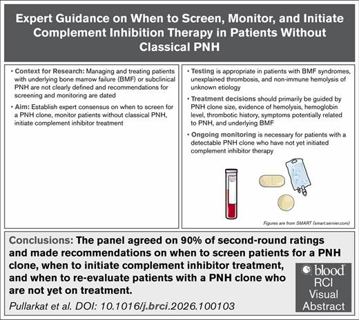
Treatment decisions should be guided by PNH clone size, hemolysis, hemoglobin level and thrombotic history, PNH-related symptoms, and BMF. Read in Blood RCI: https://t.co/hfXuAQxEMj
This landmark paper, published in 1988, was the first to describe the use of all-trans retinoic acid (ATRA) in the treatment of acute promyelocytic leukemia (APL). Read "Use of all-trans retinoic acid in the treatment of acute promyelocytic leukemia" here: https://t.co/QfOAd7k5Qh
Analysis of longitudinal blood samples shows that #CD19#CARTs can be detected at least 10 years post-treatment in patients with #lymphoma, with deeper profiling from a single patient showing that these CARTs have a dominant double negative activated effector memory-like phenotype.
@MarcoRuella@ruellalab@pennmedicine@paruzzol
https://t.co/AKP1zWUrDH
1/THREAD
Shortness of breath is a known side effect of the antiplatelet medication ticagrelor.
This association surprised me when I first learned about it.
Why would a drug that inhibits platelet function also lead to dyspnea?
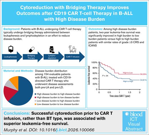
Successful cytoreduction before CD19-directed CAR T-cell therapy improves LFS in high DB B-ALL. Read the full study in Blood ICT: https://t.co/WoBERgew21 #B-ALL #BloodICT
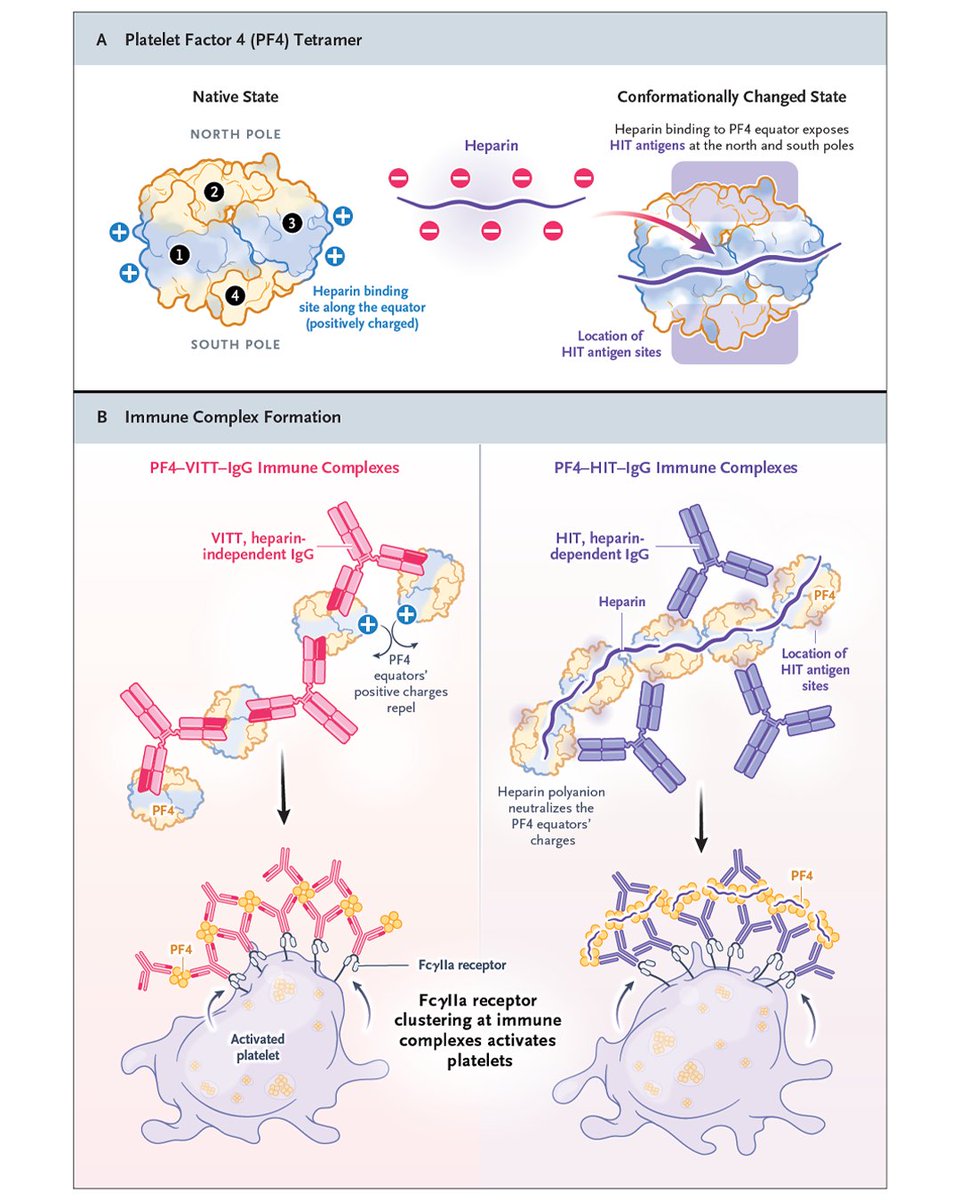
𝗥𝗲𝘃𝗶𝗲𝘄 𝗔𝗿𝘁𝗶𝗰𝗹𝗲: 𝗣𝗹𝗮𝘁𝗲𝗹𝗲𝘁-𝗔𝗰𝘁𝗶𝘃𝗮𝘁𝗶𝗻𝗴 𝗔𝗻𝘁𝗶–𝗣𝗹𝗮𝘁𝗲𝗹𝗲𝘁 𝗙𝗮𝗰𝘁𝗼𝗿 𝟰 𝗗𝗶𝘀𝗼𝗿𝗱𝗲𝗿𝘀
Platelet-activating antibodies against platelet factor 4 (PF4) cause highly prothrombotic disorders with reduced platelet counts. In heparin-induced thrombocytopenia (HIT), these antibodies bind PF4–heparin complexes, causing heparin-dependent platelet activation. Less common autoimmune and spontaneous HIT variants that are triggered by heparin and nonpharmacologic polyanions, respectively, have atypical clinical features and antibodies with additional heparin-independent platelet-activating properties. Vaccine-induced immune thrombocytopenia and thrombosis (VITT) antibodies directly target PF4. Initially, VITT was linked to adenoviral vector–based coronavirus disease 2019 vaccines, but in rare cases, an immune thrombocytopenia and thrombosis disorder that is clinically nearly identical to VITT can be caused by infection resulting from natural exposure to viruses, especially adenovirus. In persons with the IGLV3-21*02/*03 gene, anti–adenovirus protein VII antibody specificity shifts to PF4 by way of a specific somatic hypermutation (K31E) that creates VITT antibodies. In VITT-like monoclonal gammopathy of thrombotic significance, monoclonal anti-PF4 antibodies cause chronic prothrombotic conditions. Accurate diagnosis relies on distinct assays for HIT and VITT antibodies. Beyond anticoagulation, inhibition of FcγIIa receptor–mediated platelet activation may be needed for anti-PF4 disorders with heparin-independent reactivity (e.g., high-dose immune globulin in acute disease manifestations and Bruton’s tyrosine kinase inhibitors in chronic manifestations).
Learn more in the Review Article “Platelet-Activating Anti–Platelet Factor 4 Disorders” by Theodore E. Warkentin, MD, and Andreas Greinacher, MD: https://t.co/5mnvrPal1Z
Today I learned that the most common bacterial infections ate different between blood vs platelet Transfusions:
RBCs (stored 4°C): Yersinia enterocolitica (thrives in cold!)
Platelets (stored 20-24°C): Staph aureus/epidermidis (love room temp!)
Temperature = key determinant
A jar of marinara sauce taught me that, at the end of the day, what patients really want to know is: 1) Can I trust you? and 2) Am I going to be okay?
https://t.co/WwxqeDaBRM
Sign up for email alerts-- access is free, with no subscription to @nejm required :)
1/2
WHAT DOES BLOOD FEEL LIKE?
A hematologist writing in 1905 described freshly drawn blood as “slippery” and “greasy.”
As coagulation progressed, that sensation disappeared and was replaced by “viscosity, or stickiness.”
1/3
MANUAL OR AUTOMATED DIFF?
A CBC reports two very different kinds of white cell data:
1) Manual differential
• Reports percentages
• Separates bands from mature neutrophils
• Can identify promyelocytes, myelocytes, and metamyelocytes