This article reviews the appearance of various abnormalities that affect the lumbar intervertebral disk and diskovertebral segment through anatomic-pathologic correlation in cadavers.
https://t.co/TU6UQayGfZ
#FOAMrad#neurorad
Prominent Inferior Intercavernous Sinus (IICS): A Sign of Intracranial Hypotension (SIH)
Dilatation of the IICS is frequently associated with SIH, although it can also be found in the healthy adult as a normal anatomic variant.
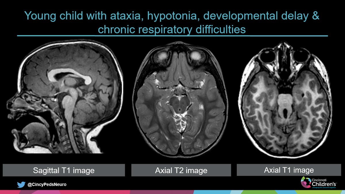
#PediNeuroRad
Case for the Weekend
Slide 1: History & images at presentation
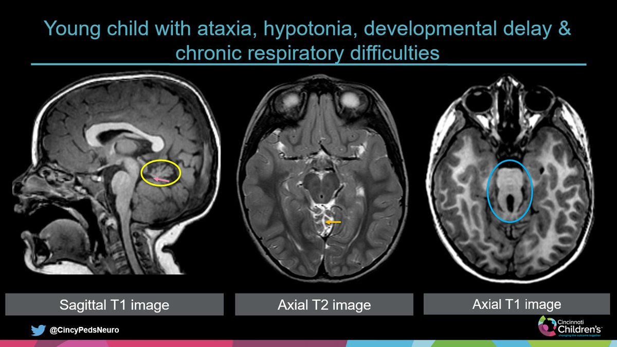
Slide 2: Annotated images
Slide 3: Answer & key points
Have a great weekend and hope to see/meet some of you at #ASPNR20 in Miami next weekend!
#PedsRad#NeuroRad#RadRes#FOAMrad
CSF leak classification
The syndrome of spontaneous intracranial hypotension (SIH) is generally caused by leakage of CSF from the thecal sac within or along the spinal canal
The authors categorized the CSF leaks into 4 types based on the morphology and distance from the midline
Looking for ways to improve the clarity and quality of your radiology reports?
@RSNA2019 electronic exhibit "How to Create a Great Radiology Report" has the answers!
CME discussion in the Learning Center today 1245-115!
#RSNA2019@frankgaillard @JeffreyKanneMD
The Floating Meniscus! Torn meniscocapsular ligaments (meniscofemoral and meniscotibial/coronary). Avulsed posterior root of the lateral meniscus. #mskrad@UTHealthSA
MRI nonhemorrhagic markers of Cerebral Small Vessel Diseases
Arteriolosclerosis:
Severe (>20) enlarged perivascular spaces (EPVS) located in the basal ganglia
Peribasal ganglia pattern of WMH following the peripheral outline of the basal ganglia
Lacunes in the basal ganglia
#FOAMrad#neurorad#radres#MS#MultipleSclerosis
Green & Red Flags MRI in MS (IV)
Green flags (typical)
Gadolinium-enhancing
(A) nodular
(B) open-ring
(C) closed-ring
(D) spinal cord nodular enhancement
Where is Broca's area? to learn more Neuroradiology fundamentals... I have added some great clinical correlation cases to https://t.co/dnjL1TkULo #meded