Sharing our state-of-the-art review on beta blockers just published in @JACCJournals! Thanks @rcstarling@DLBHATTMD@WilfriedMullens@AndreaRussoEP@MAbdeljawadmd for your expertise!
Key points:
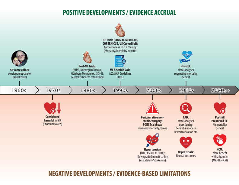
1. Beta-blockers should no longer be prescribed by reflex. The modern question is: Which beta-blocker, for which patient, for which indication, and for how long? We propose an indication- and phenotype-based approach.
2. Beta-blockers do more than slow the heart rate. Blocking cardiac β1 receptors reduces heart rate, contractility and oxygen demand, while blocking β1 receptors in the kidney’s juxtaglomerular apparatus suppresses renin release.
🫀📡 Can echocardiography really “see” LVAD complications?
A new study answers a key clinical question:
👉 How accurate is TTE in detecting outflow graft abnormalities?
✨ Key findings:
🎯 Specificity = 100% → if you see it, it’s real
⚠️ Sensitivity = 61% → but you may miss cases
📍 Much better for:
👉 Distal graft (71% sensitivity)
❗ Limited for:
👉 Proximal graft (only 33%)
📊 As shown in the graphical abstract (page 2):
👉 TTE can identify:
🔁 Kinking (≥90° bend)
🚫 Stenosis (≥50%)
👉 Doppler clue:
⚡ Vmax >2.0 m/s suggests obstruction
💡 Clinical message:
🩺 TTE = non-invasive, bedside, repeatable
🧠 Great for screening & follow-up
🧪 But CT remains gold standard for diagnosis
🚨 Why it matters:
Outflow graft complications →
❗ HF symptoms
❗ low pump flow
❗ device alarms
🎯 Big take-home:
From “echo vs CT”
➡️ to
“echo for screening, CT for confirmation”
📄 Read the full paper:
👉 🔗 DOI: 10.1093/ehjimp/qyag017
#Cardiology #Echocardiography #LVAD #HeartFailure #CardioTwitter #Imaging #CriticalCare #MedTech #Ultrasound #PrecisionMedicine 🩺📊
Our deep dive into stress testing from the ISCHEMIA trial showed that there was significant variability between severity determination between the sites and the core labs for each stress testing modality.
Nearly 1 in 4 site-classified moderate/severely ischemic studies were reclassified as showing no/mild ischemia by the core labs.
The lowest variability was seen with PET, contrast echo, and CMR.
My takeaways are that we need:
1. Modern hardware/software
2. Physician subspecialization and practice dedicated to stress test interpretation
3. Longitudinal education and quality improvement for those reading stress tests
https://t.co/Xpy9aq4s4h
Congrats star @MidAmericaHeart fellow Evan O'Keefe for leading this!
@jspertus@DRJamesOKeefe@LPhillipsMD@lesleejshaw@SripalBangalore@TimBateman6@MyASNC@SCMRorg@ASE360
The risk of new onset atrial fibrillation in patients with nonischemic cardiomyopathy (NICM) has a strong polygenic component. #NICM patients referred for cardiogenetic testing may benefit from screening for AF risk through PRSs. https://t.co/p0PWleFSQT
#JACCCEP#AFib
🫀📡 PET myocardial perfusion is not just imaging… it’s precision physiology.
A new EACVI consensus statement redefines how we perform and interpret PET/CT myocardial perfusion imaging (MPI) 👇
✨ Key insights:
🥇 PET = most advanced non-invasive tool for CAD assessment
🔍 Detects:
👉 ischaemia
👉 viability
👉 scar
⚡ Goes beyond anatomy → provides quantitative myocardial blood flow (MBF)
📊 MBF reserve <2.0 = abnormal → powerful for risk stratification
💡 From the figures (pages 8–9):
👉 Integration is key: perfusion + function + MBF + CT calcium
👉 17-segment model + polar maps = standardized interpretation
🔥 Game changers:
🧠 Quantitative MBF improves detection even when perfusion looks “normal”
⚖️ PET allows true physiological assessment of coronary disease
🧑⚕️ Strong focus on patient-centric protocols & preparation
⚠️ Important practical points:
☕ Avoid caffeine before test
💊 Medications may need adjustment
🧪 Choice of tracer = crucial (half-life, availability, accuracy)
🎯 Big take-home:
From “is there a stenosis?”
➡️ to
“is this myocardium truly underperfused?”
📄 Read the full paper:
👉
🔗 DOI: 10.1093/ehjimp/qyaf157
#Cardiology #CardiacPET #NuclearCardiology #MyocardialPerfusion #PrecisionMedicine #CardioTwitter #Imaging #CAD #MedTech #EACVI 🩺📊
🫀 CardioNugget™:
Speed Displacement (Propagation Velocity) Artifact
🧠 Mechanism: Ultrasound assumes sound travels at 1540 m/s. If tissue conducts sound at a different speed (e.g., fat ≈1450 m/s), the system assigns the wrong depth to returning echoes.
🎯 Classic TEE Board Example:
Lipomatous hypertrophy of the interatrial septum → fat slows sound → false deeper (distal) image of the septum.
Fossa ovalis is spared of fat, so no displacement artifact is seen there.
💡 Board Pearl:
Different propagation speed = incorrect depth = misplaced (not true duplicate) image.
#EchoBoards #CardioNuggets #MedEd
Among patients with multivessel coronary artery disease who were in stable condition 1 year after stenting, an additional year of DAPT led to a lower risk of ischemic events than aspirin alone, without increased bleeding. Full DAPT-MVD trial results: https://t.co/iHRM4Gv3ND
Editorial: Balancing the Scales — Extended DAPT in Coronary Artery Disease https://t.co/Nxqwkd4Trr
🫀📊 Should we screen asymptomatic patients for coronary artery disease with imaging?
A new systematic review of international guidelines gives a clear (and surprising) answer 👇
✨ Key takeaways:
🚫 No imaging for population screening of asymptomatic CAD
🚫 Not recommended even in high-risk individuals
🧮 Risk scores remain the foundation of primary prevention
🟡 CAC scoring = useful ONLY in selected cases
👉 when risk is uncertain or borderline
🤝 Strong role in shared decision-making with patients
📊 As shown in the graphical summary (page 2):
✅ Agreement → CAC as a risk modifier
⚠️ Disagreement → thresholds & re-testing intervals
❓ Gaps → outcomes, cost-effectiveness, alternative imaging
💡 Big message:
👉 Imaging is not a screening tool
👉 It’s a decision-making tool
🚀 Future direction:
Large trials (e.g. ROBINSCA, CorCal) may finally tell us
if imaging-guided prevention improves outcomes
📄 Read the full paper:
👉
🔗 DOI: 10.1093/ehjimp/qyag034
#Cardiology #Prevention #CAD #CACscore #Guidelines #CardioTwitter #PrimaryPrevention #Imaging #EvidenceBasedMedicine #MedEd 🩺📊
🧬🫀 We image the scar… but do we really understand it?
A new review explores how molecular contrast agents are redefining imaging after myocardial infarction 👇
✨ Key insights:
❤️ Post-MI remodelling drives heart failure & mortality
📊 Conventional imaging (CMR, PET) shows:
👉 infarct size
👉 function
❌ but NOT the underlying biology
🔬 Molecular imaging targets the real drivers:
👉 inflammation
👉 fibroblast activation
👉 scar formation
💡 As shown in the graphical abstract (page 2):
👉 Conventional imaging = what we see
👉 Molecular imaging = what is happening
🔥 Game changer:
Detect disease earlier (before structural changes)
Identify stage of remodelling (inflammatory → proliferative → fibrotic)
Guide targeted therapies (e.g. anti-inflammatory, anti-fibrotic)
🚀 Examples of next-gen tracers:
🧲 SPIONs → macrophage activity (MRI)
🔥 FDG / DOTATATE → inflammation (PET)
🧵 FAPI → fibroblast activation
🧱 Collagen/elastin tracers → scar maturation
🎯 Big take-home:
From “how big is the infarct?”
➡️ to
“what biology is driving the remodelling?”
📄 Read the full paper:
👉
🔗 DOI: 10.1093/ehjimp/qyag019
#Cardiology #CMR #CardiacPET #MolecularImaging #HeartFailure #PrecisionMedicine #CardioTwitter #Research #MedTech #PostMI 🧠💓
CardioNugget™ 🫀
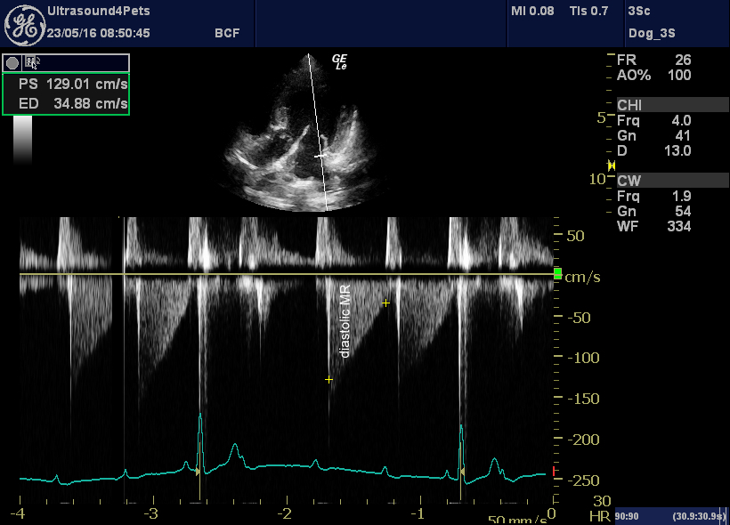
Why does diastolic MR occur in acute severe Aortic Regurgitation?
1️⃣ Acute severe AR → rapid filling of a noncompliant LV
2️⃣ LVEDP rises dramatically
3️⃣ LV diastolic pressure exceeds LA pressure → premature mitral valve closure
4️⃣ LV pressure continues to rise in late diastole → blood is forced back across the closed mitral valve = diastolic MR
Often accompanied by premature MV closure and a normal-sized LV in acute severe AR.
#CardioNugget™ #EchoBoards #EchoFirst #ASEcho #Cardiology #FOAMed #BoardPrep #AorticRegurgitation
The primary endpoint and 1-year outcomes of the ENCIRCLE MAC Registry were presented as an LBCT at New York Valves (June 25, 2026). This is the first pivotal evaluation of fully percutaneous transseptal TMVR in patients with MAC-related mitral valve dysfunction using a dedicated mitral THV. Patients with symptomatic MAC-related mitral valve dysfunction treated with transseptal TMVR using the SAPIEN M3 system had 30-day mortality lower than predicted by the STS score, durable MR reduction to ≤1+, sustained improvements in symptoms and quality of life, and stable THV performance through 1 year. These findings support consideration for transseptal TMVR with SAPIEN M3 as a treatment option for appropriately selected patients with symptomatic MAC-related mitral valve dysfunction.
@MayoClinicCV #NYValves #TMVR #StructuralHeart #MitralValve #MAC #SAPIENM3
European Journal of Heart Failure 2026
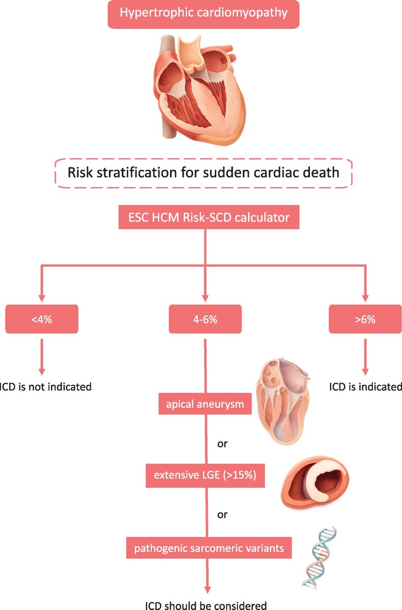
"EJHF expert consensus statement on the diagnosis and management of hypertrophic cardiomyopathy"
https://t.co/jpahvHVbgD
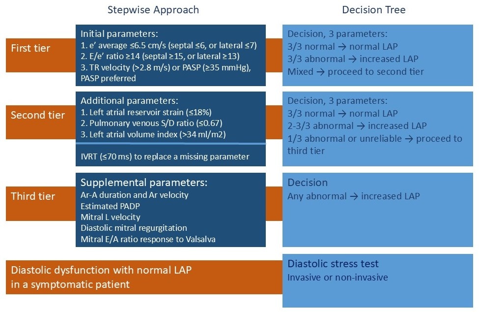
Struggling with diastolic function assessment? This practical 3-tier algorithm for estimating left ventricular filling pressures simplifies the approach. Tier 2 has been refined to reduce dependence on left atrial volume index alone.
https://t.co/FSnXDYKnKQ
Echocardiographic Drag Effect Index and Its Association with Response to Mavacamten in Obstructive Hypertrophic Cardiomyopathy
Drag-based echocardiographic indices offered high discriminatory performance for mavacamten response
@MasriAhmadMD
https://t.co/du1rM1235Y
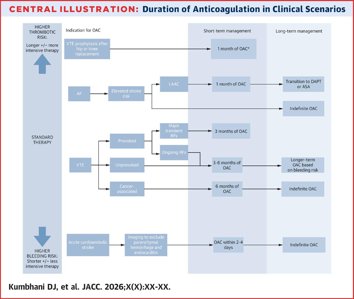
Despite strong evidence for their efficacy & safety, DOACs remain underused & at times inappropriately dosed, especially in high-risk pts. New ACC Scientific Statement provides guidance for primary & secondary prevention of thrombotic events https://t.co/l1Y0FmToIL #JACC#cvPrev
LVEF is not a linear marker.
There is a U-shaped relationship between LVEF and outcomes.
It is well established:
👉🏻Risk is high at low levels and lowest between 60% and 70%.
However:
👉🏻 Risk increases again when LVEF is ≥ 70%.
Not always “higher is better." Chang, EJHF 2025.
📄 2025 ESC/EACTS Valvular Guidelines: imaging perspective
🔗 DOI: https://t.co/X7MTuO2DZF
🫀 Core message
The new guidelines mark a major shift:
👉 from a single decision (surgery vs transcatheter)
👉 to a lifetime management strategy
➡️ In this framework, imaging becomes central to Heart Team decision-making.
🔑 Key clinical updates
Aortic stenosis:
👉 Age threshold for TAVI lowered to ~70 years
👉 Decisions now based on lifetime strategy (redo options, coronary access, anatomy)
Primary mitral regurgitation:
👉 Surgery remains the gold standard
👉 Earlier intervention recommended in selected asymptomatic patients
Atrial secondary MR:
👉 Recognised as a distinct entity
👉 New indications for surgery and transcatheter therapies
Tricuspid regurgitation:
👉 Transcatheter therapies enter mainstream guidelines
👉 Strongly dependent on RV function and pulmonary hypertension
🧠 New role of imaging
👉 No longer just to “confirm severity”
👉 But to shape the entire clinical pathway
Three key shifts:
From single parameters → integrated phenotyping
Right modality at the right time
Standardisation and reproducibility
🖥️ Role of imaging modalities
Echocardiography: first-line, now expected to be integrative (3D, stress)
CT: crucial for anatomy, feasibility, and lifetime planning
CMR: reference for volumes, regurgitation, and myocardial damage
Multimodality imaging: essential for complex decision-making
🔄 Paradigm shift
👉 Imaging-driven pathway:
DEFINE → SELECT → DELIVER → TRACK
➡️ Moving from thresholds → personalised, predictive care
🤖 Future: AI in imaging
👉 Not just automation, but:
standardisation
detection of discordance
guideline-based decision support
⚠️ Must remain transparent, validated, and clinically accountable
🚨 Bottom line
👉 Imaging is now:
a continuous, central, decision-making tool in valve disease
👉 Not just measuring valves
➡️ but guiding lifelong patient management strategies