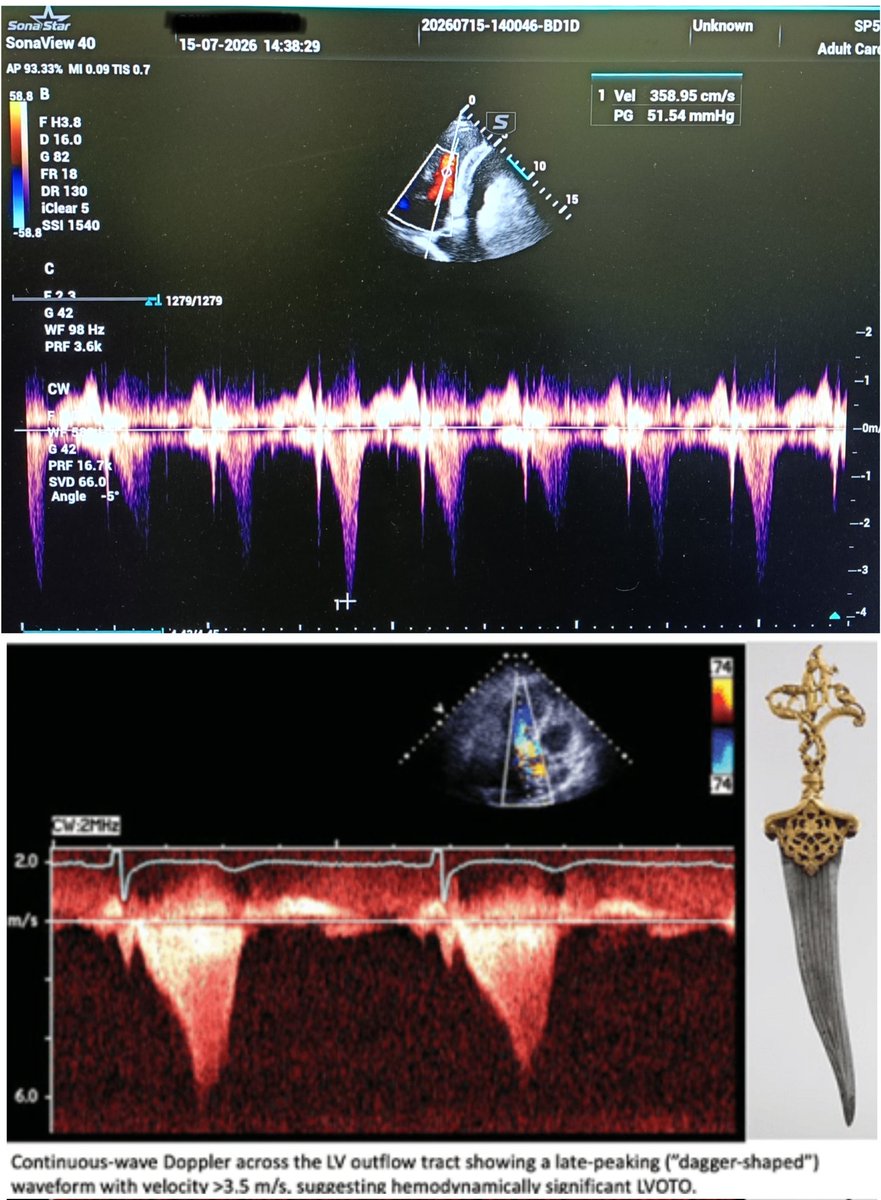
Significant Mid cavitary gradient in LVH, Hypovolemic State, severe tachycardia produced in CW has a late systolic peaking.
Don't confuse with LVOT gradient in HOCM.
1st pic - mid cavitary gradient of 51 mmHg in a patient with concentric LVH.
2nd pic - 🗡️ shaped CW,in HOCM.
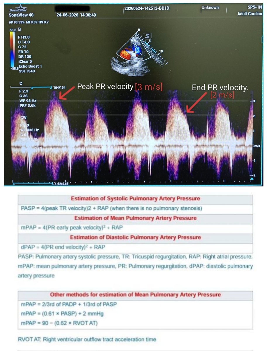
Using PR jet velocity for estimating mean Pulmonary artery pressure (meanPAP).
Here RAP = 15 .
mPAP= 4(3)² + 15 = 51 mmHg.
Can be used for estimating mPAP when TR velocity is less.
Here PR is Severe - Dense jet,steep deceleration & early termination of diastolic flow.
#echofirst
🟣 Purple urine bag syndrome - PUBS is the result of UTIs with certain bacteria which produce sulphatases and phosphatases -> tryptophan metabolism to produce indigo (blue colour) and indirubin (red colour).
Can be seen in patients with long standing urinary catheter.
#MedTwitter
Male,35 Y, having fever,jaundice,prerenal AKI,farmer by profession.
Now c/o B/L subconjunctival hemorrhage.
Lepto IgM (+).
Classical Weil's Disease.
#MedTwitter
Methotrexate (MTX)-cornerstone in treatment of RA,to be taken once weekly with Folic acid supplementation.But unfortunately few patients take it on regular basis leading to MTX toxicity.
MTX mucositis is the presentation.
T/t:1.) Stop MTX.
2.)Inj. Leucovorin.
3.) Hydration.
Case of massive Pericardial effusion with impending cardiac tamponade.....see the swinging heart inside Pericardial effusion...this is k/c/o - Double output Right ventricle (DORV).
#POCUS#echofirst
Left Atrial Appendage clot can be missed in routine 2D ECHO....TEE is the investigation of choice to rule out LAA clot.
Here a patient with Post partum cardiomyopathy,EF ≈20%,LAA clot can be seen in 2D ECHO...echogenic pedunculated mass attached with LA Appendage.
#POCUS#ECHO
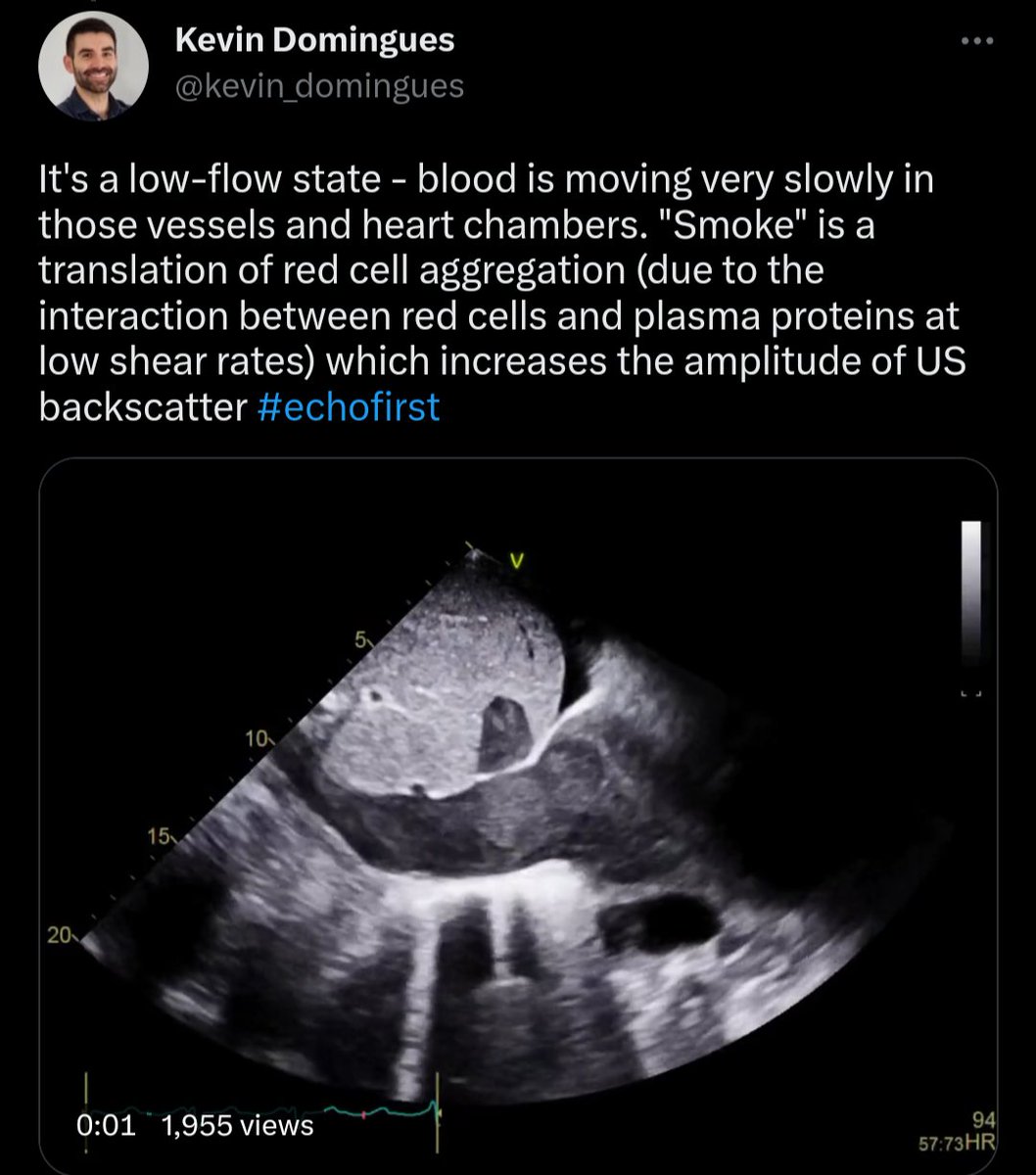
Look at the IVC....
The echogenic things floating within the IVC. It’s called particulate matter and is often observed in patients with no known cardiac pathology.
This is a normal finding.
#echofirst#POCUS
Bicuspid Aortic Valve.
28 Y/Male patient.
PXAS view showing RCC & LCC fused with raphe.
Valve opening and closing properly.
There was no COA.
No AR ,No AS.
Ascending aorta was not dilated.
#POCUS#echofirst
Classical windsock deformity of ruptured sinus of Valsalva.
This patient had H/O previous intracardiac repair of VSD with Aortic Valve replacement.
#MedX#ECHO#POCUS#RSOV