If robots could dream of microtubules, how would they look like? An amazing story by Alon Saguy, @ShechtmanLab and colleagues. Proud we could contribute to it
https://t.co/0DnDSxu248
Thrilled to share OM2Seq! We applied #AI methods to #Genomics, improving the speed and accuracy of optical genome mapping for fast diagnostics of diseases. @DanielleSapir, Tahir Detinis Zur, Nir Weinberger, @boknilev, @hagenom, @ShechtmanLab
🔬🚀
We used the Incucyte system by @SartoriusGlobal. All this and more in our latest paper: https://t.co/SvA8joKlpH. Big thanks and congrats to all co-authors and collaborators: Nadav, @EliasNehme9 Noam, Ilana, Reut, Boris, Paul, @AlaloufOnit , @SartoriusGlobal !
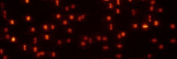
PSF engineering typically requires a 4-f system, but this is not always feasible, e.g. due to size constraints. Nadav Opatovski attached a phase mask directly to the objective lens, see beads defocusing here. Tetrapod mask shown here (wait for the end):
This enables PSF engineering in space-limited microscopy systems, e.g. that fit into incubators. Uses include extended depth-of-field and snapshot 3D spheroid from 2D, using algorithms by @EliasNehme9. Bonus: Obtaining training images is easy with a high-throughput microscope.
@Ella_Maru@naturemethods@AlonSaguy@AlaloufOnit@HeilemannLab Thanks!
Re simultaneous processes - there will be a complexity-performance tradeoff that would be interesting to check (TBD). Of course if the processes are labeled orthogonally (e.g. different colors) then each one will just be solved separately.
New prepint just out! Live cell single molecule localization microscopy at >3 fps. The information is mostly there - just spread out in space and time :)
We use a recurrent neural net (LSTM) + some assumptions to extract it. All details here:
https://t.co/0IYdFunUlJ
One of these super-resolution microtubule images was captured by a microscope and the other one was generated by a diffusion model. Can you tell which is which? Try more here: https://t.co/ANBQ5grOAp
1/4
Alon Saguy et al. trained a diffusion model to generate synthetic images of super-resolution microscopy images using data of only a few images from ShareLoc (https://t.co/x8DhXmcIsQ).
2/4
This is a very ongoing project, with many open questions: what are the performance limits? How much can we really gain beyond the information in the training data? and of course: what would be the best uses of something like this in super-resolution microscopy?
4/4
@christlet Interesting. Do you also get blinking-pairs mistaken for a vertically-elongated PSF (high Z)? I would expect to see a similar number of those.