Every year, America's Poison Centers publishes their annual report, and among the treasure trove of data, is the emerging trends section- This year, they highlighted the roll of ECMO in poisoning
https://t.co/0aigIusbk9
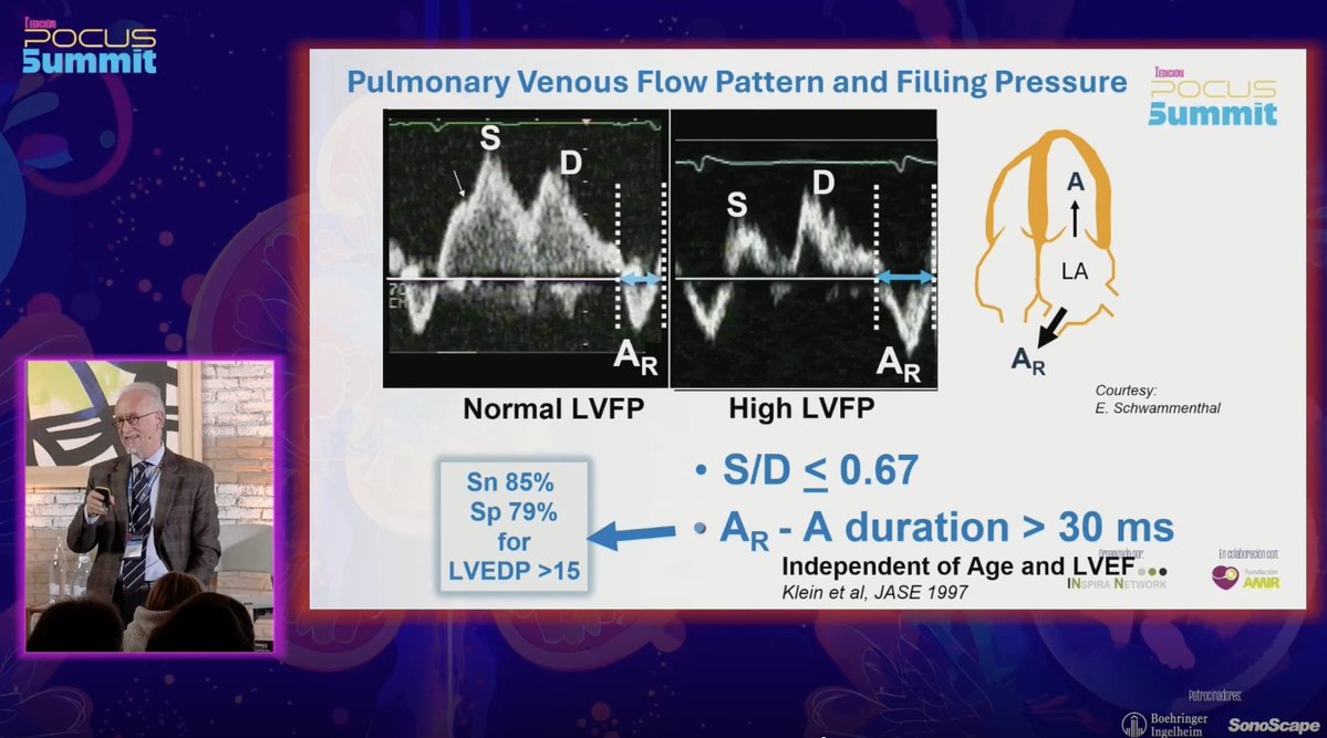
🔍 Detecting Volume Intolerance Through Filling Pressures
Brilliant presentation by Prof. @AleksandarNesk6 at the #PoCUSummit highlighting the value of pulmonary venous Doppler. Patterns such as S/D ≤ 0.67 and prolonged AR–A duration reliably reflect elevated LV filling pressures (LVEDP >15 mmHg), independent of age or LVEF. A powerful, underused window for diastolic and hemodynamic assessment. 🫀🔍 @NephroP@ArgaizR@EchoSoliman@EACVIPresident@ERAkidney@SENefrologia@nefrocat
#PoCUS #CardioRenal #Echocardiography #Hemodynamics
Recommend #POCUS reading of the day.
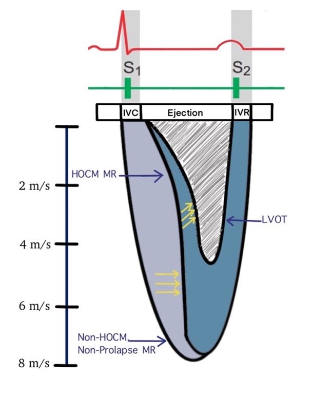
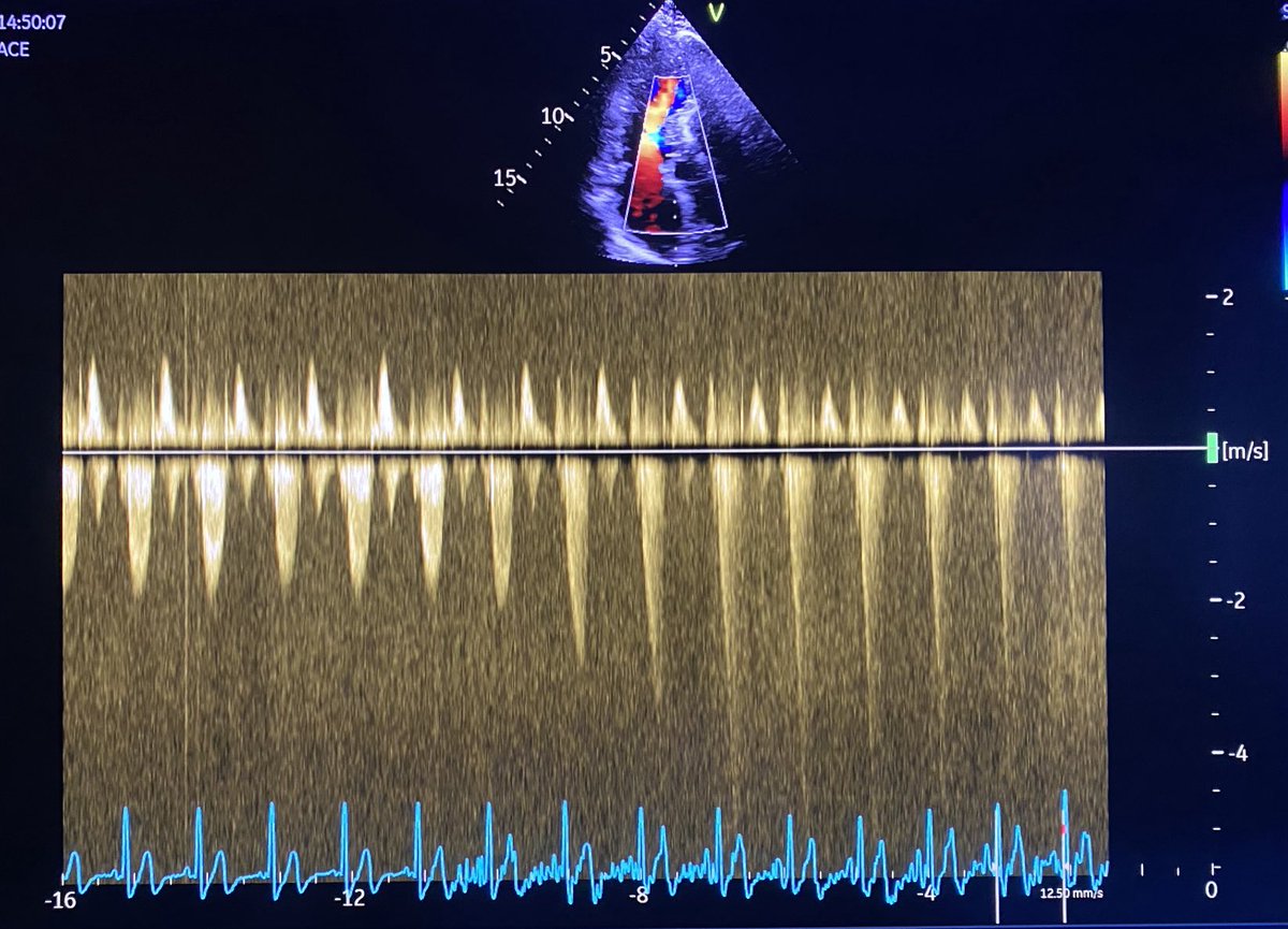
Measuring Left Ventricular Outflow Tract Signal Gradient in Hypertrophic Cardiomyopathy.
❓MR Doppler signal vs LVOT ❓
#FOAMed#echofirst
🔗 https://t.co/QQRH0xvJgM
Assessment of LVEDP Using AR–A Duration Difference
■ How to measure
Obtain pulmonary venous flow (usually from the right upper pulmonary vein) using pulsed-wave Doppler in the apical 4-chamber view.
■ Identify:
S wave (systolic forward flow)
D wave (diastolic forward flow)
AR wave (atrial reversal)
■ Measure:
AR duration = time from onset to end of the atrial reversal wave.
Compare it with mitral A-wave duration (from transmitral inflow Doppler).
If AR–A duration difference > 30 ms, → elevated LVEDP
If ≤ 30 ms, → normal LVEDP
■ What it indicates
● Normal: In a healthy heart, the PV AR duration is shorter than the mitral A duration, with a small or negative difference.
● Elevated LVEDP/Diastolic Dysfunction: When the left ventricle is stiff or relaxes poorly (diastolic dysfunction), it can't accept blood quickly enough during the A-wave, causing the A-wave to shorten and the PV AR wave to extend later into diastole. This results in a longer PV AR duration and a positive AR/A duration.
》Cutoff: A difference of >30-35 ms suggests elevated left atrial pressure (LAP) and LVEDP.
■ Notes:
Best used in sinus rhythm (needs identifiable A-wave).
Less reliable in AF, significant MR, or pulmonary venous stenosis.
Should be interpreted along with other diastolic indices (E/e’, LA size, etc.).
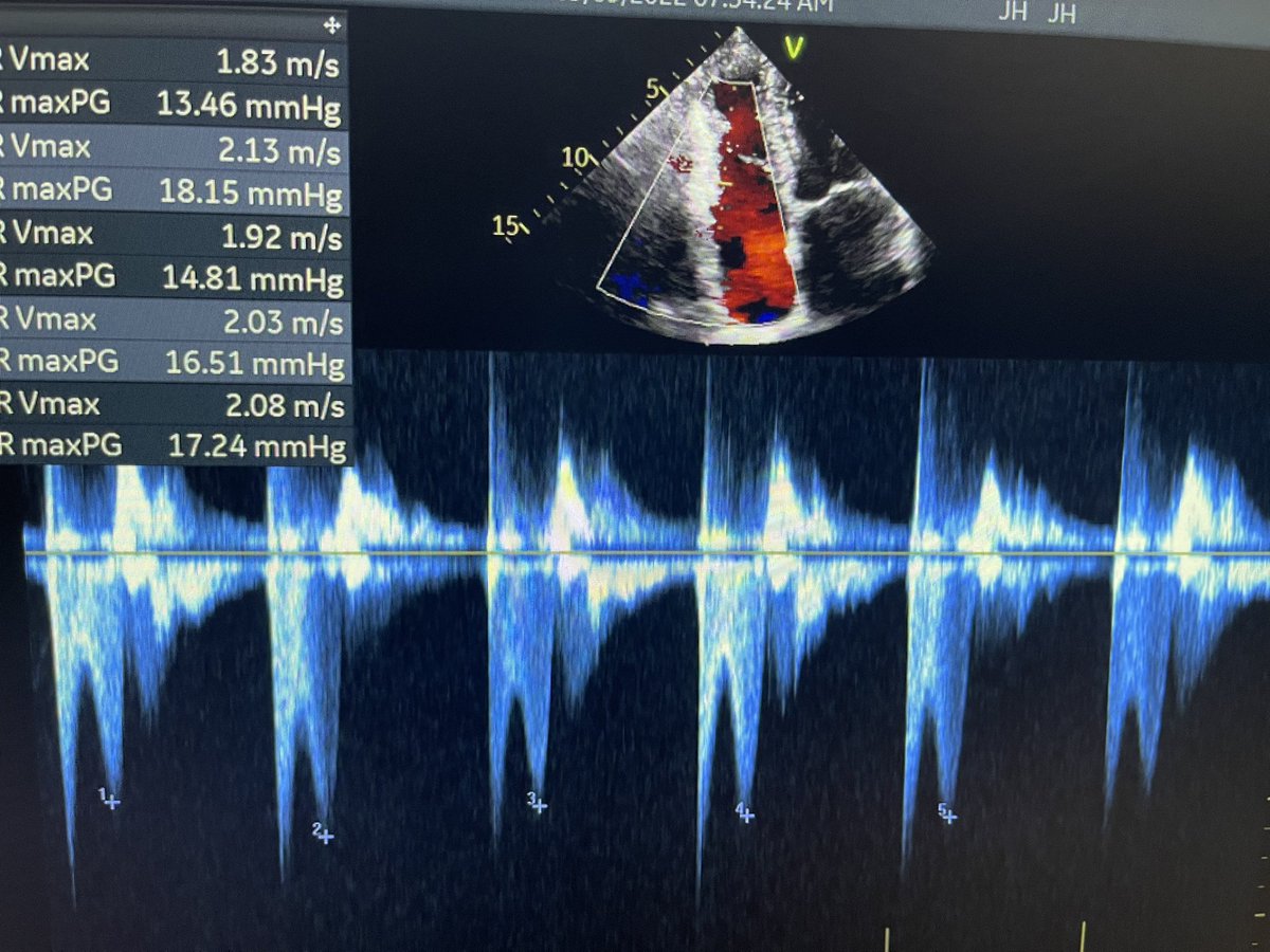
The diastolic pulmonary artery pressure (DPAP) & The mean pulmonary artery pressure (mPAP)
》The DIASTOLIC pulmonary artery pressure (DPAP) can be estimated from the velocity of the END-diastolic pulmonary regurgitant velocity using the modified Bernoulli equation.
DPAP = 4V(END-diastolic pulmonary regurgitation velocity)² + RA pressure.
》The MEAN pulmonary artery pressure (mPAP) can be estimated from the EARLY PEAK pulmonary regurgitation velocity using the modified Bernoulli equation.
mPAP = 4V(early peak pulmonary regurgitation velocity)² + RA pressure.
(1/x) High quality CPR, early defibrillation, and treating the underlying cause saves lives in cardiac arrest, but what else can we do?
Here are 7 advanced therapies for cardiac arrest👇
A🧵
Caution: Bleeding edge - evidence 'light' zone.
What is the correlation between echocardiographic & invasive assessment of hemodynamics in acute decompensated heart failure-related cardiogenic shock (ADHF-CS)?
ICU Hemodynamics - Literature Review:
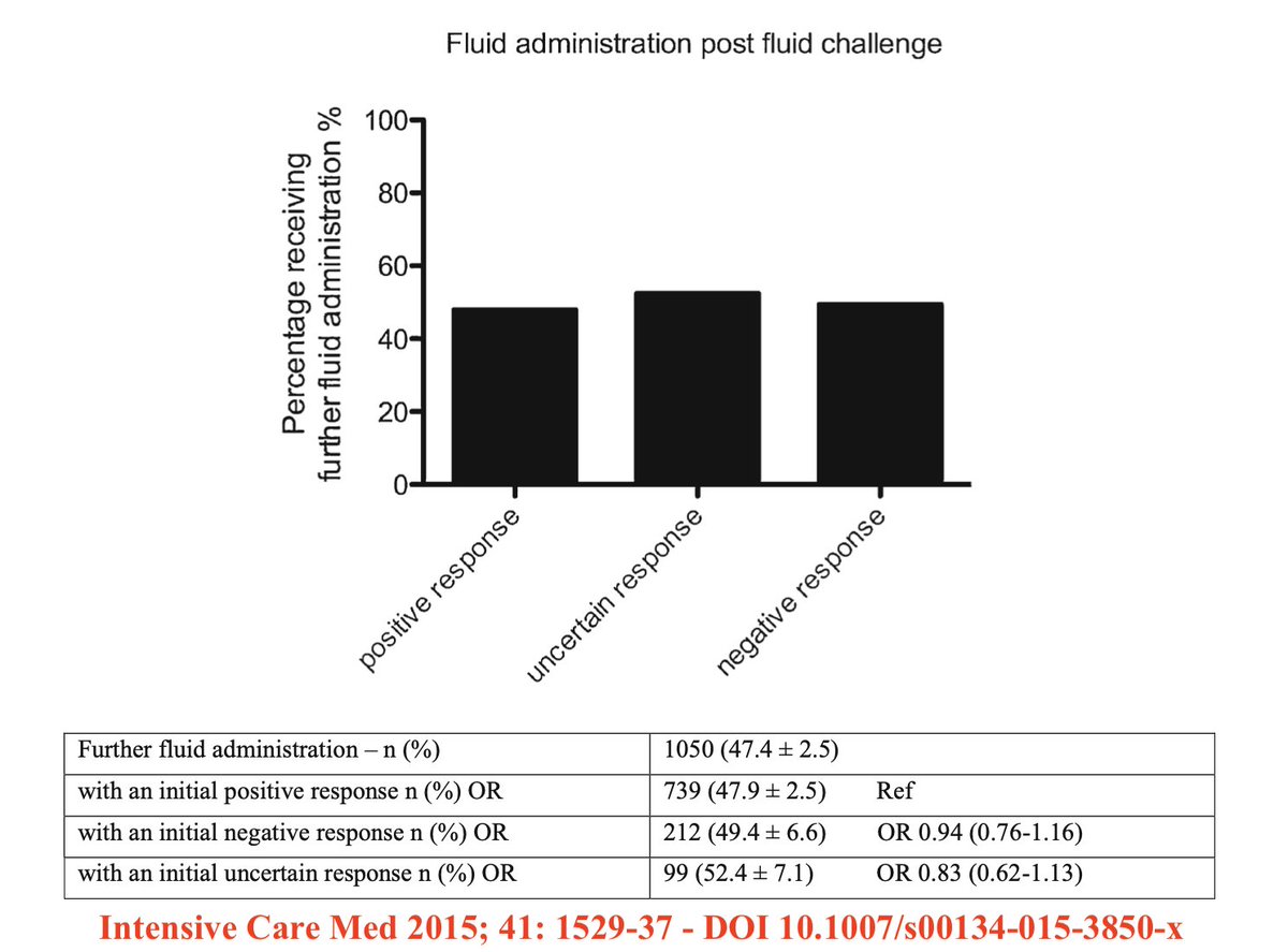
Speaking about fluid responsiveness...
This was my favorite figure from FENICE, a multicenter international observational study of 2,213 pts aiming to evaluate the practice of fluid challenge (FC) in critical care settings (@DrMCecconi):
including a study from this paper's authors, that "impaired RV function and RV-PA coupling are associated with severe pulmonary congestion on admission, and less resolution of pulmonary congestion during hospital stay". This may be due to a number of factors:
@khaycock2 Pardon my ignorance, but how do you use pre ejection time and total ejection time to help with your management? Any resources to read about it?