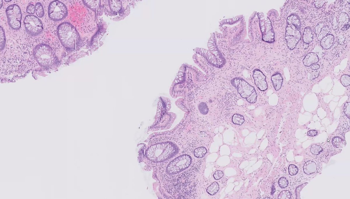
34F with multiple colon polyps, macrocephaly, and trichilemmomas on face and neck. Colon biopsy is shown below.
What is the most common mutation seen in this syndrome?
@KMirza#TwitterHomework
Kinyoun stain (cold stain- no heating like Ziehl-Neelsen), was developed to stain acid-fast species. The high concentration of phenol in the stain serves to "dissolve" lipid material in the cell wall, allowing penetration of the dye without heating. @KMirza#TwitterHomework
Most phyllodes tumors are benign, but up to 10% can be malignant. Pictured is a tumor with marked atypia and malignant heterologous elements. Loss of epithelial interaction in stromal component is believed to lead to malignant progression @KMirza#TwitterHomework
Endomyocardial biopsy showing Giant Cell Myocarditis (GCM). Notice the diffuse heterogenous infiltrate composed of lymphs, prominent giant cells and occasional plasma cells. One key feature to distinguish GCM is the lack of granulomas! @KMirza#TwitterHomework
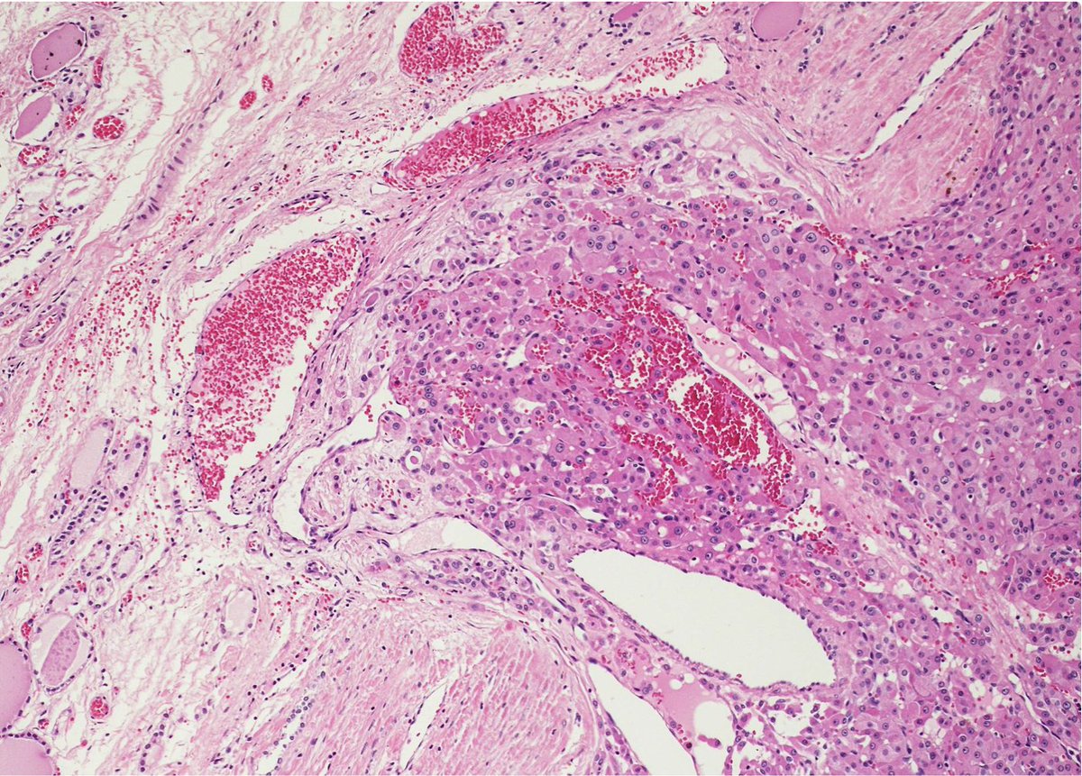
Follicular carcinoma can resemble adenoma at low power. Very important to assess presence of capsular or vascular invasion. Cellular atypia and necrosis, while potentially present, are not sufficient to make the diagnosis @KMirza#TwitterHomework
Architectural features of chronicity in a GI biopsy include: crypt distortion, crypt loss, crypt atrophy, basal plasmacytosis & Paneth cell metaplasia @KMirza#TwitterHomework
When evaluating a breast biopsy, what feature of usual ductal hyperplasia helps to differentiate it from low-grade DCIS?
Cellular maturation. The cells condense as they move from the basal layer toward the center of proliferation @KMirza#TwitterHomework
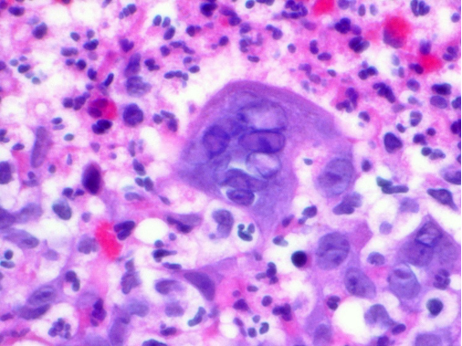
Herpes simplex virus (HSV) can be identified histologically with the 3M's. Multinucleation, nuclear Molding and Margination (of chromatin to the periphery of the nucleus). @KMirza#TwitterHomework
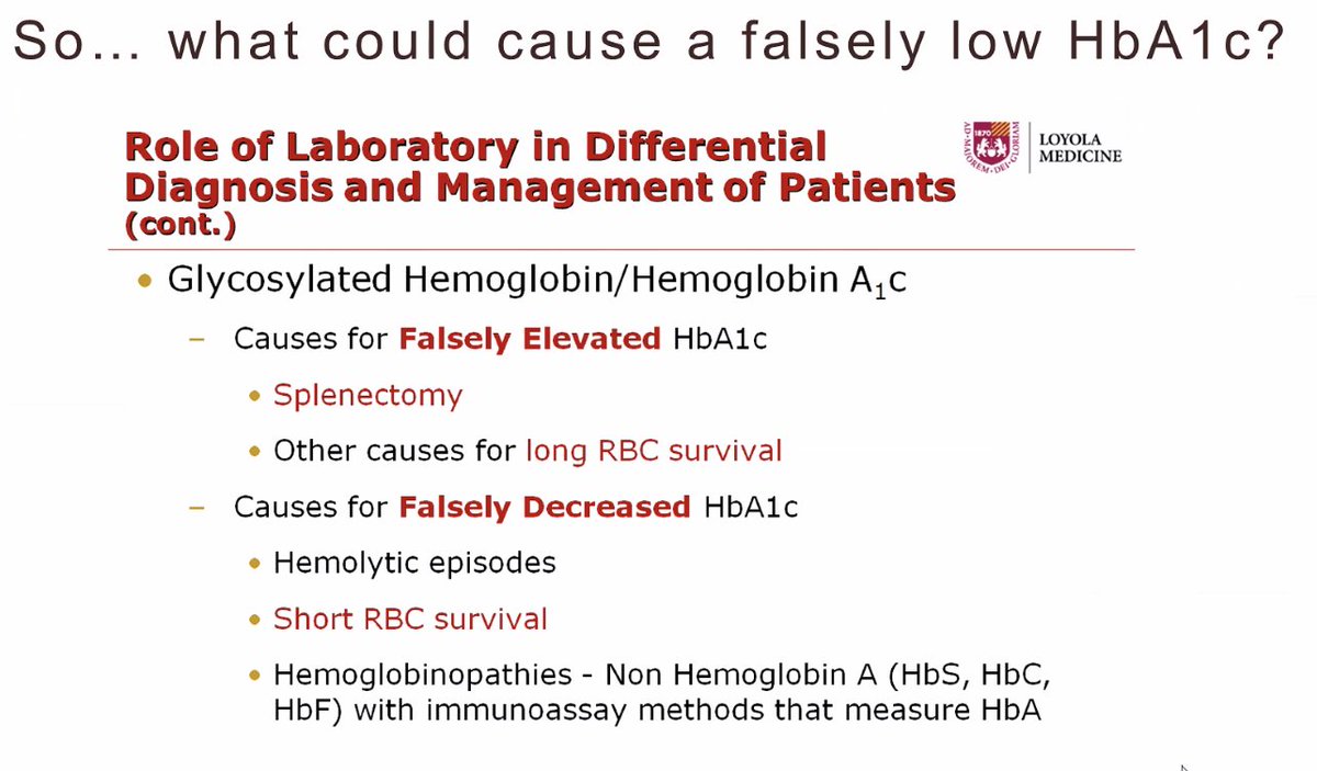
Great clinical chemistry lecture @loyolapathology. Always important to consider and evaluate etiologies of falsely low or elevated A1C levels @KMirza#TwitterHomework
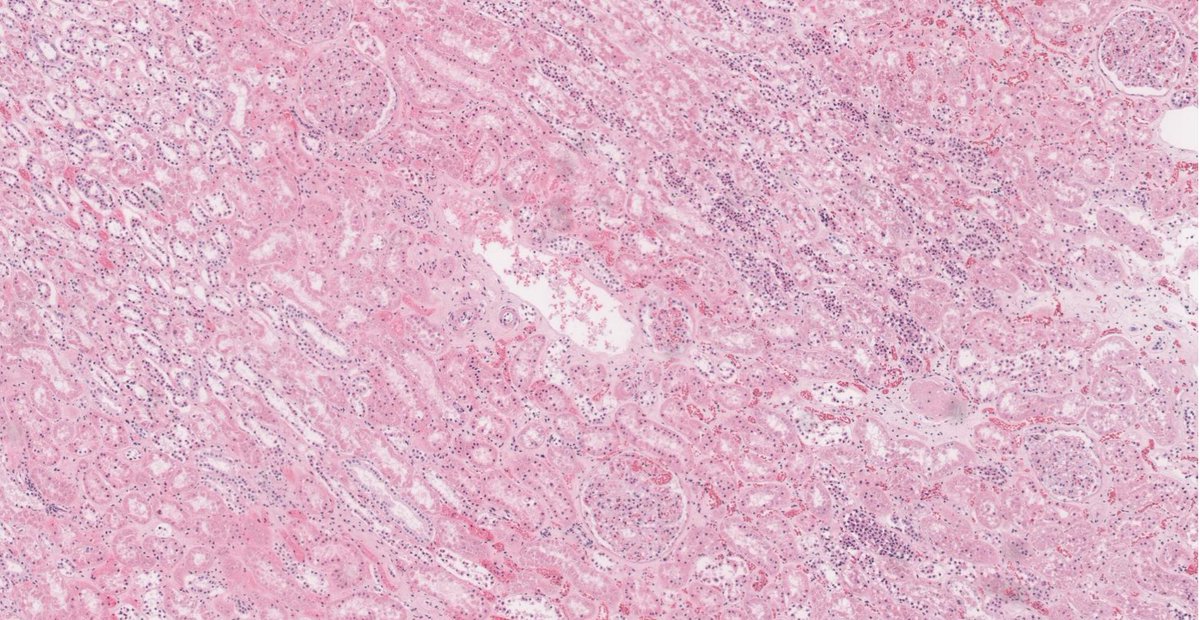
When evaluating a kidney biopsy, cells of the proximal tubules appear eosinophilic and granular (due to the high mitochondrial content, lots of energy being used for transport!). The distal tubules have a flatter appearance with less cytoplasm @KMirza#TwitterHomework