The ALMF is a part of the Center for Open Research Resources and Equipment at UConn. We provide microscopy expertise and access to cutting edge instruments.
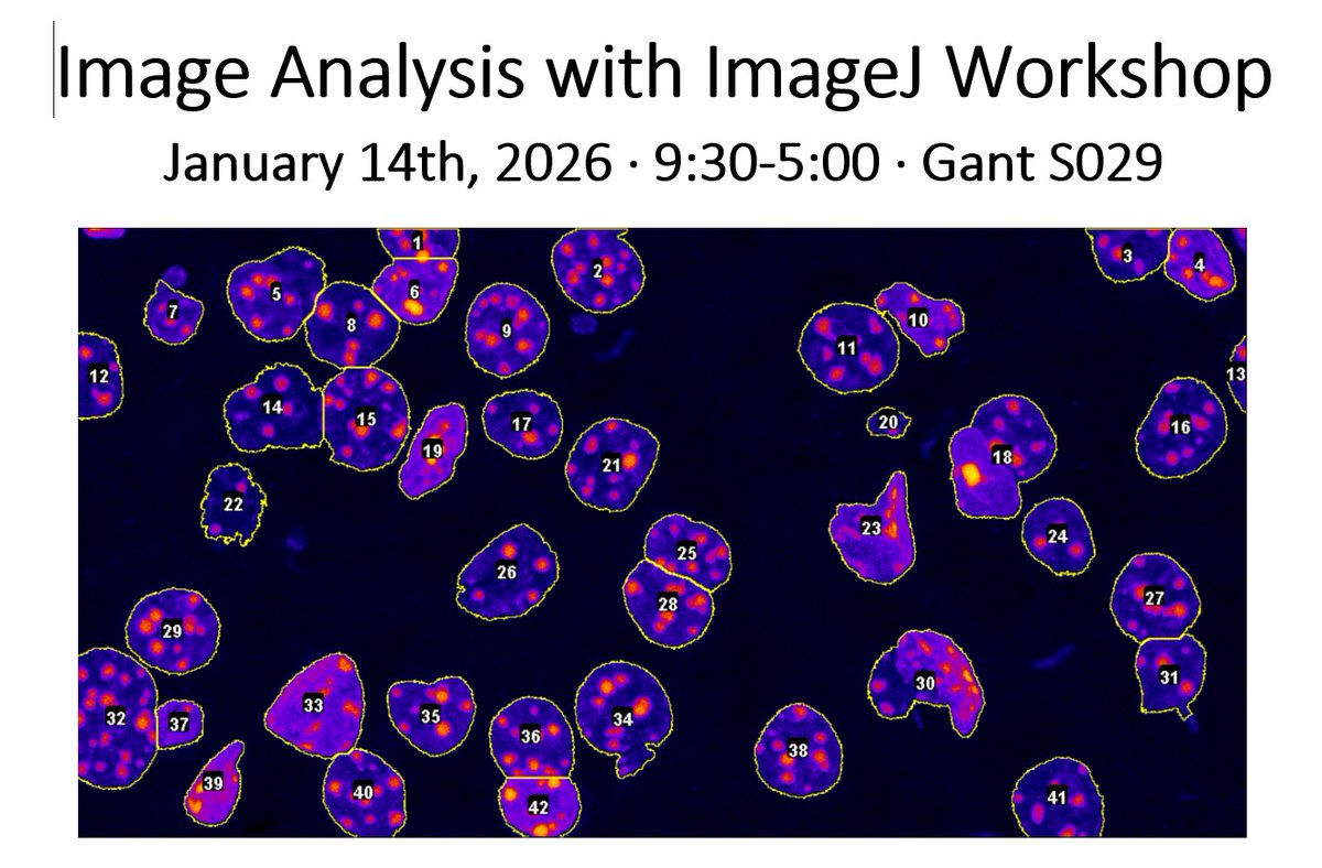
The UConn Advanced Light Microscopy Facility will be holding a one-day workshop, "Image Analysis with ImageJ" on Wednesday, January 14th. Attendees will learn practical workflows and practice on real datasets. Info and registration at https://t.co/zH0ZfqUQUm
According to Dr. Chen, “we developed a nanoparticle-based therapeutic for treating osteoarthritis. Our nanoparticles have a unique nano-rod shape, enabling better penetration into articular cartilage compared to traditional spherical nanoparticles.
Want to see more of the core facilities that UConn Storrs and Health have to offer? Register for the Open House! Free lunch with registration, and vendors will be here too!
Mark your calendars for the first ever COR²E Open House & Vendor Show on Mon, Oct 2 (11am-1pm) in the Student Union Grand Ballroom! We will highlight the service and analysis capabilities available in COR²E. Free lunch too! Registration and details here: https://t.co/r3XpHNjkRU
Happy #FluorescenceFriday! Autofluorescent plant cell wall structures were captured on our @NikonInst AXR #confocal and denoised. ImageJ was used for processing, displaying it in the magma LUT and rotating it to create this fun little GIF. Depth coding from NIS Elements below👇
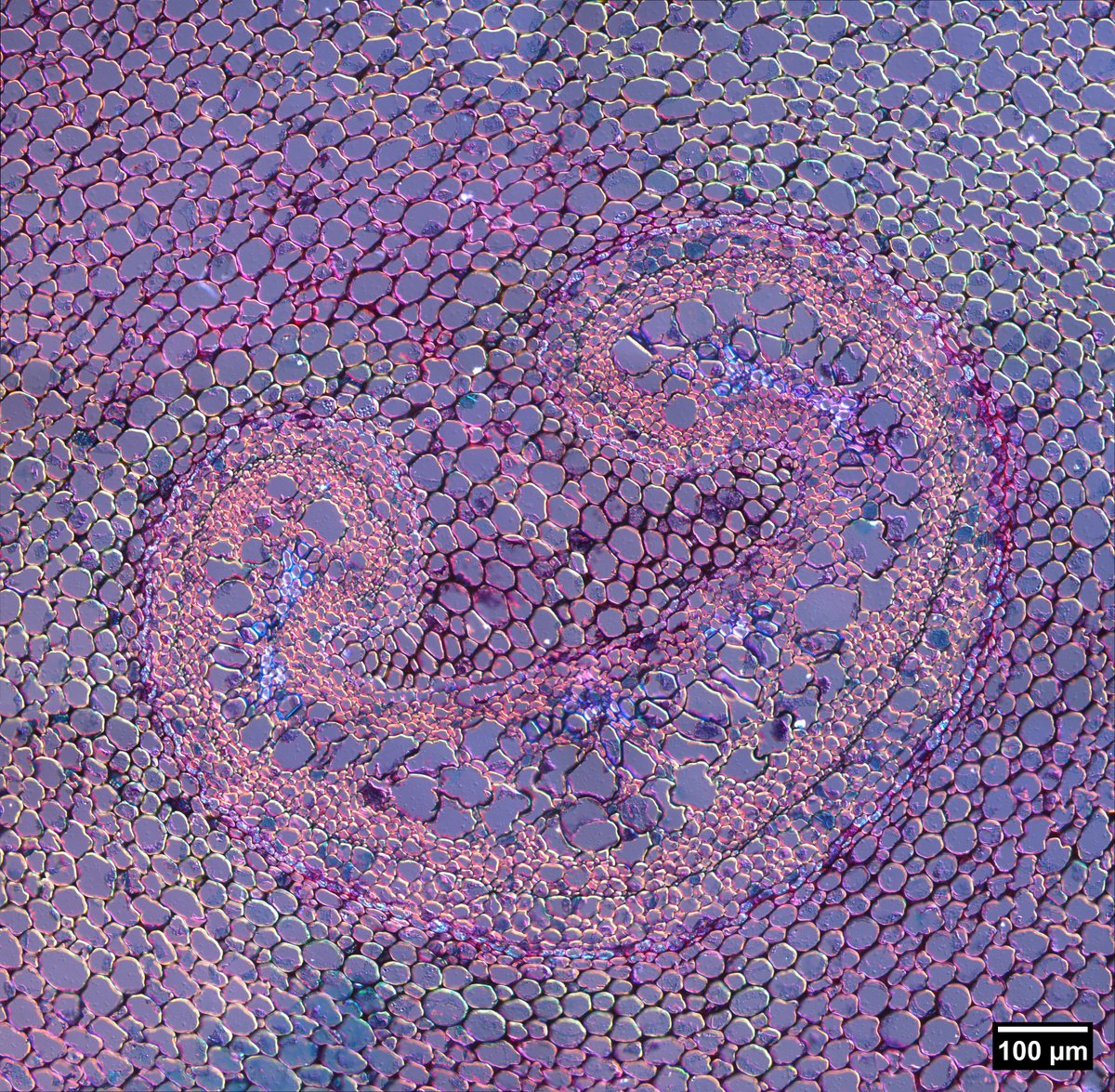
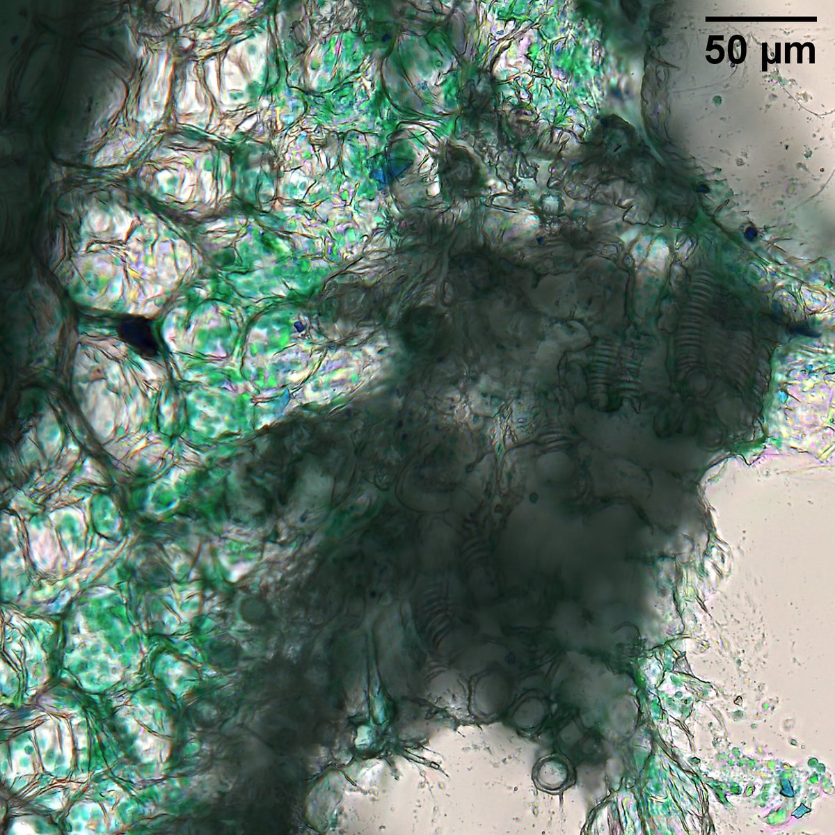
Double dipping on this same stellate trichome (left) for #microscopymonday! This image is using the @LeicaMicro Thunder K5C color camera and applying extended depth of focus. We also imaged part of the stem itself (right) and found other interesting structures!
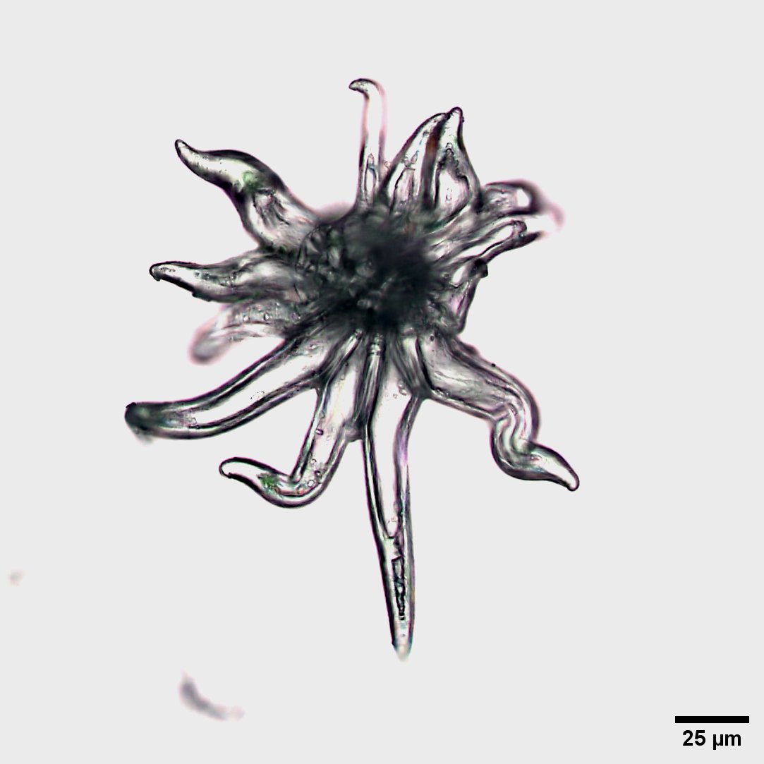
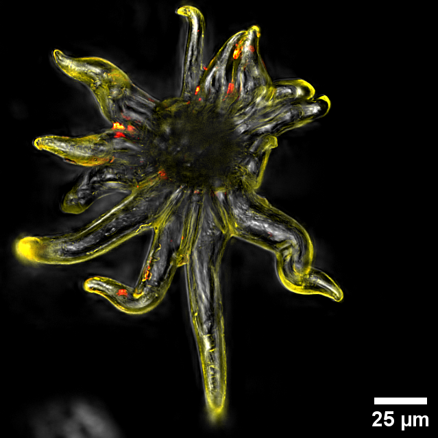
This #FluorescenceFriday we bring you some fun #sciart of a "microscopic sunflower," found from a hibiscus sample! Captured using green and far red fluorescence (yellow and orange here, respectively) and DIC on our @LeicaMicro Thunder. Any #plantsci people able to ID this?
@jasonmileskirk @LeicaMicro That's amazing!! Looking into trichomes more because they're fascinating. So many morphologies, too! Thank you! https://t.co/8WDwinZ4pA
This #FluorescenceFriday we bring you some fun #sciart of a "microscopic sunflower," found from a hibiscus sample! Captured using green and far red fluorescence (yellow and orange here, respectively) and DIC on our @LeicaMicro Thunder. Any #plantsci people able to ID this?
If you're a lab at @UConn or @UConnHealth there's still a few weeks left to apply for internal funds if you want to try out one of the UConn Core facilities!
Want to incorporate new technology in your research program @UConn or @UConnHealth but low on grant funds?💵COR²E and OVPR announce a new funding opportunity for research projects that specifically engage UConn's world-class and diverse core facilities! 💰https://t.co/UHQKdY5qa3
Happy #MicroscopyMonday! Here we have a fern rhizome imaged with a color camera on a @LeicaMicro Thunder without (Left) and with (Right) a DIC prism in the lightpath, creating interference that results in other colors.
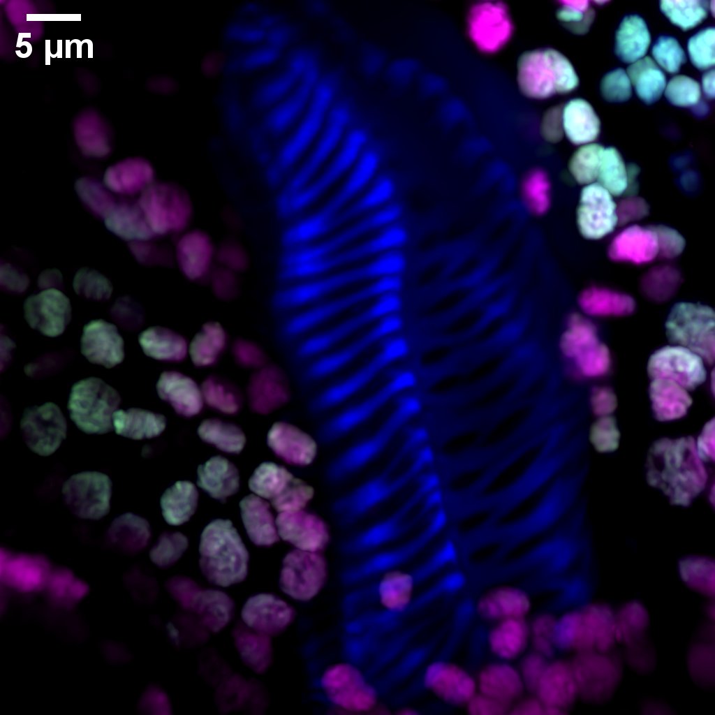
@NikonInst The maximum intensity projection shows the quality much better! Chlorophyll are in green and magenta, and what we believe is the cell wall is in blue. If we see different secondary cell wall structures here, that would be fun!
Where are the #plantsci people this #FluorescenceFriday? We found what appears to be secondary xylem cell wall structure (lignin?) in a hibiscus leaf, shown here in blue among chlorophyll. Imaged on our @NikonInst AXR confocal and processed with the NIS movie maker.
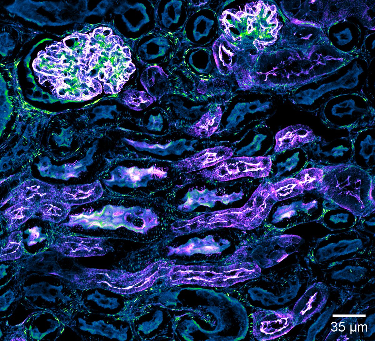
Happy #FluorescenceFriday! We've been playing around with large image + z-stack using JOBS on the @NikonInst AXR, which is super useful! Image 1 is a 2D overview, and 2 & 3 are max projections from select regions of this kidney section at 40X.
The value of machine learning using @zeiss_arivis software for segmentation becomes clear in a sample with high dynamic range, such as this mitotic spindle (bottom). Most of the dimmer astral microtubules are lost with traditional segmentation algorithms (top).
More specifically, this protocol describes use of PKMO to resolve inner membrane dynamics with neuroblastoma model SH-SY5Y cells and primary rat hippocampal neurons, along with some example analyses. Enjoy!
🔬📃Publication alert! A protocol with the @alderlab using our @Abberior STED was published in @JoVEJournal: https://t.co/WxJ7NucpZm
New mitochondrial inner membrane dyes allow researchers to take greater advantage of live cell STED imaging, showcased here 👇
Having fun with our new @NikonInst AXR confocal and using the NIS Elements movie maker function. This is a 3D reconstruction of the crypts in a mouse intestine section showing goblet cells (blue), nuclei (green), and actin (red).#confocal
COR²E has many important updates to share with the UConn research community: recently completed projects, state-of-the-art instrumentation installations, and new people! Read all about them in our brand new Summer 2023 COR²E newsletter here:
https://t.co/WsQqSx6ZfR