We're hosting a #tissueclearing Lunch & Learn with the Advanced Imaging Facility at @ucdavisvetmed!
Join us on 🗓️ Tue, Feb 18, 2025, from 11:30 am - 12:30 pm, to learn what tissue clearing is and how it's transforming biological research!
RSVP details below👇
[1/2]
Learn best practices, ask questions, and dive into hands-on demos. A great opportunity for UC Davis researchers & students interested in #LightSheetImaging and #3DHistology.
RSVP for the event:
https://t.co/CXAZidq07f
[2/2]
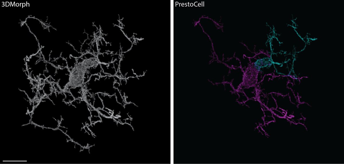
Interested in microglia analysis from light microscopy data, but think the standard ways of segmentation are missing things?
Online now is our manuscript where we created a new tool called PrestoCell at PlosONE https://t.co/rJI1tBiWD3
Happy #FluorescenceFriday! We are finally getting to play with the light sheet using chick embryos. 😍 Can’t wait to do more imaging! Thanks to the @ucdavisvetmed Advanced Imaging Facility and to Julia Godinez who took this image during her rotation in our lab!
Thanks to @zeiss_micro for a great (in person!) seminar on the uses of light sheet microscopy and the amazing things that can be done on the LS7 that is installed in @ucdavisvetmed here at @ucdavis.
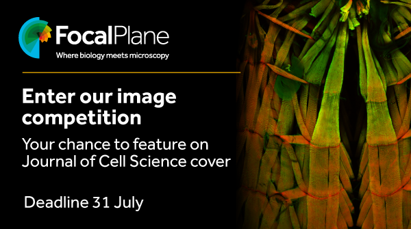
Enter our image competition to have your image featured on the cover of Journal of Cell Science @J_Cell_Sci.
Deadline to submit your image is 31 July
https://t.co/oV42vRp27W
Out today! Best practices and tools for reporting reproducible fluorescence microscopy methods!! These are the among the most comprehensive guidelines we've seen, and we hope they become second nature to microscopists and improve rigor and reproducibility. https://t.co/nChItWilot
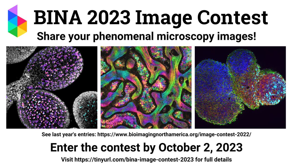
With #StayAtHome, we noticed there aren't too many puzzles related to microscopy. Let's change that. Send us your best/fav image you took with a microscope and we'll choose our favorite to make into a 1000 pc puzzle and send it to you! Here's how to enter. #echopuzzlecontest


![BioTranslucence's tweet photo. We're hosting a #tissueclearing Lunch & Learn with the Advanced Imaging Facility at @ucdavisvetmed!
Join us on 🗓️ Tue, Feb 18, 2025, from 11:30 am - 12:30 pm, to learn what tissue clearing is and how it's transforming biological research!
RSVP details below👇
[1/2] https://t.co/hvb9lPMkbt](https://pbs.twimg.com/media/Gh1zypSWoAEOXA5.jpg)