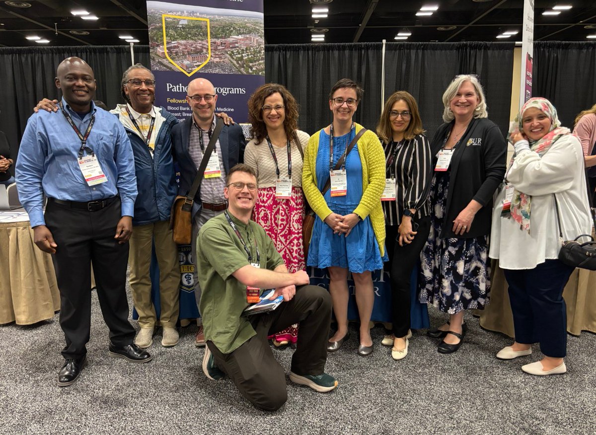
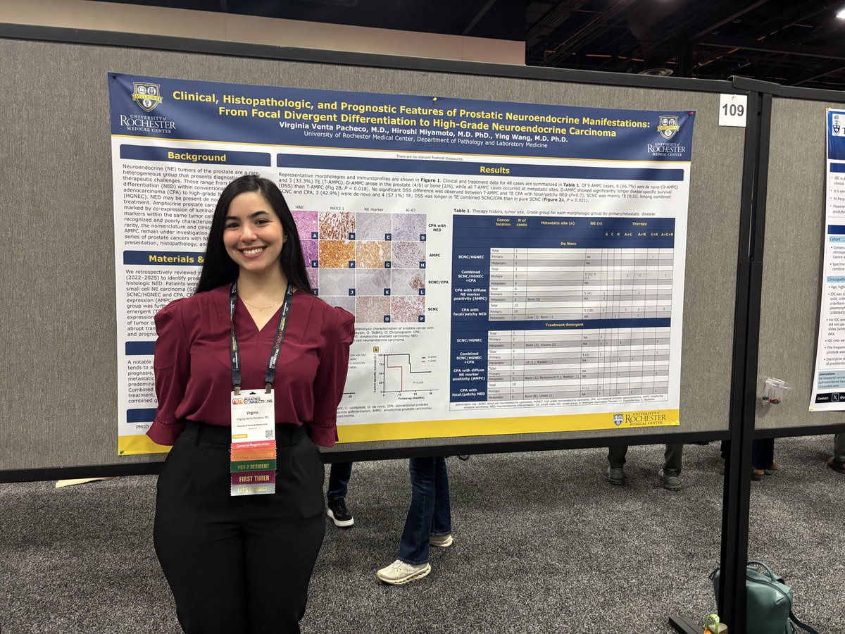
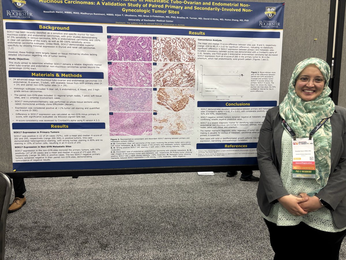
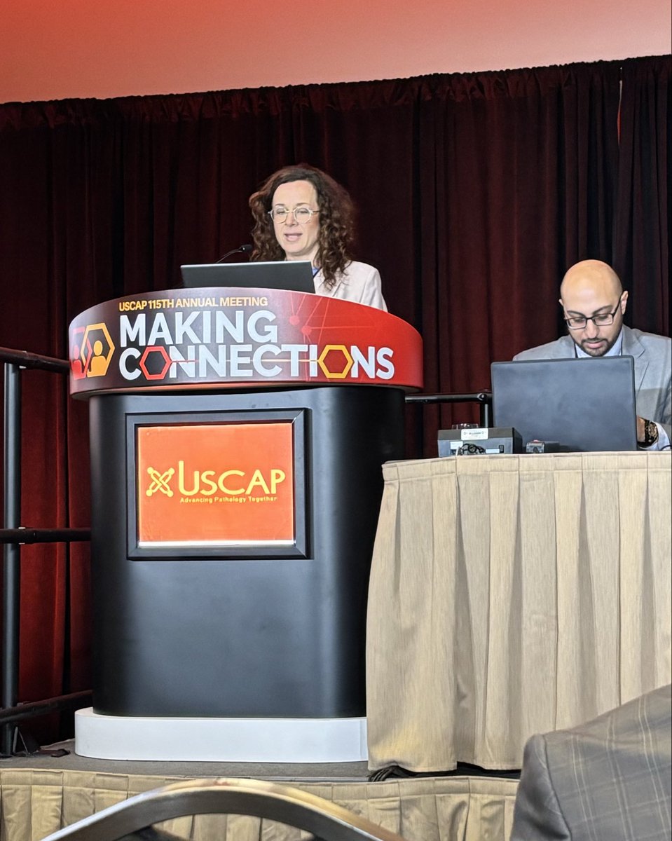
Don't miss Friday's "Clotting Disorders—Doing Without the 'Yellow,'" delivered by URochester Medicine's Majed Refaai, MD, director of Transfusion Medicine/Blood Bank. 8 to 10 a.m. EST. @bmssociety#bloodlessmedecineandsurgeryweek@URochester_SMD https://t.co/mOpCJJ6Tvn
Interesting variants of mucoepidermoid carcinoma. Part 1
- They can be ciliated
- They can have acinar differentiation (it is not acinar cell carcinoma)
- They can be spindled (usually keratin positive)
- They can be oncocytic (these were the old oncocytic carcinomas described in salivary glands which no longer exist)
Dr. Bishop #USCAP2026 #pathology #PathX #PathTwitter
Today was @PathologyUrmc Research Day, with presentations by our residents and graduate students. The keynote speaker was @smlungpathguy, who spoke on granulomatous diseases.
Seen here with @jacob_leonce and yours truly.
Today was @PathologyUrmc Research Day, with presentations by our residents and graduate students. The keynote speaker was @smlungpathguy, who spoke on granulomatous diseases.
Seen here with @jacob_leonce and yours truly.
Great start at URMC Pathology Residency Research Day! 🧬
Impressed by the residents’ innovative work and dedication to advancing patient care. Proud to be part of this community. 👩⚕️🔬
@Pathology_URMC
Great presentation on myxoid liposarcoma—an excellent final talk as a resident by our outstanding soon-to-be GI pathology fellow 👏 Dr. Olivia Sagan @Pathology_URMC
It's always an honor and a privilege during National Medical Laboratory Professionals Week to celebrate the extraordinary work of our staff, trainees, and faculty. Your dedication to accuracy, innovation, and people-centered care sets the standard for excellence. Meliora.
Our GME PDs & APDs along with our UME leadership are key to the success of our @NU_Pathology Education Programs. Thank you for your dedication and hard-work! Glad to celebrate you tonight.
Your diagnosis? An elderly male with a history of ulcerative colitis and prior right hemicolectomy was found to have an enhancing nodule in the posterior wall of the gastric antrum. EGD with endoscopic ultrasound demonstrated a mural gastric lesion. https://t.co/AFuVOkwUIH