If you didn't have a chance to go to the #ACR2023 in San Diego or didn't have time to stop by to see our work 👀, we made a video summary. We explored the attitudes, knowledge and practices of young Rheumatologists regarding the use of ultrasound in clinical practice. Thanks @ReumaJoven for your wonderful contribution! #FOAMed #Rheumatology #ultrasound #MSKUS @ColMexReuma@ACRheum@TecdeMonterrey
Double Contour sign #GOUT#Knee#cartilage
1️⃣Change insonation angle📐
2️⃣Go as parallel as you can: DC doesn’t disappear
3️⃣Remember: Look for DC sign moving together with subchondral bone.
#POCUS helps diagnose & implement early treatment.
OMERACT 2019 Definition👇
STENER MADE EASY
IP flexion (with fixed MCP) shows Add. Pollicis aponeurosis sliding over UCL when intact. It slides directly over bone when Stener has occurred.
Pimped up Clip from my participation at 2020 @RadiologiaChile Congress🇨🇱
#msk#ultrasound
The double contour (DC) sign in gout and CPPD has different dynamic behaviour. In gout the superficial margin moves together with the subchondral bone (DC sign), whereas in CPPD it moves in the opposite direction (pseudo DC sign).
FREE ARTICLE:
https://t.co/WMy2zvEWDq
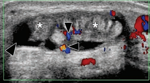
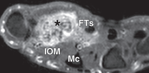
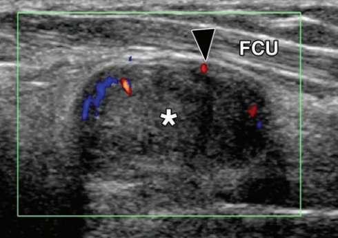
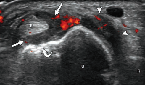
US in Rheumatology: The joint alterations in adult-onset Still's disease are comparable to those of rheumatoid arthritis, with synovial proliferation, power Doppler signal, and formation of bone erosions. @pompermarioMD@USMERoficial@MGH_MSUS@MSKUS_PT_KSA@drmariochavez
Ultrasound quiz: when examining this AC joint, you see an hyperechoic line in the deep, with an anechoic region above it. What is this?
The answer will follow tomorrow