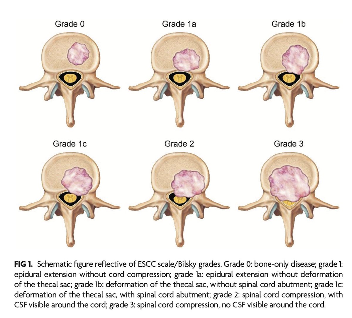
"Spinal Metastasis Reporting: Evidence-Based Recommendation on Behalf of the American Society of Spine Radiology Education and Standards Committee"
https://t.co/Ei4zhI4b5S
Anatomy Pearl 2: Simplify your posterior skull base evaluation by identifying the "Jugular Foramen" & "Hypoglossal Canal" at the CORONAL LEVEL OF the easily identifiable odontoid process (DENS) of C2 vertebra.
Look for the "Double-Eagle" on the "Mountain" top.
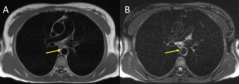
32 year old white woman presenting with a cold arm. Non-contrast MR imaging of the chest with axial T1 darkblood (A) and axial T2 darkblood fat-suppressed sequences. Note subtle cuff of aortic wall thickening on T1 sequence (yellow arrow) with associated T2 hyperintensity. Finding raises concern for large-vessel vasculitis (Takayasu or Giant cell) or possibly medium-vessel (i.e. Kawasaki) or variable-vessel vasculitis (Bechet's) that also affects the aorta. Takayasu affects women in 80-90 percent of cases, with onset between 10-40 years. Giant-cell affects the aorta and branches but almost never occurs before 50 years old. Kawasaki disease rarely occurs in adults.
From "Imaging Review of Vasculitis"
https://t.co/aTe6NG94vg
🦠 Hantavirus Awareness
Hantavirus is a rare but potentially life threatening viral infection spread mainly through contact with infected rodents, especially their urine, saliva, or droppings. People can become infected after breathing in contaminated airborne particles.
Mortality rate can be as high as 40%.
⚠️ Early symptoms may feel like the flu: • Fever
• Chills
• Muscle aches
• Headache
• Nausea or vomiting
• Stomach pain
As the illness progresses, severe breathing difficulty and lung involvement can occur, known as Hantavirus Pulmonary Syndrome (HPS), which carries a significant mortality risk.
🚨 Seek urgent medical attention if flu-like symptoms develop after rodent exposure, especially if breathing problems begin.
🛡️ Prevention Tips:
• Keep homes and workspaces clean
• Seal holes where rodents can enter
• Store food securely
• Avoid dry sweeping rodent droppings
• Spray contaminated areas with disinfectant before cleaning
❌ There is currently no specific licensed antiviral treatment or vaccine for hantavirus. Early recognition and supportive medical care can save lives.
IVERMECTIN DOESN'T TREAT HANTAVIRUS.
Public health awareness and accurate medical information matter. Stay informed, not alarmed.
#Hantavirüs I #MedTwitter
🟢. Causes of Short 4th/5th Metacarpals (Knuckle Dimple Sign) & How to Differentiate
1. Pseudohypoparathyroidism (PHP)
• Genetic resistance to PTH.
• Features:
• Short 4th/5th metacarpals (knuckle dimple sign)
• Round face, short stature, obesity
• Subcutaneous calcifications, developmental delay
• Lab: Hypocalcemia, Hyperphosphatemia, ↑ PTH
2. Pseudo-pseudohypoparathyroidism (PPHP)
• Same phenotype as PHP (Albright Hereditary Osteodystrophy) but normal labs.
• Key differentiation: Normal calcium, phosphate, and PTH.
3. Turner Syndrome (45,XO)
• Girls with short 4th metacarpal.
• Other features: Short stature, webbed neck, shield chest, gonadal dysgenesis, streak ovaries, infertility.
• Labs: Low estrogen, ↑ FSH/LH.
4. McCune–Albright Syndrome
• Triad:
1. Café-au-lait spots (irregular borders)
2. Polyostotic fibrous dysplasia (bone lesions, deformities)
3. Precocious puberty (esp. in girls)
• May also have endocrine overactivity (thyroid, pituitary, adrenal).
• Short metacarpals may be seen due to bony dysplasia.
5. Kallmann Syndrome
• Hypogonadotropic hypogonadism with anosmia.
• Main signs: Delayed puberty, infertility, small testes, absent secondary sexual characteristics.
• No typical metacarpal shortening → if present, it’s coincidental, not a hallmark feature.
6. Sickle Cell Anemia
• Repeated bone infarctions can cause skeletal deformities and shortened metacarpals/metatarsals.
• Other features: Hemolytic anemia, dactylitis in children, avascular necrosis, crises.
7. Osteomyelitis (chronic, childhood)
• Can damage growth plate and cause localized shortening/deformity of affected bones.
• History of infection, pain, swelling, systemic features differentiate it.
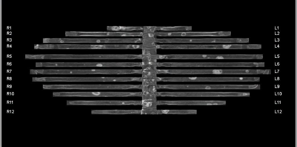
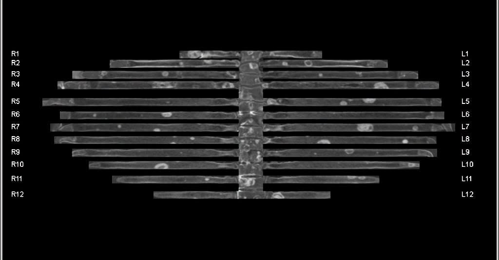
State-of-the-art Diagnosis and Treatment of Metastatic Melanoma: MDCT Protocol Optimization to Identify Metastases in Unusual Locations and Review of Innovative Therapeutic Agents
https://t.co/lB2CiJM6RN
1/Do radiologists sound like they are speaking a different language when they talk about MRI?
T1 shortening what? T2 prolongation who?
Here’s a translation w/an introductory thread to MRI.
1/Does your ability to remember temporal lobe anatomy seem, well, temporary?
Or are you feeling temporally challenged when it comes to this complex region?
Here’s a thread to help you remember the structures of the temporal lobe!
Uterine Artery (UtA) Doppler assessment procedure at 11-14 week
A 3.5–5‑MHz curvilinear TAUS transducer is utilized for this examination.
This procedure is performed preferably with the maternal bladder empty.
The probe is pressed at the level of the iliac fossa, either left or right, to obtain a midsagittal section of the uterus. At this stage, the cervical canal is identified to assist in further imaging.
Once the cervical canal is visualized, the probe is tilted laterally while maintaining medial angulation. This maneuver continues until the paracervical vascular plexus becomes visible.
Color Doppler is activated to identify the UtA as it turns cranially, ascending toward the uterine body.
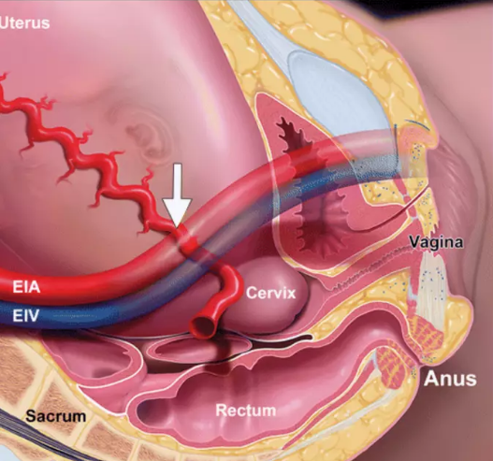
During pregnancy, the UtA and EIA may appear to cross paths, a phenomenon caused by increased uterine growth and the resulting lateral shift of both arteries.
After the UtA is identified, PW Doppler is applied with the sampling gate set to 2 mm to encompass the entire vessel. The angle of insonation is adjusted to less than 30º for accuracy.
PSV should be greater than 60 cm/s to confirm that the uterine artery, rather than the arcuate artery, is being assessed.
Measurements are taken at the point just before the uterine artery branches into the arcuate arteries.
The same process is repeated on the opposite side to ensure a thorough evaluation of both uterine arteries.
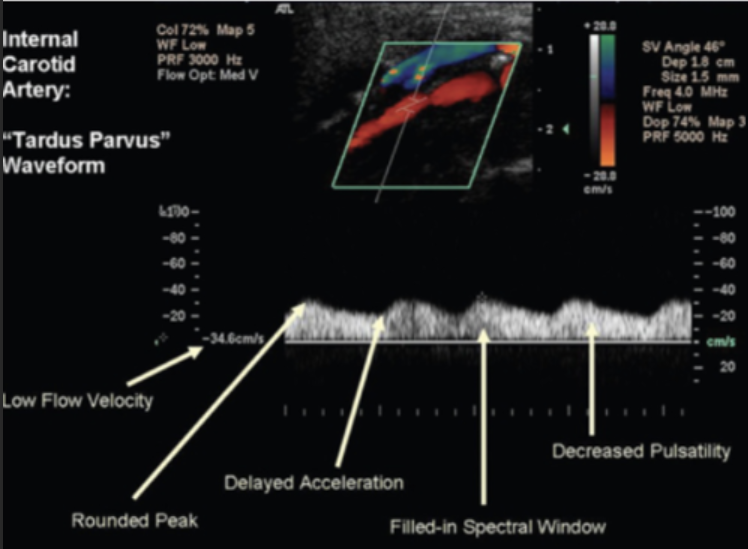
Prolonged upstroke vs Dampened waveform!
Prolonged upstroke (Delayed systolic acceleration/increased acceleration time)
(The tardus part of tardus‑parvus).
Definition.
Slow, delayed rise to systolic peak.
The systolic “upstroke” is not sharp.
Waveform appearance
Sluggish systolic rise.
Rounded peak.
Increased acceleration time (> 140 ms)
PSV may be normal or low.
Commonest causes
Proximal arterial stenosis.
2. Dampened waveform (low pulsatility).
(The parvus part of the of tardus‑parvus).
Definition.
Low‑energy, blunted Doppler.
Reduced amplitude and pulsatility
Waveform appearance
Low PSV
Blunted, rounded systolic peak
Narrow systolic envelope
Reduced pulsatility index
Commonest cause is proximal stenosis
Prolonged upstroke + Dampened waveform = The classic tardus‑parvus pattern.
Turdus-Parvus is characterized by a delayed and reduced pulse upstroke, resulting in a pulse that is both late (tardus) and small (parvus)!
The Tardus Parvus 👇👇