St. Francis Health Care Services located in Njeru Town Council Buikwe District brings free Pelvic Organ Prolapse Surgery for women with vault, rectal, uterus or bladder protruding through the vagina. The Free Service ends in June. Please reach out and get sorted. Thank you
During a routine C-section, doctors witnessed something incredibly rare.
The baby emerged still enclosed inside the amniotic sac, this moment is so uncommon that many medical professionals never see it in their careers.
Diagnosis: Kartagener Syndrome
(A clinical triad within the spectrum of Primary Ciliary Dyskinesia)
1. Clinical Presentation
The case presents a classic diagnostic triad that aligns with the patient’s history of recurrent infections:
Situs Inversus Totalis: Complete transposition of thoracic and abdominal viscera.
Chronic Sinusitis: Resulting from impaired mucociliary clearance in the paranasal sinuses.
Bronchiectasis: Permanent dilation of the bronchi due to chronic, recurrent chest infections.
2. Radiographic Analysis (CXR)
The provided Chest X-ray exhibits several pathognomonic features:
Dextrocardia: The cardiac silhouette is oriented toward the right hemithorax (the apex points to the right).
Gastric Bubble: Visible under the right hemidiaphragm instead of the left.
Liver Shadow: Positioned on the left side of the abdomen.
Bronchiectatic Changes: Increased bronchovascular markings and "tram-track" opacities, particularly in the lower zones, indicating chronic airway remodeling.
3. Pathophysiology
The condition is an autosomal recessive disorder characterized by a structural defect in the dynein arms of the cilia.
In Utero: Normal ciliary beating is required for the proper rotation of internal organs during embryonic development. Lack of movement results in a 50% chance of random organ orientation (Situs Inversus).
Post-birth: Non-functional cilia cannot clear inhaled pathogens or mucus from the respiratory tract, leading to chronic colonization, inflammation, and eventual lung tissue damage.
4. Diagnostic Confirmatory Tests
Beyond the radiological evidence, the following are used to confirm the diagnosis:
Nasal Nitric Oxide (nNO): Typically shows extremely low levels in these patients.
Ciliary Function Analysis: High-speed video microscopy to observe the ciliary beat frequency and pattern.
Genetic Testing: Identifying mutations in genes such as DNAH5 or DNAI1.
Semen Analysis: In males, this often reveals infertility due to immotile spermatozoa (which share the same structural microtubule defects
St. Francis Health Care Services located in Njeru Town Council Buikwe District brings free Pelvic Organ Prolapse Surgery for women with vault, rectal, uterus or bladder protruding through the vagina. The Free Service ends in June. Please reach out and get sorted. Thank you
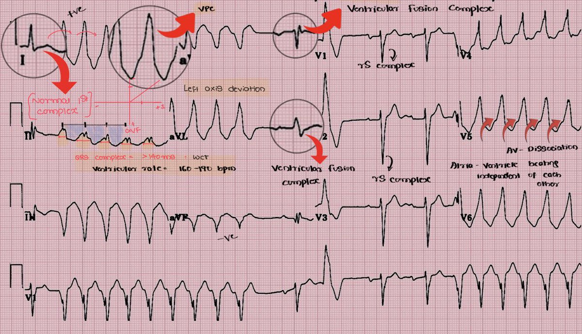
🚨 Breakdown of this ECG
1️⃣ Lead I
The first complex in Lead I is a normal narrow QRS, indicating an initial sinus conducted beat.
Immediately afterward, wide complexes begin, marking the onset of AV dissociation with ventricular activity.
2️⃣ AV Dissociation in Lead I
During the tachycardia, atria and ventricles appear to depolarize independently.
At the end of Lead I, a normal narrow QRS complex reappears, representing a sinus capture beat.
Capture beats are strong diagnostic evidence of ventricular tachycardia.
3️⃣ Lead V1 findings
In Lead V1, the sequence of ventricular premature complexes (VPCs) abruptly terminates.
This is followed by rS complexes, which reflect normal ventricular activation via the His-Purkinje system.
4️⃣ Fusion Complex in V1
Following the rS complexes, a ventricular fusion beat is seen.
Fusion complexes occur when simultaneous sinus and ventricular impulses depolarize the ventricles, producing a hybrid QRS morphology.
This finding further supports ventricular tachycardia.
5️⃣ Run of Ventricular Complexes
Multiple wide complex ventricular beats occur consecutively, forming a run of ventricular tachycardia.
The complexes are broad and uniform, consistent with monomorphic ventricular activity.
6️⃣ Abrupt Sinus Capture
After the ventricular run, there is sudden sinus capture, demonstrated by the appearance of normal narrow QRS complexes.
This indicates temporary restoration of normal conduction through the AV node and His-Purkinje system.
7️⃣ Key Diagnostic Features Observed
✔ Initial sinus beat
✔ Sudden onset wide complex tachycardia
✔ AV dissociation
✔ Sinus capture beats
✔ Ventricular fusion complexes
✔ Abrrupt termination with normal QRS complexes
8️⃣ Conclusion
A sequence of ≥3 consecutive ventricular beats with a rate >100 bpm that terminates spontaneously (<30 seconds) fulfills the definition of:
➡️ Non-Sustained Ventricular Tachycardia (NSVT)
The presence of AV dissociation, capture beats, and fusion complexes makes this a classic ECG demonstration of NSVT.
#MedTwitter #MedX #ECG
Dr Joe Njagi Turns Theatre into Fun: Challenges Patient to Singing Contest as She Drifts into Anesthesia. Makofi kwa Daktari. He makes this ease the tension.
We are now counting hours to the biggest surgical camp in the country. We cannot wait to be part and contribute to our best abilities.
Members of MUSS will be present thanks to @ASOU_Official leadership for their support and inclusion.
#globalsurgery#CommunityHealth