@fringeblog I’d say so. The inspiration is flowing like a firehouse. I have a small natural grooming company called Three Vikings and a Goat. One day screenwriter brain sat down and asked if I’d ever thought…and the kid’s show was born. The pilot script is almost done.
🧠 COVID-19 is not just a respiratory illness—it is a vascular and neurological threat that compounds with every single infection.
Here is what researchers are seeing as infections add up:
🔴 1st Infection (Mild to Moderate Damage): Acute neural strain, leading to the onset of "brain fog," fatigue, headaches, and a temporary loss of smell or taste.
🔴 3rd Infection (Multiplied Strain): The biological risk multiplies. Systemic and vascular damage compounds, resulting in severe cognitive struggles, persistent "brain fog," and autonomic dysfunction (dysautonomia).
🔴 5th Infection (Severe Chronic Damage): Deep, long-term neurological injury. Studies show severe memory impairment, chronic fatigue, a vastly increased risk of micro-clots and strokes, and cognitive decline resembling up to 20 years of accelerated brain aging.
Every infection rolls the dice again. Protect your brain and take steps to avoid reinfection. 😷👇
#LongCovid #COVID19 #BrainHealth #Neurology #COVIDIsAirborne #NeurologicalDamage
🔬🧬 THE DEEP DIVE: What is "Spikeopathy" and How Does Covid's Spike Protein Impact the Body? 🔬🧬
Have you heard the term “spikeopathy” lately?
As researchers continue to study the long-term impacts of COVID-19 and its viral components, a growing body of scientific evidence has been looking closely at the spike protein—the part of the SARS-CoV-2 virus that allows it to bind to human cells—and its independent effects on our health.
Here is what the latest 2025–2026 medical reviews and studies say about "spikeopathy" and how it's shaping our understanding of Covid's post-viral complications:
🔍 1. What is "Spikeopathy"?
"Spikeopathy" refers to the study of the pathogenic (damage-causing) effects of the SARS-CoV-2 spike protein itself, independent of the active replication of the whole virus.
Researchers are investigating how these loose, circulating, or persistent spike proteins interact with human tissue over time, particularly in those with Long COVID (PASC).
🧬 2. The Mechanisms of Impact
Recent studies have highlighted a few primary pathways through which the spike protein might affect the body:
• Cellular Binding & Barrier Penetration:
The spike protein targets ACE2 receptors found on various human cells, which can trigger cellular dysfunction.
Notably, peer-reviewed research has shown that free-floating spike proteins can cross the blood-brain barrier, offering clues into neurological symptoms like "brain fog" and fatigue.
• Chronic Inflammation:
A 2026 UCLA-led study revealed that the immune system can break down spike proteins into "fragment" pieces.
These fragments can inadvertently mimic human signaling molecules, target cells with specific "spiky" shapes, and drive prolonged inflammatory responses.
• Vascular and Microvascular Effects:
The spike protein can interact directly with endothelial cells (which line our blood vessels) and platelets.
This can potentially contribute to micro-clotting, vascular inflammation, and "microvascular" issues that are frequently reported in post-viral conditions.
• Persistent Reservoirs:
One of the most discussed theories behind Long COVID is "viral persistence."
This is the idea that fragment pieces of the virus—specifically the highly durable spike protein—can linger in tissue reservoirs long after the acute infection has passed, keeping the immune system on high alert.
🏥 The Path Forward
Understanding spikeopathy is helping researchers transition from simply treating acute infections to developing targeted therapies for chronic inflammation, autoimmune-like responses, and microvascular issues.
Recent trials continue to evaluate therapeutic approaches that help the body clear persistent proteins and restore immune balance.
Knowledge is power!
Staying informed about the biology of post-viral conditions is the first step toward better treatments and recovery. 💪👇
Check out the infographic below to see how these spike proteins interact with the body's systems!
#CovidResearch #LongCovid #Spikeopathy #ScienceUpdate #MedicalResearch #HealthEducation
@insomnxcguy Brought to you by the same queerbaiting writers who have jerked our chains for 9 out of 10 seasons. Brought to you by the obsequious gay bootlickers who kowtow to the mad king who governs our country.
6 entire posts on Covid 19s effects on the brain according to the most recent information available and no response?
So much for my career as a content creator.
🧠 HOW COVID-19 AFFECTS YOUR BRAIN (1/6): THE FRONTAL LOBE
The Frontal Lobe (right behind your forehead) is your brain’s project manager.
It controls focus, planning, and decision-making, while its lower section—the orbitofrontal cortex—connects directly to the smell nerves in your nose.
Recent brain scans show how COVID-19 impacts this region:
• Shrinks smell-connected tissue after 1 infection: Before-and-after MRI scans taken ~4.5 months after a single infection (96% mild) showed an extra 0.2% to 2% loss of grey matter thickness in the orbitofrontal cortex compared to uninfected peers (PubMed).
• Stabilizes in size after 1 year while rewiring: Tracking patients 12 to 23 months after a single infection shows early right frontal lobe shrinkage (~1.35 mL) stops progressing after year one (DZNE 2025 Study), while frontal nerve networks actively rewire over 2 years to compensate (RWTH Aachen & Charité Berlin 2025 Study).
• Lowers planning scores after 1 infection: Testing over 112,000 adults linked a single resolved mild infection to an average reasoning and planning drop equal to 3 IQ points—rising to 6 IQ points with lingering symptoms and 9 IQ points after ICU care (CIDRAP NEJM Summary).
• Compounds cognitive loss with multiple infections: Catching COVID-19 a second or third time adds nearly 2 extra IQ-equivalent points of cognitive loss (CIDRAP NEJM Summary) and makes long-term brain disorders 1.6x (60%) more likely than after a single infection (WashU Medicine).
📚 Citations & Links:
• Oxford UK Biobank Study (Nature): https://t.co/WRzGN6YZTs
• DZNE 12-Month AI MRI Study (Diagnostics, 2025): https://t.co/a0aEUauPB6
• Aachen & Berlin 2-Year MRI Study (2025): https://t.co/sH6QqmrKni
• Imperial College NEJM Study Summary (CIDRAP): https://t.co/4NrnwFk3wZ
• WashU Medicine Reinfection Study (Nature Medicine): https://t.co/08YUp2jo0C
#COVID19 #BrainHealth #FrontalLobe #BrainFog #LongCOVID #Neuroscience #ReinfectionRisk
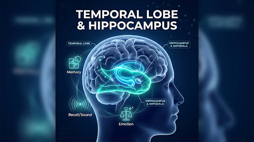
🧠 HOW COVID-19 AFFECTS YOUR BRAIN (2/6): THE TEMPORAL LOBE & HIPPOCAMPUS
Tucked behind your temples, the Temporal Lobe and seahorse-shaped Hippocampus act as your brain’s filing cabinet.
They record new memories, help you find words, and work with the amygdala to regulate emotions and anxiety.
Long-term MRI tracking shows why memory is hit so hard by COVID-19:
• Thins memory bridges after 1 mild infection: UK Biobank scans found up to 2% extra grey matter loss in the parahippocampal gyrus—the bridge feeding sensory memories into the hippocampus—within 4.5 months of a single mild case (University of Oxford).
• Continues shrinking progressively at 12 months: While other brain areas stabilize, a December 2025 AI-MRI study found delayed, ongoing shrinkage in the right hippocampus a full year after a single infection—dropping 3.0% to 5.6% after mild cases and 7.6% to 8.6% after severe cases (DZNE 2025 PDF).
• Triggers 2-to-3-year memory and work struggles: Testing 2 to 3 years after a severe first infection showed an average 10-IQ-point-equivalent attention and memory deficit, with 1 in 4 survivors reporting worsening memory and 1 in 4 changing jobs due to cognitive demands (University of Oxford PHOSP-COVID Study).
• Worsens memory encoding with multiple infections: COVID-19 specifically impairs how the brain records new memories (Riot IQ NEJM Breakdown), and repeat infections raise neurological and memory disorder risk by 1.6x compared to a single infection (WashU Medicine) while adding nearly 2 extra IQ-equivalent points of decline (CIDRAP NEJM Summary).
📚 Citations & Links:
• DZNE 12-Month AI MRI Study (Diagnostics, Dec 2025): https://t.co/WosTlFI48d
• Oxford 3-Year PHOSP-COVID Study (Lancet Psychiatry): https://t.co/xHx3dXGUrr
• Texas A&M Grey Matter Breakdown: https://t.co/kLGSOyoKYb
• NEJM Memory & IQ Analysis (Riot IQ): https://t.co/FWbR64Wl8k
• WashU Medicine Reinfection Study (Nature Medicine): https://t.co/vnmz4W9z2S
#COVID19 #Hippocampus #MemoryLoss #TemporalLobe #LongCOVID #BrainHealth #Neurology
🧠 HOW COVID-19 AFFECTS YOUR BRAIN (PART 3): THE PARIETAL LOBE
Sitting near the upper crown of your head, the Parietal Lobe (paired with the deep relay station called the thalamus) is your brain’s sensory and navigation hub. It processes physical touch and pain, tells you where your body is in space, and helps you juggle divided attention when two things happen at once.
Recent two-year MRI tracking shows how the parietal lobe responds and adapts after COVID-19:
Reduces sensory-strip volume early after a single infection: Patients scanned after a single COVID-19 infection showed lower volume in the postcentral gyrus—the parietal lobe's main sensory strip—alongside early volume drops across the parietal cortex and right thalamus (RWTH Aachen & Charité Berlin 2025 Study; DZNE 2025 Study).
Compensates between left and right sides over two years: Over a 23-month follow-up after a single infection, the left postcentral sensory strip continued to shrink slightly while the right postcentral strip actually regrew volume to compensate—yet between one-third and nearly half of post-COVID patients still struggled with divided attention and alertness at the two-year mark (RWTH Aachen & Charité Berlin 2025 Study).
Stabilizes overall lobe size after 12 months: One-year AI volumetric scans confirm that after the initial hit from a single infection, overall parietal lobe volume changes level off to match normal aging rates (DZNE 2025 PDF).
Escalates sensory and neurological risks with multiple infections: Each repeat COVID-19 infection stacks new neurological risk on top of the first—making patients with two or more infections 1.6 times more likely to suffer brain, sensory, and attention disorders than those infected only once (WashU Medicine), and compounding attention and reasoning drops by nearly 2 extra IQ-equivalent points (CIDRAP NEJM Summary).
📚 Citations & Links:
RWTH Aachen & Charité Berlin 2-Year MRI Study (Psychiatry & Clinical Neurosciences, 2025): Two-year impact of COVID-19: Longitudinal MRI brain changes and neuropsychiatric trajectories
University Hospital Bonn & DZNE Study (Diagnostics, Dec 2025): Longitudinal Monitoring of Brain Volume Changes After COVID-19 Infection
Washington University School of Medicine Reinfection Study (Nature Medicine): Repeat COVID-19 infections increase risk of organ failure, death
Imperial College London REACT Study Summary (CIDRAP / NEJM, 2024): Even fully recovered survivors of mild COVID can lose IQ points
#COVID19 #ParietalLobe #BrainFog #AttentionDeficit #LongCOVID #Neuroimaging #BrainHealth #PublicHealth
🧠 HOW COVID-19 AFFECTS YOUR BRAIN (4/6): THE OCCIPITAL LOBE
At the very back of your head, the Occipital Lobe is your brain’s visual processing center.
It turns signals from your eyes into reading, depth perception, face recognition, and mental 3D shape rotation.
Recent imaging shows how COVID-19 affects vision and visual attention:
• Spares physical tissue size after 1 infection: Twelve-month AI MRI tracking shows that unlike the memory center, the occipital lobe’s physical grey matter volume is largely spared from shrinkage both early on and 1 year after a single infection (DZNE 2025 PDF).
• Alters visual network wiring over 2 years: Even without tissue shrinkage, functional MRI scans taken 7 to 23 months after a single infection show disrupted signaling between the occipital lobe and other brain hubs, directly tracking with slower divided visual attention (RWTH Aachen & Charité Berlin 2025 Study).
• Hinders 3D mental rotation after severe single cases: In the 112,964-adult NEJM cognitive study, survivors of severe single infections showed a sharp drop in visuospatial performance—specifically when mentally rotating 3D shapes (Riot IQ NEJM Breakdown).
• Compounds visual-cognitive drops with multiple infections: Repeat infections add nearly 2 extra IQ-equivalent points of global cognitive loss across visual, memory, and reasoning tasks (CIDRAP NEJM Summary) and increase long-term neurological disorder risk by 60% compared to one infection (WashU Medicine).
📚 Citations & Links:
• DZNE 12-Month AI MRI Study (Diagnostics, Dec 2025): https://t.co/WosTlFI48d
• Aachen & Berlin 2-Year MRI Study (2025): https://t.co/EH8Cif4k6o
• NEJM Visuospatial & IQ Analysis (Riot IQ): https://t.co/FWbR64Wl8k
• Imperial College NEJM Study Summary (CIDRAP): https://t.co/4NrnwFk3wZ
• WashU Medicine Reinfection Study (Nature Medicine): https://t.co/08YUp2jo0C
#COVID19 #OccipitalLobe #VisualProcessing #BrainConnectivity #LongCOVID #BrainScience #Neurology
🧠 HOW COVID-19 AFFECTS YOUR BRAIN (5/6): THE CEREBELLUM
Tucked underneath the back of your brain, the Cerebellum ("little brain") controls physical balance, smooth coordination, and how fast your brain processes information.
Brain-imaging studies show how COVID-19 slows down this control center:
• Shrinks thinking-speed zones after 1 mild infection: Before-and-after UK Biobank MRI scans found that even a single mild infection shrinks cognitive zones of the cerebellum within 4.5 months, directly correlating with a 12.2% slower time on complex mental trail-making tasks (University of Oxford; Riot IQ NEJM Breakdown).
• Doubles grey matter loss over 12 months: One-year AI MRI tracking found cerebellar grey matter shrank by 1.64% after mild/asymptomatic COVID-19 and 1.70% after severe COVID-19—nearly double the 0.87% normal aging change in uninfected controls (DZNE 2025 PDF).
• Weakens network links up to 2 years later: Follow-up scans 11 to 23 months after a single infection reveal reduced connectivity between the cerebellum and memory hubs, plus altered cerebellar energy use even in symptom-free survivors (RWTH Aachen & Charité Berlin 2025 Study).
• Further slows processing with multiple infections: While one mild infection costs an average of 3 IQ-equivalent points on timed cognitive tasks, reinfection adds nearly 2 more points of loss (CIDRAP NEJM Summary) and increases long-term neurological risk by 1.6x compared to a single infection (WashU Medicine).
📚 Citations & Links:
• Oxford UK Biobank Study (Nature): https://t.co/WRzGN6YZTs
• Texas A&M Cerebellum & Aging Breakdown: https://t.co/kLGSOyoKYb
• DZNE 12-Month AI MRI Study (Diagnostics, Dec 2025): https://t.co/WosTlFI48d
• Aachen & Berlin 2-Year MRI Study (2025): https://t.co/EH8Cif4k6o
• WashU Medicine Reinfection Study (Nature Medicine): https://t.co/08YUp2jo0C
#COVID19 #Cerebellum #ProcessingSpeed #BrainHealth #LongCOVID #Neuroscience #MedicalResearch
🧠 HOW COVID-19 AFFECTS YOUR BRAIN (6/6): THE BRAINSTEM
Connecting your brain to your spinal cord, the Brainstem (midbrain, pons, and medulla) is your body’s automatic life-support switchboard.
It runs breathing, heart rate, blood pressure, sleep cycles, and energy levels without you thinking about it.
Ultra-powerful 7-Tesla (7T) MRI scanners at Cambridge and Oxford revealed what standard hospital MRIs couldn't see:
• Leaves microscopic inflammation months after 1 infection: Ultra-high-resolution 7T MRI scans of hospitalized patients more than 6 months after a single infection found lingering immune inflammation in the medulla, pons, and midbrain—right in the nerve clusters controlling breathing, fatigue, and anxiety (University of Cambridge 7T MRI Study; NIHR Oxford BRC).
• Elevates inflammation chemicals tied to worse mental health: A November 2025 7T brain-chemistry study found that survivors with higher brainstem levels of myo-inositol (an inflammation marker) reported significantly poorer mental wellbeing after recovery (University of Cambridge Clinical Neurosciences, Nov 2025).
• Drives fatigue and sleep issues over 2 years: Two-year MRI tracking links brainstem volume changes to severe post-COVID fatigue at 7 months (r = 0.32) and poor sleep quality at 23 months (r = 0.55), with 45% of long-COVID cohort patients still severely fatigued nearly 2 years after 1 infection (RWTH Aachen & Charité Berlin 2025 Study).
• Multiplies organ and autonomic risk with repeat infections: Because the brainstem regulates vital organs, repeat infections hit hard—making people 1.6x more likely to develop brain disorders, 3x more likely to suffer heart conditions, and 3.5x more likely to develop lung problems than after a single infection (WashU Medicine).
📚 Citations & Links:
• Cambridge & Oxford 7T MRI Study (Brain, 2024): https://t.co/c5kvg5pnT2
• Cambridge 7T Brainstem Chemistry Study (Nov 2025): https://t.co/wGUgXYkbZi
• NIHR Oxford BRC Summary: https://t.co/11cC9D6JVO
• Aachen & Berlin 2-Year MRI Study (2025): https://t.co/EH8Cif4k6o
• WashU Medicine Reinfection Study (Nature Medicine): https://t.co/08YUp2jo0C
#COVID19 #Brainstem #LongCOVID #ChronicFatigue #7TMRI #BrainInflammation #AutonomicNervousSystem
If SARS-CoV-2 was really damaging people’s frontal lobes, you’d be noticing people being more brazenly rude, hostile, bigoted, antisocial, and tribalistic. Both online & irl.
You’d notice people would be quicker to lose their temper, and unable to keep a lid on their emotions.
🧠 HOW COVID-19 AFFECTS YOUR BRAIN (5/6): THE CEREBELLUM
Tucked underneath the back of your brain, the Cerebellum ("little brain") controls physical balance, smooth coordination, and how fast your brain processes information.
Brain-imaging studies show how COVID-19 slows down this control center:
• Shrinks thinking-speed zones after 1 mild infection: Before-and-after UK Biobank MRI scans found that even a single mild infection shrinks cognitive zones of the cerebellum within 4.5 months, directly correlating with a 12.2% slower time on complex mental trail-making tasks (University of Oxford; Riot IQ NEJM Breakdown).
• Doubles grey matter loss over 12 months: One-year AI MRI tracking found cerebellar grey matter shrank by 1.64% after mild/asymptomatic COVID-19 and 1.70% after severe COVID-19—nearly double the 0.87% normal aging change in uninfected controls (DZNE 2025 PDF).
• Weakens network links up to 2 years later: Follow-up scans 11 to 23 months after a single infection reveal reduced connectivity between the cerebellum and memory hubs, plus altered cerebellar energy use even in symptom-free survivors (RWTH Aachen & Charité Berlin 2025 Study).
• Further slows processing with multiple infections: While one mild infection costs an average of 3 IQ-equivalent points on timed cognitive tasks, reinfection adds nearly 2 more points of loss (CIDRAP NEJM Summary) and increases long-term neurological risk by 1.6x compared to a single infection (WashU Medicine).
📚 Citations & Links:
• Oxford UK Biobank Study (Nature): https://t.co/WRzGN6YZTs
• Texas A&M Cerebellum & Aging Breakdown: https://t.co/kLGSOyoKYb
• DZNE 12-Month AI MRI Study (Diagnostics, Dec 2025): https://t.co/WosTlFI48d
• Aachen & Berlin 2-Year MRI Study (2025): https://t.co/EH8Cif4k6o
• WashU Medicine Reinfection Study (Nature Medicine): https://t.co/08YUp2jo0C
#COVID19 #Cerebellum #ProcessingSpeed #BrainHealth #LongCOVID #Neuroscience #MedicalResearch