Great work by Nadeem and team to explore a problem nearly every advanced endoscopist has faced:
* patient presents with obstructive jaundice
* you place a biliary stent perfectly
* several days later, the bilirubin has barely budged
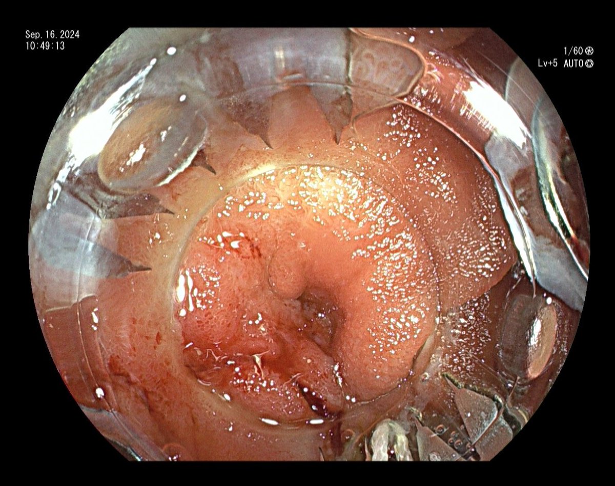
Precut Fistulotomy , Small papilla with lot of folds
Do a suction and make CBD prominent, Iron out the CBD and make a single clean cut over the impression
And Go into duct
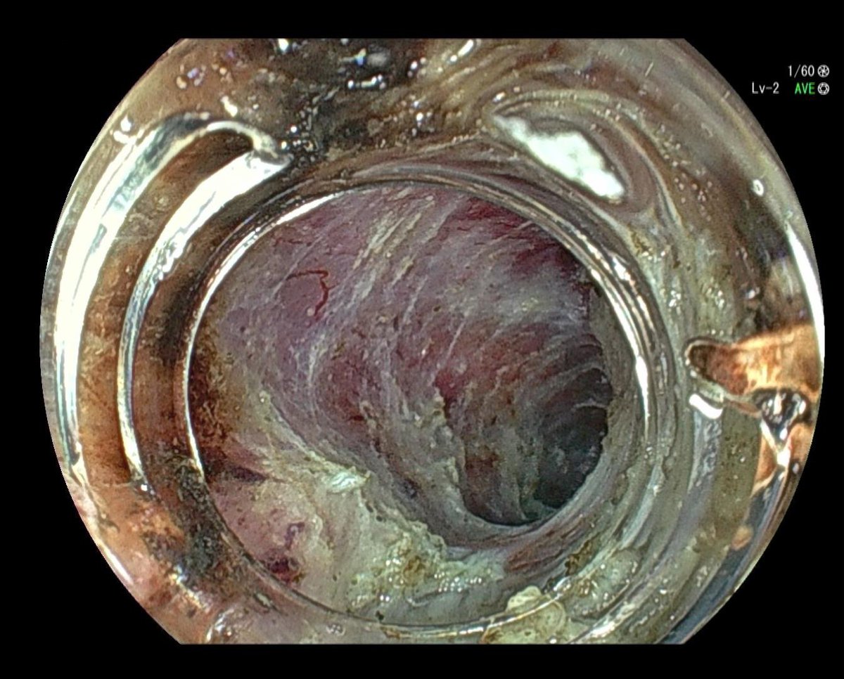
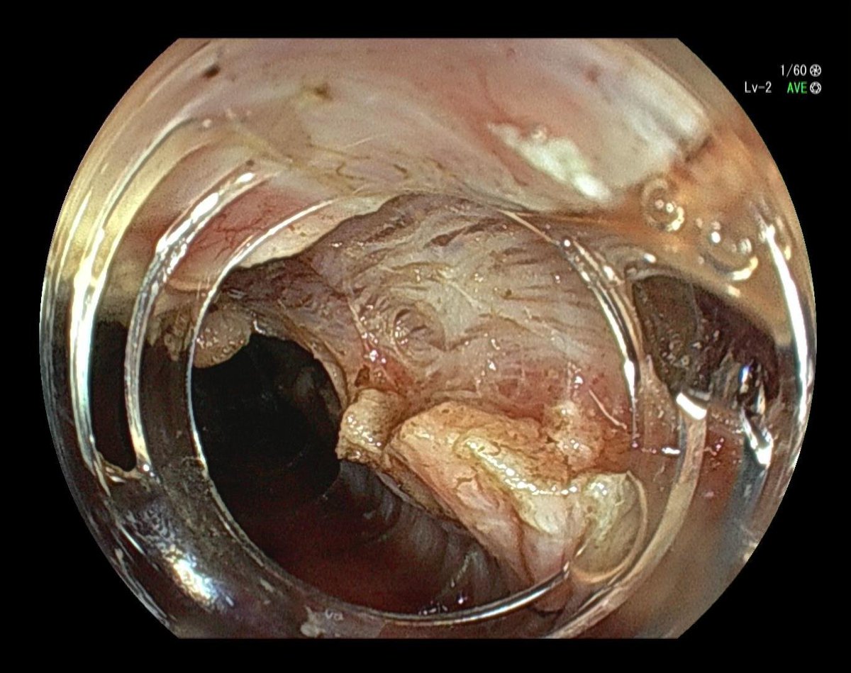
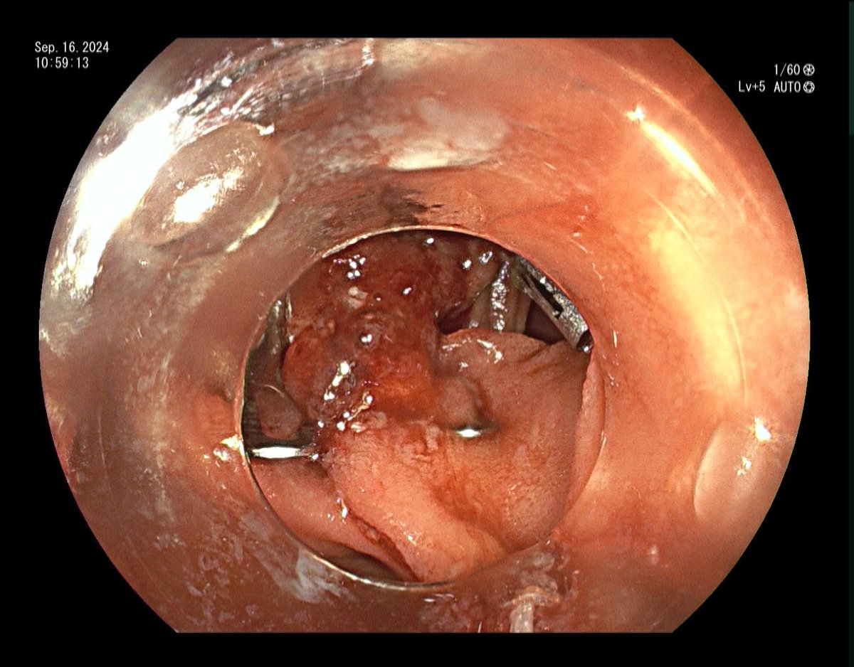
Pt with gastric adenocarcinoma s/p subtotal gastrectomy, developed J tube enterocutaneous fistula. Closed endoscopically with Ovesco OTSC. Can be hard to suction up the fistula into cap sometimes due to underlying fibrosis, what are your tricks?
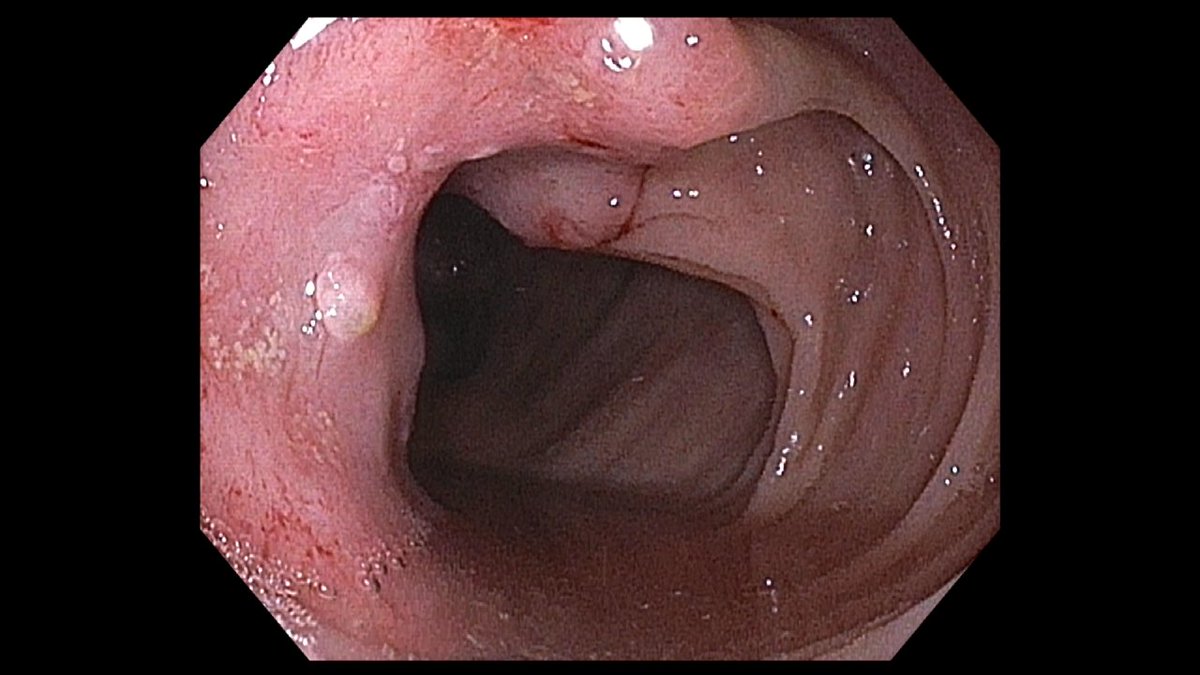
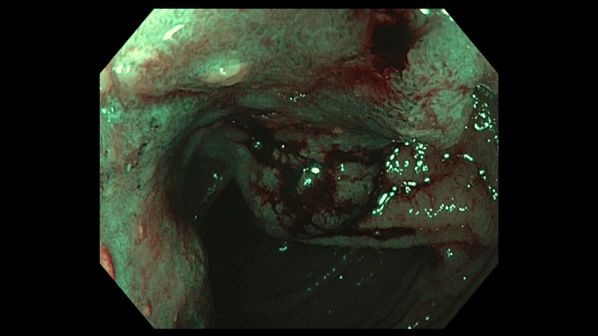
Colonoscopy performed for evaluation prior to possible colonic interpolation graft, showed sigmoid lesion with saddle feature and Kudo V pit pattern. Decision made to forego resection and biopsy. Path returned positive for mucosal mets of primary gastric cancer. (2/3)
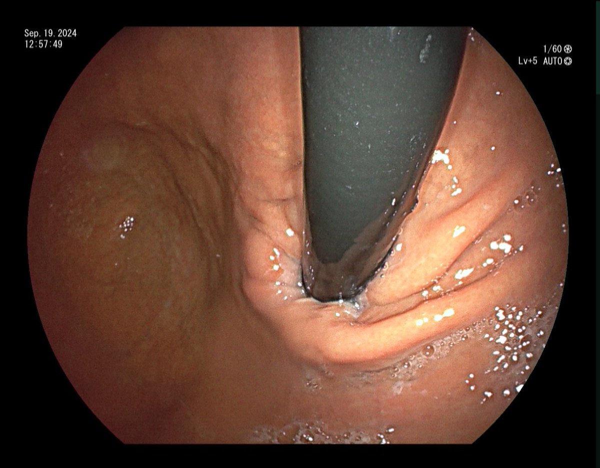
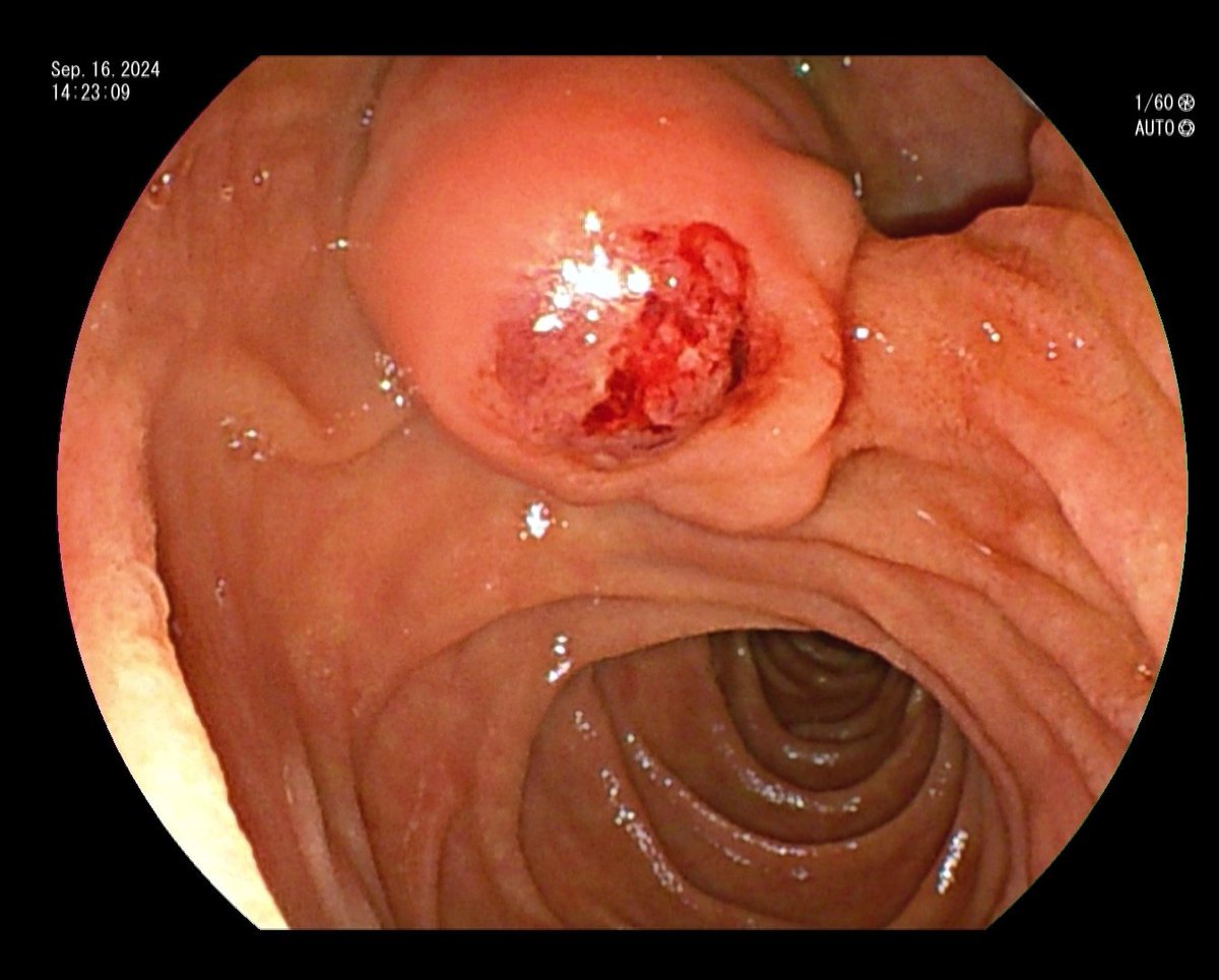
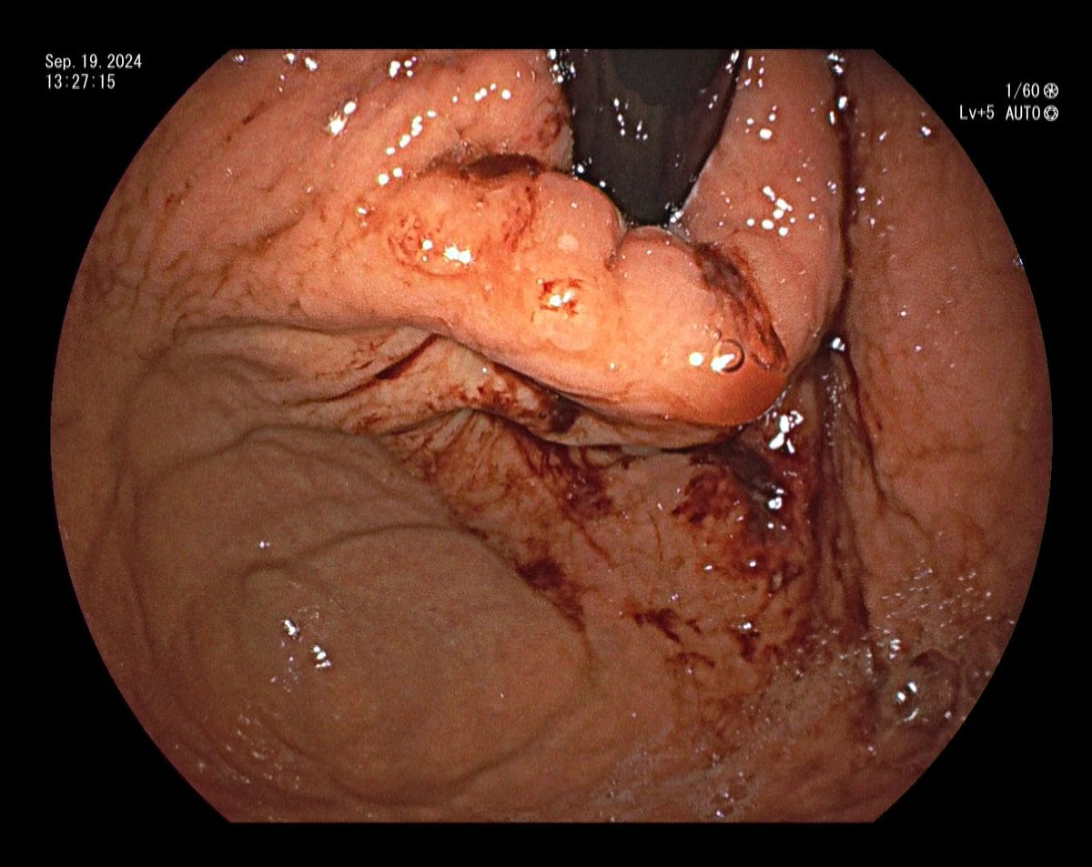
70 y/o presented with dysphagja and weight loss, found to have poorly differentiated gastric adenocarcinoma with signet ring cell features. EGD showed invasion into the esophagus with EUS showing serial involvement. (1/3)
Melena and Hgb drop after liver biopsy should raise the concern for hemobilia, clots in the bile duct can cause obstructive jaundice, abd pain, (Quincke's triad). Treatment can be covered metal stent, but more often, localizing the bleed and IR embolization.
Pt underwent liver biopsy for concerns of transplant rejection, started having melena and recurrent hemoglobin drop. Acute increase of bilirubin to 7. Initial view of the papillary showed oozing blood. . .(1/5)
A repeat cholangiogram did not show contrast extravasation, but did reveal improved opacification of the proximal ducts. Pt was started on pRBC and sent to CTA with IR consultation. (4/5)
@Nicolascole77 Very rarely is something created from a vacuum, scientists, writers, musicians, directors all borrow ideas from one another and have influences that impact their work.