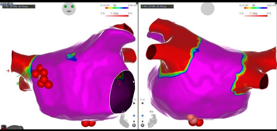
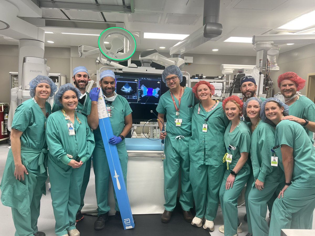
Not every persistent AF case stops at PVI ⚡
Arian Sultan maps substrate before ablation and identifies anterior + posterior wall disease before touching the veins
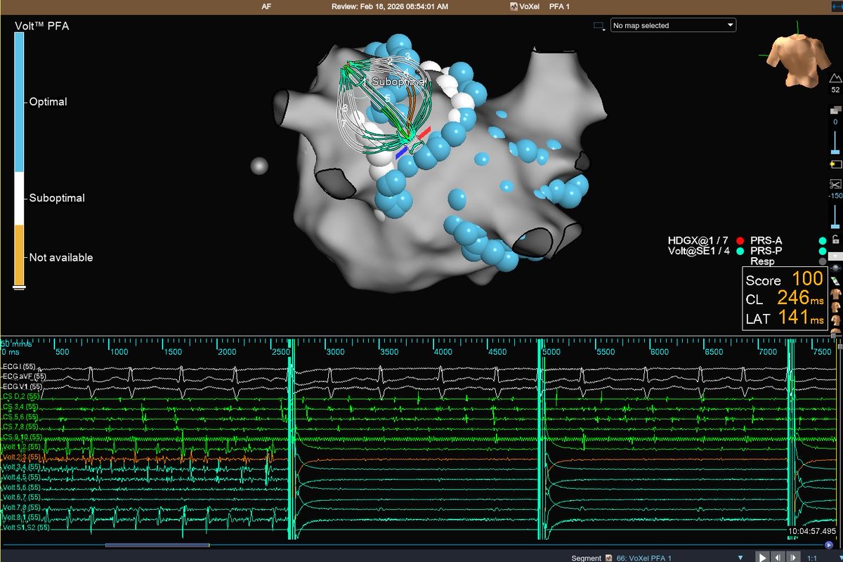
Real-time contact feedback from the Volt™ system helps guide every application
(1) We have cases where PVCs are not produced on the table, particularly after sedation. @drrakeshg1 and I were able to prove a theory I have held for years regarding the MOA of outflow tract PVCs. Our workflow allows for identification of the trigger site when PVCs are absent.
Can balloon technology do more than PVI?
Case led by Arian Sultan and discussion with Roland Tilz.
Selective electrodes, controlled overlap, and targeted energy delivery help extend ablation strategies beyond the veins.
Here they highlight a smarter way to personalise lesion sets.
#EPeeps
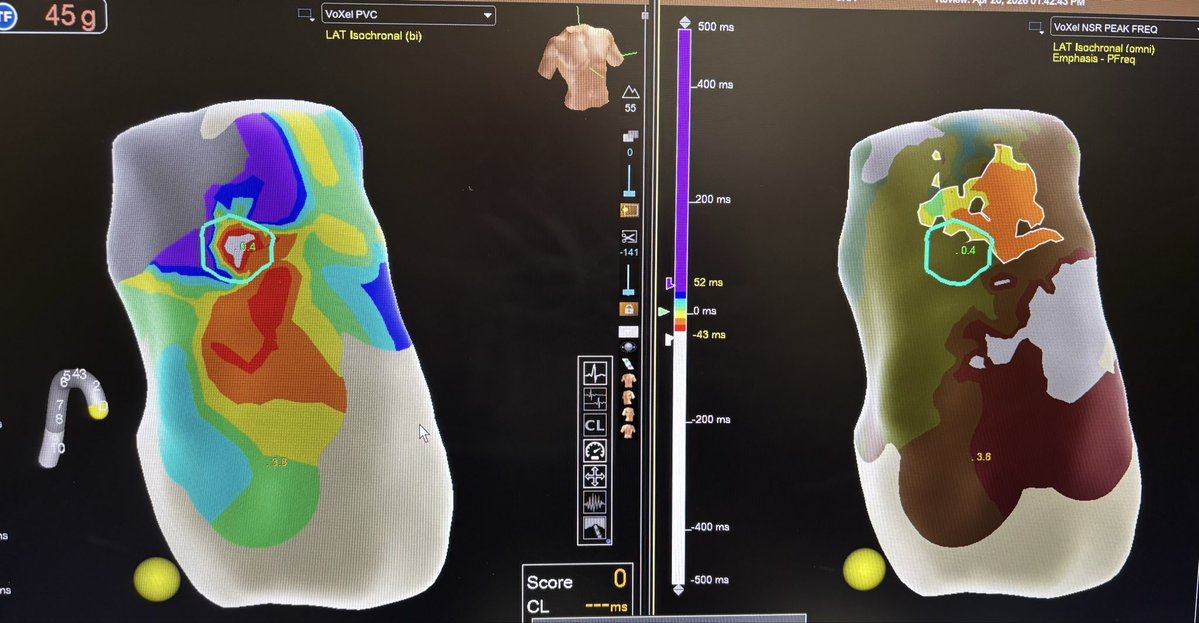
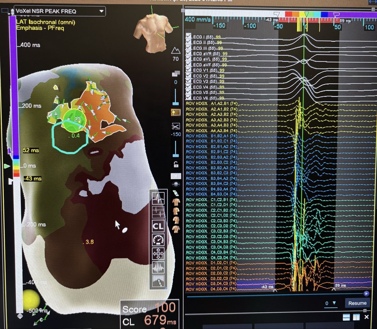
Fresh from #HRS2026 by @drrakeshg1 using Peak-frequency SR mapping to localize PVCs. We applied it on an RVOT PVC today #EnSiteX.
Pic 1: LAT map of PVC (L), Peak Freq map in SR (R)
Pic 2: Successful RF site (teal tag)
Pic 3: Reference poster
@GoArmyChris@Coops_Squigles
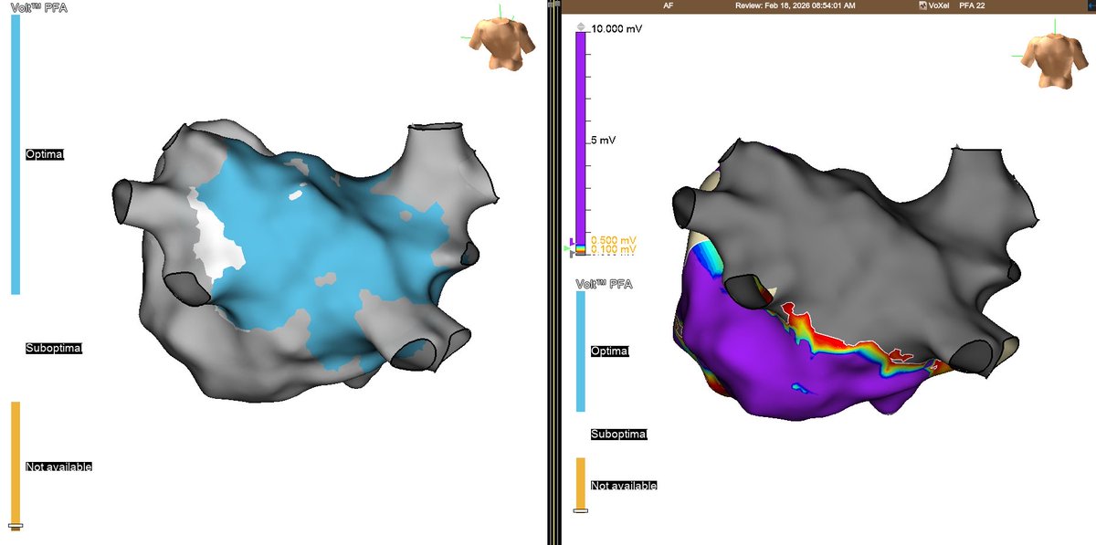
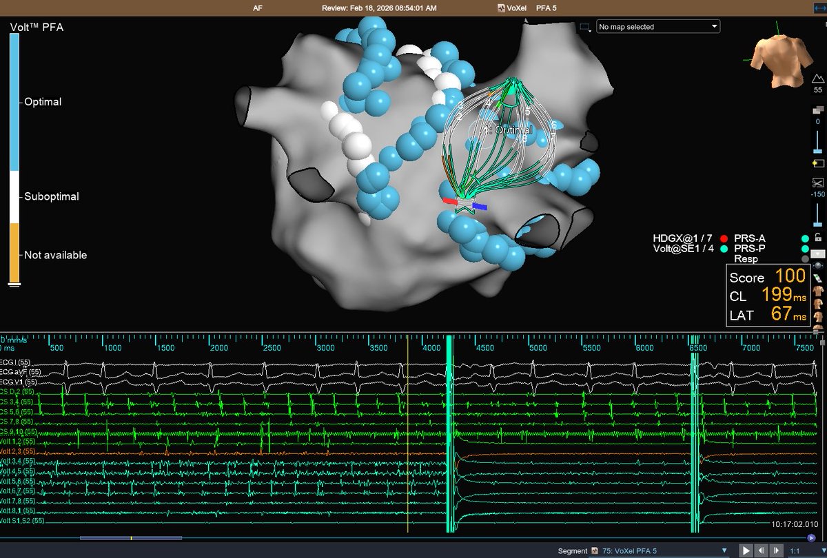
The #VoltPFA insights keep coming!
Late-breaking results on Volt PFA System PVI durability and patterns help answer the question: does the pulse field ablation system matter?
Explore the data straight from #HRS2026 ⬇️
Safety Info: https://t.co/d6tUnQ2Cuh
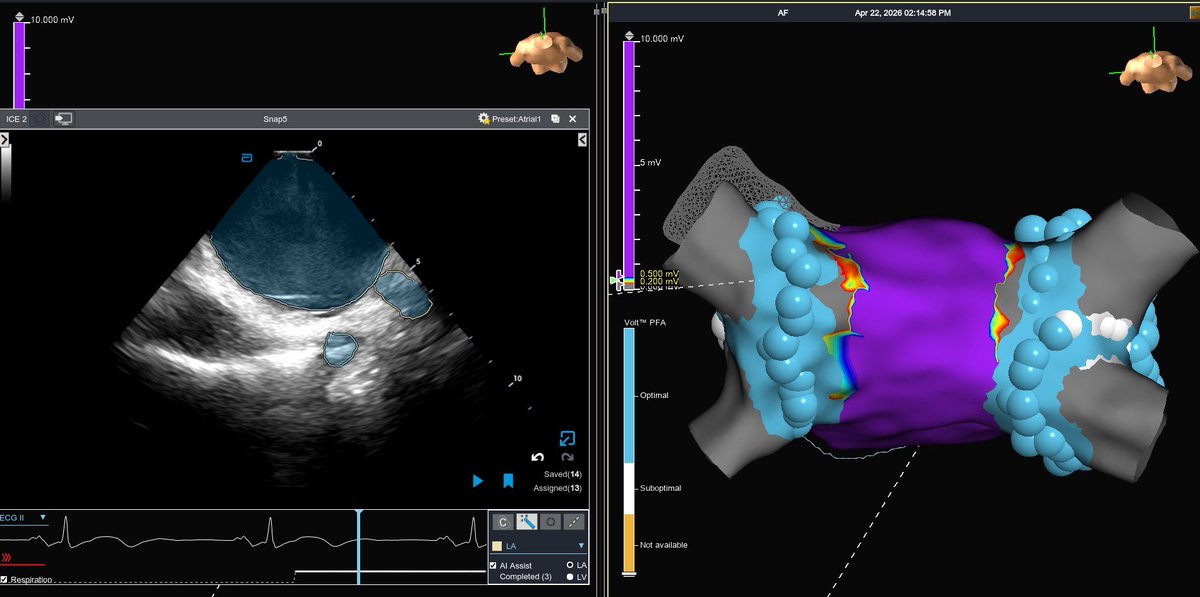
Excellent in-booth session with Dr. JRWinterfield this afternoon on Auto geo creation of the LV with #ViewFlex X ICE catheter at #HRS2026!
Safety Info:
https://t.co/PjGcZvuN8q
https://t.co/rSkvxxZF7U
#AbbottProud
Effects of Pacing Sites on Substrate Mapping Using Decrement-Evoked Potential Mapping for Scar-Related Ventricular Tachycardia #OpenAccess
https://t.co/GWYsXPKks1
Open access books can be downloaded using this QR code. Content also appears weekly in @JACCJournals Thank you to @HRSonline for making this a special annual component of HRS for all EP’s.
CLEAR-VT
Interesting results of PFA in VT.
High efficacy … but still room to improve on safety. Are we just taking what works in the atrium to the ventricle? slide on perspective taken from the comment.
An excellent first in US experience with Volt 2.0 with ICE integration. Having used this technology in trial, LMR, and now 2.0, the newest version adds excellent maneuverability to a refined, effective, and safe platform. Bravo @AbbottCardio@uyaron@JoeProvost7
EHRA 2026❗️come see Dr Javier MORENO @Arritmias_HRC and Dr Ivo ROCA @ivroca discussing their workflows using new TACTIFLEX DUO on VT ABLATION (Sunday 5-6pm) and COMPLEX ARRHYTHMIA ABLATION (Monday 12-1pm) at Tutorials area. Don´t miss out‼️we will have fun!!😀
2022: 1st AF ablation (another hospital).
2023: recurrence. Pentaray showed completely isolated PVs. CNA due to vagotonia.
2026: recurrence. HD-Grid map showed LSPV and RSPV reconnection.
Question: Did the pentaray fail to show EGMs due to bipolar blindness? #epeeps#ensitex
Dr. Dhanunjaya Lakkireddy walks through how myocardial inflammation, micro-scar formation, & Purkinje system involvement can explain PVCs.
Full discussion on the MedicalVisual Clinician Hub: https://t.co/c0KRzVwKm3
@DJ_Lakkireddy#Myocarditis#VentricularArrhythmias
As described by Wilber et al, in a subset of patients with inferior MI, "an isthmus of surviving tissue between the scar border and mitral annulus appears to constitute a critical region of slow conduction." This isthmus provides a target for VT ablation. https://t.co/ltMc1s5PHr
Refining activation maps of atrial tachycardia!
👇
Near field EGM detection with peak frequency emphasis highlighting the critical site of reentry.
#EPeeps