تم انتقالي إلى مستشفى الدكتور سليمان الحبيب الجديد في الفيحاء و اغلاق عيادتي للأشعة التداخلية في مستشفى السلامة. سنوافيكم بإذن الله في حال تم افتتاح العيادة في المكان الجديد.
الرياضة ليست مجرد ساحة للمنافسة، بل هي منصة قوية لصقل الشخصية وفتح الآفاق نحو مجتمع أكثر صحة وتماسكاً. من خلال تعزيز الروح الجماعية وتوطيد العلاقات، تصبح الأنشطة الرياضية حجر الزاوية في بناء مجتمعات مترابطة ومتفاعلة.
أظهرت الأبحاث المتقدمة أن الانخراط بالأنشطة الرياضية يمكن أن يغير بنية الدماغ ويعزز من قدراته على مواجهة الضغوط النفسية. إدراج الرياضة ضمن الروتين اليومي لا يُعد مجرد نشاط بدني بل استثمارا حقيقي في الصحة.
ضمن خطط رؤية المملكة 2030 🇸🇦 تأتي الرياضة كأحد أعمدة النمو الوطني، مع التركيز على تطوير البنية التحتية الرياضية وتحفيز المجتمع بأسره للمشاركة الفعّالة. هذا يعكس استراتيجية المملكة لتنشئة جيل يتسم بالقوة والنشاط والحيوية.
تحت مظلة رؤية 2030 تسعى المملكة لتعزيز نمط حياة صحي ونشط بين أبنائها، مع التأكيد على أن الرياضة تساهم في تعميق النسيج الاجتماعي وتعزيز الصحة العامة. هذه الخطوة تستهدف تطوير مجتمع متكامل يشارك في صنع مستقبل مزدهر.
تقف الرياضة كركيزة للتواصل الحضاري والتفاهم المجتمعي، وتوفير فرص للشباب لبناء مستقبلهم وتحقيق أحلامهم.
#الديوان_الملكي: استكمل #خادم_الحرمين_الشريفين الفحوصات الطبية التي أجريت في العيادات الملكية في قصر السلام بجدة، وقد تبين وجود التهاب في الرئة، وقرر الفريق الطبي خضوعه ـ حفظه الله ـ في قصر السلام بجدة لبرنامج علاجي عبارة عن مضاد حيوي حتى يزول الالتهاب بحول الله.
#واس
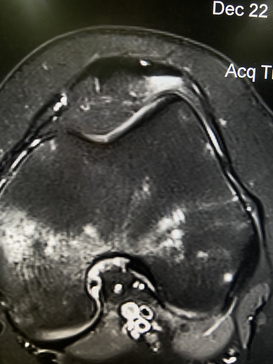
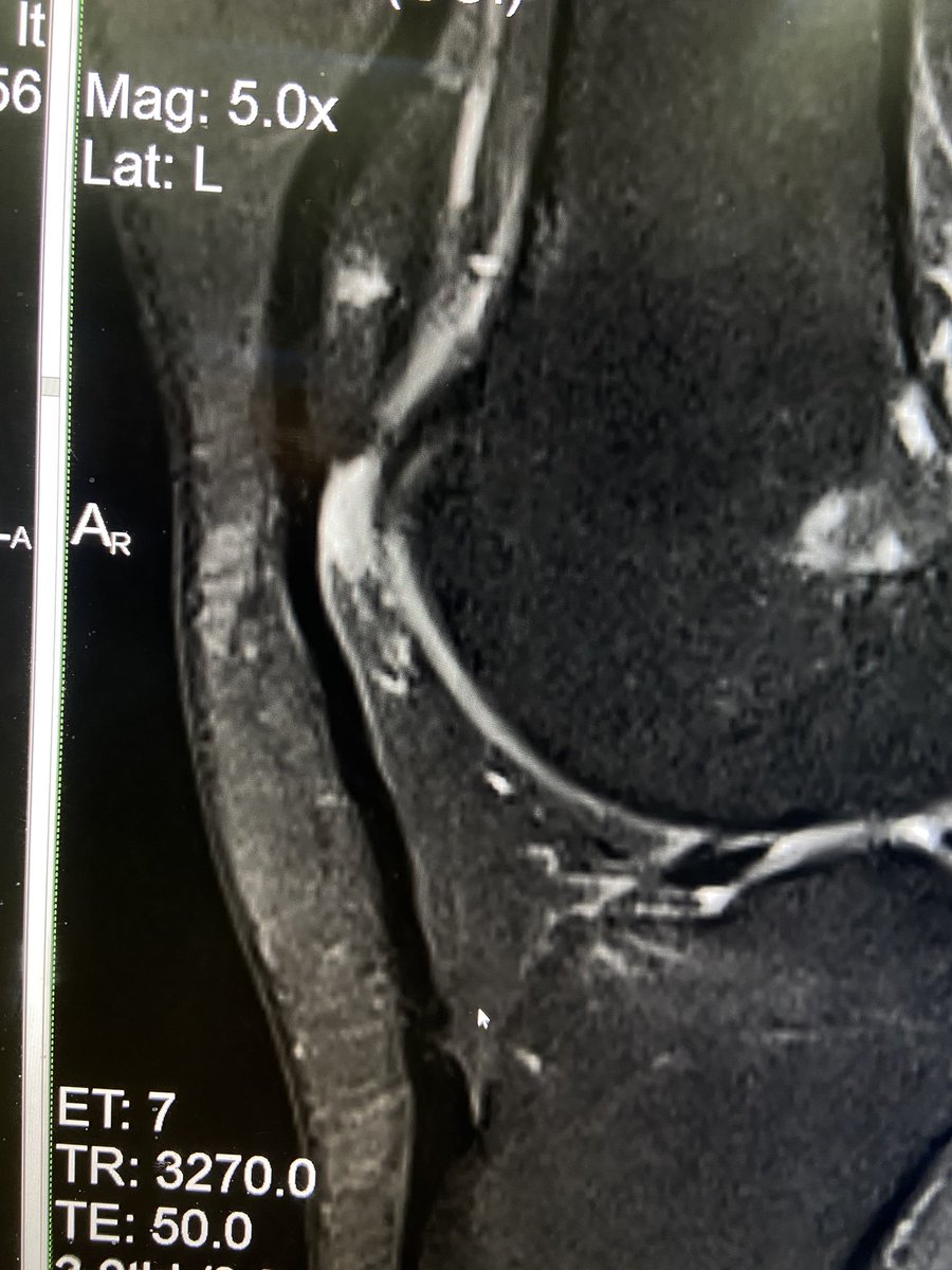
9/4/2024.🦶 Thick walled apparently chronic distal intermetatarsal bursitis in the 3rd space. The main differential: Morton´s neuroma. XRAY, MRI and US correlation. #MSKRadiology#MRI#Ultrasound#Orthopedics#MedTwitter🟢
Struggling with Musculoskeletal imaging?
Don’t worry
@OrthopedicTeam will make it easy for you 🦴
We bring you a systematic approach to master image reading of Musculoskeletal system 🩻
Presented by Dr.Thamer Alghamdi @_drthamer
Do not miss the opportunity ✨
Registration link:
https://t.co/h4oMNvqCcb
#UQUMSC2024
ان يختصك احد مراجعيك القدامى بإتصال تهنئه، تعلم حينها ان جودة حياته قد اختلفت تماما ويعلم هو يقينا بذلك ويحمل بقلبه الفضل لك بهذا التغير بعد فضل الله وكرمه، الحمدلله من قبل ومن بعد واسال الله لي وللجميع العفو والعافيه.
The scapular ‘Y’ view is excellent for detection of coracoid fracture at the shoulder!
[in this case not seen on AP internal / external rotation views]
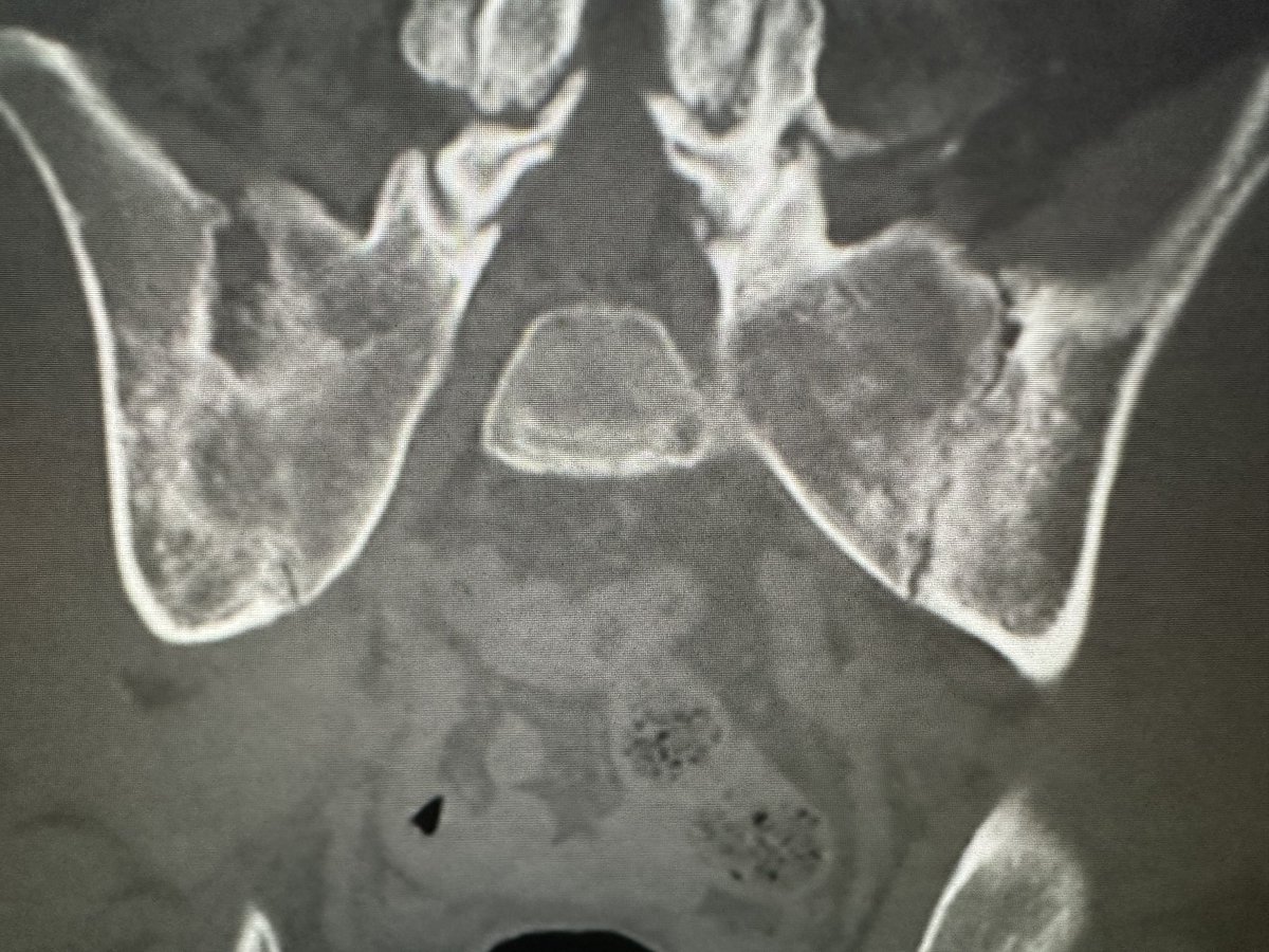
Please, Body readers who quickly skim the bones at the end and overlook sacroiliitis cases like this. This pt could have been diagnosed nearly 7 years earlier had reader not overlooked the SI joints here. Was quite advanced at that time but still.
Feel BACKED into a corner reading spine MRIs?
Looking for a better way to grade spinal stenosis?
Time to get BACK to the basics!
Want a grading system that’s both easy & reproducible? I’ve got your BACK!
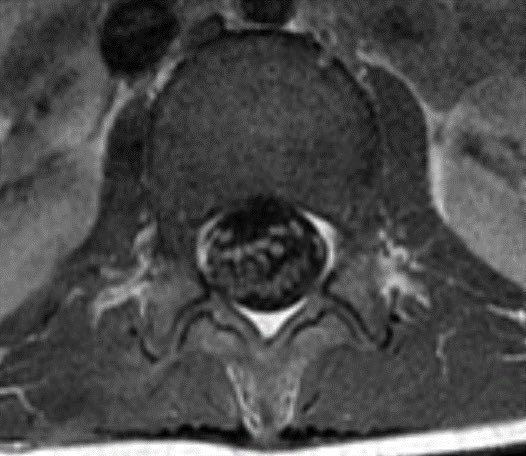
Here’s the lumbar stenosis grading system you’ve been waiting for!
➡️Think of it functionally. Nerves need to fit in their space, like you fit in clothing.
🔸Mild stenosis is like comfy clothes—no squeezing.
🔸Moderate stenosis clothing isn’t loose, but there isn’t extra room either.
🔸Severe stenosis is like too tight jeans, your body gets compressed
➡️How do we tell if the nerves have enough room—if the clothing fits loosely, tight, or too tightly?
We look at the space around them.
🔸For the canal, it is CSF—if there is enough room, the extra space will be filled by CSF.
🔸For foramina, it is fat—extra room is filled by the foraminal fat.
➡️For the canal:
🔸Mild stenosis: Mild CSF attenuation, but still plenty of around, like plenty of room in comfy sweat pants
🔸Moderate stenosis: Less CSF & nerve roots appear aggregated. Like club clothes, not much room between skin & clothing
🔸Severe stenosis: It’s too tight jeans. Canal doesn’t just hug up to the nerve roots, it compresses them.
➡️For the foramina:
🔸Mild stenosis: Lose fat on 2 sides. Still comfy bc fat is preserved on the other 2 sides.
🔸Moderate stenosis: Lose fat on all 4 sides, but nerve is not compressed. Like a sleek outfit, it shows curves, but doesn’t deform them.
🔸Severe stenosis: Fat is gone & nerve is compressed. Like trying to fit into jeans from high school--squishing in everything you can to get it to fit.
These systems both named the Lee systems. These are easy to remember, make sense, and are based in literature.
*⃣Here are the references:
🔸Lee et al. AJR Am J Roentgenol. 2010 Apr;194(4):1095-8
🔸Park et al. Br J Radiol. 2013 May;86(1025):20120180
Hopefully, now when reading lumbar spine MRIs, these grading systems will get you BACK in the groove!


![morrisonMSK's tweet photo. The scapular ‘Y’ view is excellent for detection of coracoid fracture at the shoulder!
[in this case not seen on AP internal / external rotation views] https://t.co/JENnu0ctHt](https://pbs.twimg.com/media/FfUczO9XkAAnWTV.jpg)