We should know the gravity (dependent) side of the lesion before starting resection, why?
Compare ESD to removing a door from its frame.
Which part is hardest to remove?
The side attached to the hinges.
In ESD, think of the gravity (dependent) side as the hinge.
If you start dissecting from the anti-gravity side, the specimen falls toward the dependent side—making the “hinged” portion progressively harder to access.
🏦So remove the screws first.
🔑 Start your mucosal incision/dissection from the GRAVITY side.
As the lesion becomes free, gravity can then work for you rather than against you.
So after the correct incision, more of the submucosa will be exposed.
PS: This is my understanding, not endorsed by anyone (including ChatGPT)
Methylene blue is the best agent for gastric #chromoendoscopy. It gets absorbed by the intestinal metaplastic cells and cannot be washed out, unlike in the colon.
Because it also gets absorbed by normal intestinal cells, patients will pee blue that day💧
#medtwitter#gitwitter
✨ إنقاذ حياة لهذا الإسبوع:
🧓 مريضة مسنّة.
التاريخ الطبي: تليف الكبد وسرطان الخلايا الكبدية 🦂 (Child C، MELD 18).
عانت من فقر الدم، ونزف معوي تغوطي (أعزكم الله)، وقيء دموي🩸.
أظهر التصوير المقطعي دوالي المعده متضخمة.
كشف المنظار عن نزيف نشط في الجهاز الهضمي العلوي 💢.
تم إحالتها إلى الأشعة التداخلية والقسطرة ☢️، وتم إجراء💉 غلق وريدي عكسي بإستخدام البالون الطبي ولفيفة معدنية والحقن بالصمغ الطبي🧪 (BRTO/CARTO) بنجاح.
توقف النزيف من الجهاز الهضمي العلوي في اليوم التالي بشكل كامل ⚕️.
خرجت المريضة إلى المنزل بصحة جيدة 🪩.
يستطيع تخصص الأشعة التداخلية المساعدة دائما❗
#الأشعة_التداخلية #سرطان_الكبد #دوالي #المعده #المناظير
To cover a bleeding point (varices) or perforation, sinply deploy at cardia , and pull back scope in 1:1 fashion. Don’t even need to touch the catheter.
At TSGE: checkout the @bostonsci Agile TTS FCSEM stent.
5 years ago new covered metal stents were made to pass through the scope (tx gastroscope 3.7 mm), allowing for direct visualization , no need for fluoro! Great for refractory variceal bleeding @ ICU!
#GITwitter
Mastering ERCP technique:
A critical anatomical review.
• Pancreatic duct stent integrity confirmed
• No sphincter laceration observed
• Key anatomical structures:
- Bile duct
- Pancreatic duct
- Septum
Optimal positioning: Duodenum to left on screen (anatomic right) for accurate cannulation and imaging correlation.
The @STERISEndoscopy Raptor grasping forcep can rotate. If you place a 30 degree “bend” it rotates/ reaches more angles than before in endosocpy.
Laparoscopic instruments have this bend and for good reason! Look to your surgical colleagues for innovation!
#GITwitter
I always encourage the gen GI fellows to join during ESDs as there’s a lot of tools/tricks to learn
Example: Control of active bleeding w coag grasper
✅ Irrigate continuously to locate the spot
✅ Grasp - bleeding should stop if you’ve got the vessel
✅ Pull away gently & burn
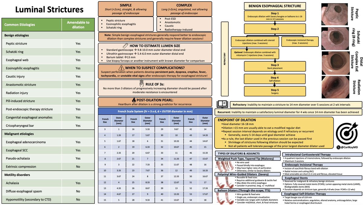
Luminal Strictures
▪️Benign vs. malignant
▪️Simple vs. complex
▪️How to estimate lumen size
▪️When to suspect complications
▪️“Rule of 3s”
▪️👀post-dilation heartburn
▪️French scale
▪️Definition of refractory & recurrent
▪️Endpoints 🥅
▪️Types of dilators & adjuncts
#GITwitter